Abstract
First Person is a series of interviews with the first authors of a selection of papers published in Disease Models & Mechanisms, helping early-career researchers promote themselves alongside their papers. Chady Hakim is first author on ‘ Extensor carpi ulnaris muscle shows unexpected slow-to-fast fiber-type switch in Duchenne muscular dystrophy dogs’, published in DMM. Chady is a Research Assistant Professor in the lab of Dongsheng Duan at the University of Missouri, Colombia, MO, USA, investigating the preclinical development of gene therapy for Duchenne muscular dystrophy (DMD), with a particular interest in using the canine DMD model.

Chady Hakim
How would you explain the main findings of your paper to non-scientific family and friends?
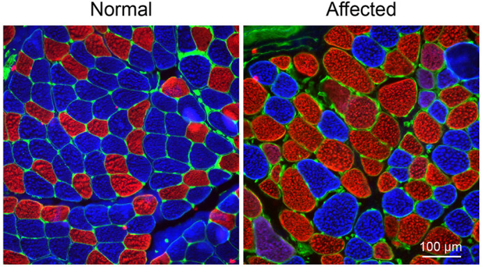
DMD is a severe muscle disease caused by dystrophin deficiency. Loss of dystrophin leads to muscle degeneration and remodeling, and eventually to muscle death and replacement by fatty and fibrotic tissues. The canine DMD model shares clinical and pathophysiological similarities to that of human patients. Therefore, studies performed with the canine model provide critical insight into understanding muscle disease in DMD. In this study, we were first interested in developing a force assay platform to evaluate the contractile force and characterize the kinetic properties of a single muscle in the canine DMD model. We focused on the extensor carpi ulnaris (ECU) muscle from the forelimb muscle group. As expected, we saw a loss of muscle force in affected dogs. Surprisingly, we observed an unexpected contractile kinetic profile. It has been well established that the dystrophic muscle undergoes a fast-to-slow fiber-type switch. This led us to predict that the affected muscle would exhibit slow contraction and relaxation. Surprisingly, we saw just the opposite. There was a decrease in the time taken to reach peak tension and relax the affected ECU muscle, indicating a faster contraction and relaxation. Additional characterization of myofiber-type composition in the normal and affected ECU muscle confirmed the kinetic assay results.
“[…] studies performed with the canine model provide critical insight into understanding muscle disease in DMD.”
What are the potential implications of these results for your field of research?
The unexpected slow-to-fast myofiber-type switch highlights the complexity of muscle remodeling in dystrophic large mammals and paves the way for better utilizing dystrophic canines as a preclinical model in the study of DMD pathogenesis. Additionally, the fiber-type switch phenomenon offers a unique entry point for (1) investigating the molecular mechanism(s) that lead to this phenomenon and how it directly correlates to the loss of dystrophin, and (2) evaluating the pathophysiological implications for muscle strength and in determining whether this is unique to canine muscle. Most importantly, these results have significant implications for therapeutic approaches to DMD, such as gene replacement and editing, and evaluating their efficacy in correcting the fiber-type switch.
What are the main advantages and drawbacks of the model system you have used as it relates to the disease you are investigating?
When initiating this study, our goal was to evaluate muscle strength in the affected dogs. To achieve this goal, we developed an all-in-one automated in situ force assay platform. This novel platform has several advantages. First, we designed all the components to be adjustable to meet the need for studying muscles at different anatomic locations or with different sizes. Our design was also made with the consideration to adopt the platform to accommodate other large animal models besides the canine. Second, we developed a detailed protocol to optimize the stimulation parameters, allowing the muscle to reach its optimal force during contraction. This allowed the comprehensive evaluation of the contractile and kinetic properties of a single muscle. Together, this novel platform offers a unique ability to correlate the physiological findings with the molecular, cellular, biochemical and histological changes in a single muscle. This ability is critical for evaluating preclinical intervention studies. Unfortunately, this is a terminal assay, limiting the investigators to follow disease progression and therapeutic response in the same animal over time.
What has surprised you the most while conducting your research?
It is well established that the dystrophic muscle undergoes a fast-to-slow, rather than a slow-to-fast, transition in fiber type. In this study, we observed the opposite in affected canine muscles as they were mainly composed of the fast fiber type. The underlying mechanism of this fiber-type switch needs to be further investigated so it can be determined whether it is unique to canine muscle. Furthermore, muscles that are mainly composed of the fast fiber type are characterized by a higher force, higher contraction and relaxation rate, and less time needed to achieve full contraction and relaxation. In affected dogs, we noticed a reduction in the time taken for contraction and relaxation. Surprisingly, the force, contraction rate and relaxation rate were significantly reduced in the affected muscle compared to the normal muscle.

Representative myosin heavy chain isoform immunostaining photomicrographs of a normal (left) and an affected (right) ECU muscle. Blue, type I myofiber; red, type IIa myofiber; magenta, type I/IIa hybrid myofiber; green, laminin immunostaining
Describe what you think is the most significant challenge impacting your research at this time and how will this be addressed over the next 10 years?
Unlike other genetic diseases, DMD is very challenging to treat. First, it is caused by mutations in the second largest gene in the body. The large size of the dystrophin gene makes it impossible to replace it through the gene replacement approach unless a truncated gene with a similar function to the full-length gene is used. Second, DMD affects every muscle type in the body, making a whole-body treatment necessary to achieve a complete cure. Gene therapy using adeno-associated virus (AAV)-mediated CRISPR/Cas9 gene editing shows much promise for treating DMD. It allows for the restoration of a near full-length dystrophin protein without the need for using a highly truncated microgene that can only result in limited function rescue (Hakim et al. 2018). We recently showed that AAV CRISPR therapy resulted in efficient dystrophin restoration in affected dogs but resulted in a Cas9-specific immune response that eliminated the edited cell. This unfortunate response is a critical barrier to advancing CRISPR therapy into clinics. With further advances in gene therapy, combined with advances in the understanding of CRISPR genome editing and how to evade the Cas9-specific T-cell response, AAV CRISPR therapy will be suitable for treating DMD.
What changes do you think could improve the professional lives of early-career scientists?
I believe it is critical for early-career scientists to collaborate and interact with other related research fields. This empowers and extends their knowledge, and also has a positive influence on their research focus. Before I started my PhD, I had gained some knowledge about muscle physiology. I wanted to extend this knowledge in my PhD studies by building a bridge between muscle physiology and molecular biology, and the perfect application was the field of DMD gene therapy. Through my previous experience, I was able to develop tools, such as the platform presented in this study, to answer critical molecular questions in the field of gene therapy. As a matter of fact, the outcome observation of the fiber-type switch was a result of analyzing the kinetic properties of the affected muscle force. This observation will now become an important biomarker in the evaluation of the efficacy of novel therapy.
“I believe it is critical for early-career scientists to collaborate and interact with other related research fields.”
What's next for you?
With the novelty of the data presented in this study, I'm excited about finding out whether gene therapy approaches would correct the fiber-type switch and reverse the remodeling observed in the affected canine muscle. I'm currently working with my mentor Dr Dongsheng Duan to evaluate fiber-type composition-affected canine muscles treated with gene therapy.
References
- Hakim, C. H., Yang, H. T., Burke, M. J., Teixeira, J., Jenkins, G. J., Yang, N. N., Yao, G. and Duan, D. (2021). Extensor carpi ulnaris muscle shows unexpected slow-to-fast fiber-type switch in Duchenne muscular dystrophy dogs. Dis. Model. Mech. 14, dmm049006. 10.1242/dmm.049006 [DOI] [PMC free article] [PubMed] [Google Scholar]
- Hakim, C. H., Wasala, N. B., Nelson, C. E., Wasala, L. P., Yue, Y., Louderman, J. A., Lessa, T. B., Dai, A., Zhang, K., Jenkins, G. J.et al. (2018). AAV CRISPR editing rescues cardiac and muscle function for 18 months in dystrophic mice. JCI Insight 3, 124297. 10.1172/jci.insight.124297 [DOI] [PMC free article] [PubMed] [Google Scholar]


