Abstract
Autologous fat grafting is commonly used for soft tissue augmentation and reconstruction, this technique is limited by a high rate of graft absorption. The stromal vascular fraction gel (SVF-gel) grafting for facial volume augmentation can exert a positive effect on skin rejuvenation, but its major limitation is the low rate of conversion of Coleman fat. The purpose of our study was to investigate a novel surgery using performing high-density fat in combination with SVF-gel in the treatment of hemifacial atrophy, or Romberg diseases. From October 2017 to October 2019, 13 patients with hemifacial atrophy underwent high-density fat transfer with SVF-gel injection. The outcome was determined by the difference in presurgery and postsurgery FACE-Q modules (FACE-Q conceptual framework: 1, Satisfaction with Facial Appearance; 2, Health-Related Quality of Life; 3, Negative Sequelae; 4, Satisfaction with Process of Care), which were designed as patient-reported outcome instrument to evaluate the unique outcomes of patients undergoing facial cosmetic procedures.
The excellent cosmetic results were observed during follow-up periods, with no adverse events was seen in the treatment group. All patients showed improvements in facial augmentation and contour. In patients with facial volume loss, high-density fat transfer with SVF-gel facial injection resulted in significantly higher improvement scores and better patient satisfaction. The patient-reported FACE-Q modules presurgery and postsurgery results showed statistically significant improvement (P < 0.05). This high-density fat in combination with SVF-gel is an effective method of correcting the facial volume loss that leave no complications during follow-up, having a satisfactory volumization effect. This could largely facilitate the clinical utilization of fat.
Keywords: Facial deformities, high-density fat, stromal vascular fraction gel
Autologous fat grafting has been 1 of the most frequently and effectively performed procedures for obtaining the desired symmetrical facial appearance.1,2 It is available in easily accessible subcutaneous depots but also can be molded to reconstruct defects, especially in terms of patients with mild to moderate forms.3 Traditional fat transplantation often has unreliable long-term results because of absorption and volume loss.4 As a result, several different techniques of lipofilling have been developed in recent years.5
Clearly, how to determine an easy and reproducible method that can yield higher concentrations of preserved adipocytes and guarantee long lasting results is the crux. The recognition that fat contains multipotent stem cells that can be harvested through liposuction without altering their viability has driven further examination into the potential uses of fat and its adipose-derived stem cells (ADSCs).6,7 The application of stromal vascular fraction gel (SVF-gel), which has a high density of ADSCs, was effective in skin rejuvenation. Stromal vascular fraction -enriched lipotransfer technique was applied in the treatment of the face, but its major limitation is the low rate of conversion.8 Patients with mild to moderate deformity were typically treated with serial fat grafting procedures in our patient group. In this article, the authors introduce high-density fat grafting procedure associated with SVF-gel technique in the management of volumetric deficit of the face in volume loss.
PATIENTS AND METHODS
The high-density fat grafting procedure associated with SVF-gel technique method was performed 13 patients with hemifacial atrophy (9 females and 4 males, with a mean age of 33 years) between 2017 and 2019. They are 10 mild and 3 moderates. Before the treatment, all patients were counseled that they may need multiple fat graft procedures to achieve satisfactory effect and the follow-up period were 7 to 15 months.
All patients were clearly informed of the benefits, risks, operative complications, and postoperative care and gave their informed consent to the intervention. Furthermore, all patients were submitted to the surgery in hospital environment. The clinical final results were assessed by observation, examination with palpation, and preoperative and postoperative photographs.
SURGERY DESIGN AND PROCEDURE
Surgical Procedure
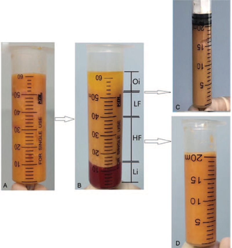
The patients were placed under general anesthesia after thorough preoperative evaluation and counseling. Human abdominal lipoaspirates were obtained from abdominal lipoaspirates after informed consent and our hospital ethics committee approval. The high-density fat and SVF-gel were prepared as the manufacturer's instruction. Briefly, liposuction was performed with a 3 mm multiport cannula. The liquid portion was then discarded and the fat layer was then centrifuged at 1200 g for 3 minutes to generate Coleman fat. After this, the inferior layer, composed mostly of blood, was thrown out and the remaining fat was the Coleman fat. We regard the upper as low-density fat and lower layers as high-density fat. The low-density fat then transferred between 2 syringes (6–8 times) at a rate of 20 mL/s until the fat converted into a uniform emulsion. After processing, the fat turned into an emulsion and was then processed by centrifugation at 1600 g for 3 minutes, was identified as SVF-gel. After discarding the superior and inferior layers of washed lipoaspirate, the middle layer, which is routinely used for adipose tissue graft, was collected for use (Fig. 1).
FIGURE 1.
Procedure for processing high-density fat and SVF-gel. (A) Sediment fat. (B) Coleman fat. (C) SVF-gel. (D) High-density fat. HF, high-density fat; LF, low-density fat. Li, liquid; Oi, oil; SVF-gel, stromal vascular fraction gel.
In cases of hemifacial atrophy, a solution of saline and 0.5% lidocaine with 1:200,000 epinephrine was injected subcutaneously fanwise around the areas to be treated with lipofilling. In undermined areas of the midface, lipofilling was injected in the sub-superficial musculoaponeurotic system layer, at the same surgical time. In the intact areas, lipofilling was executed in subcutaneous layer. Aimed at carrying out the fat implantation, small scalp incisions were made with an 18G needle and the mechanically high-density fat was injected into the target sites. The fat was injected using a blunt-tipped Coleman cannula connected to syringe, using a multichannel technique, retrogradely in each movement. Fat was homogeneously deposited in the subcutaneous level in multiple layers to achieve volume augmentation in these cases, always avoiding overcorrection. The SVF-gel injection was performed intra- and/or subdermally and injected into the dermal layer with 27-gauge needles.
Statistical Analysis
The SPSS 19.0 Data Analysis System was used for data analysis. Normal distribution and homogeneity tests were performed on all data. Data are expressed as mean ± standard deviation. Statistical significance was calculated using the paired t test. Two-sided values of P < 0.05 were considered statistically significant.
RESULTS
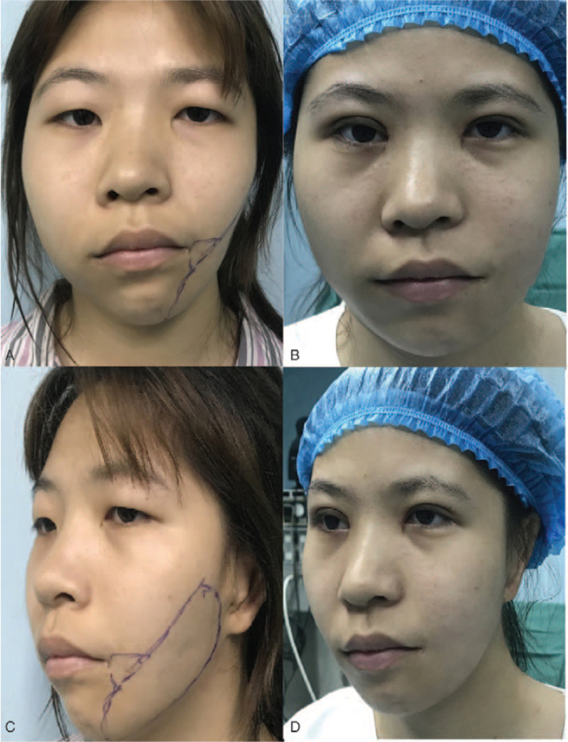
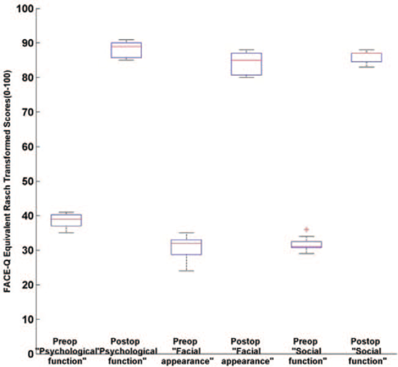
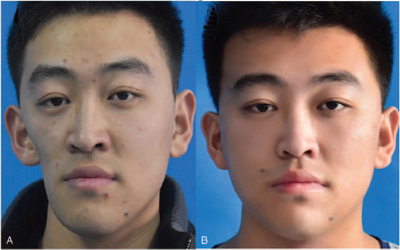
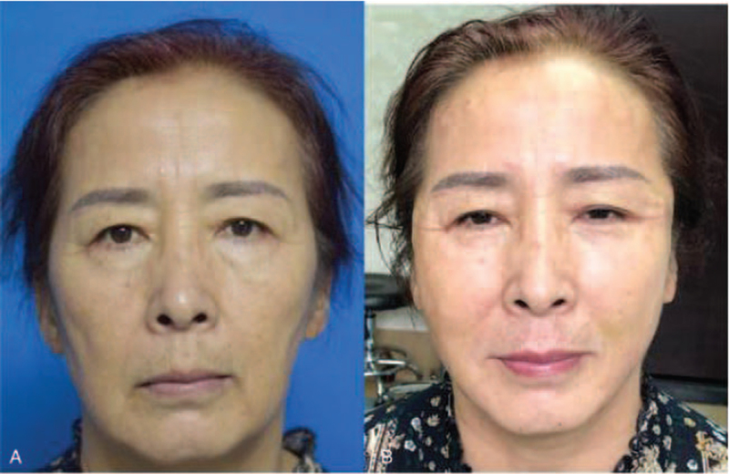
A total of 13 patients (9 females and 4 males, with a mean age of 33 years) were included in the study. The average follow-up period was 10.3 ± 2.6 months (range 7–15 months). Slight bruising and swelling was seen during the first week after surgery but no obvious contour irregularity. No further complications were evidenced during follow-up (Figs. 2-4). In the cases of hemifacial atrophy, the mean volumes amount of high-density fat and SVF-gel for face used overall were 35.6 and 8.4 mL, thus, the ratio was approximately 4.2:1. Satisfaction with overall facial appearance improved from scores of 30.7 ± 3.1 to 84.2 ± 3.0 (P < 0.05) whereas satisfaction with psychological function improved from scores of 38.5 ± 2.1 to 88.3 ± 2.4 and social function from scores of 31.8 ± 1.9 to 85.9 ± 1.8, respectively (for both, P < 0.05) (Fig. 5). Both patients and the surgeon were more satisfied with the cosmetic results of fat graft. After 2 years, 2 patients needed a second fat graft in order to improve the results.
FIGURE 2.
A woman patient with hemifacial atrophy underwent high-density fat grafting procedure with SVF-gel technique. (A-C) Front view, oblique view of preoperative face. (B-D) Front view, oblique view of the face 10 months after the surgery. The patient was without a second fat graft. SVF-gel, stromal vascular fraction gel.
FIGURE 5.
The result of FACE-Q scores comparing preoperative and postoperative psychological function, facial appearance and social function (P < 0.05). Data represent group means with minimum and maximum scores.
FIGURE 3.
Patient with hemifacial atrophy had high-density fat in combination with SVF-gel for the cheek filling. (A) Front view of preoperative face. (B) Front view of the face 14 months after the surgery. The patient was without a second fat graft. SVF-gel, stromal vascular fraction gel.
FIGURE 4.
Patient with mild hemifacial atrophy had high-density fat in combination with SVF-gel for the fullface filling. (A) Front view of preoperative face. (B) Front view of the face 12 months after the surgery. The patient was without a second fat graft. SVF-gel, stromal vascular fraction gel.
DISCUSSION
Romberg syndrome is an acquired facial deformity characterized by slowly progressive, unilateral facial atrophy involving the skin, subcutaneous tissues, fat, muscle, and underlying osseous framework.9,10 Restoration of facial contour is the main goal of patients with progressive facial hemiatrophy.
Various techniques have been used to restore symmetrical facial contours, including autologous grafting of fatty tissue, the use of pedicled flaps, and free tissue transfer based on microvascular anastomoses.11–13 Among these techniques, fat grafting is currently 1 of the most often used, especially for mild to moderate asymmetry.14,15 However, the disadvantage of fat grafting is the unpredictable resorption rate that often necessitates repetitive procedures, which in turn may have an impact on the morbidity.16 Many studies have attempted to develop a safe, rapid, cheap, and simple fat grafting procedure with a low clinical complication rate.17–19
With the continued application of fat grafting in plastic surgery, many studies have focused on various factors to improve maintenance of the fat graft volume, such as the SVF-gel. Most clinical studies and comparative animal experiments seem to demonstrate that the SVF-gel increased maintenance of fat graft volume, as few studies reported negative results.20,14 In contrast to conventional fat grafting, SVF-gel grafting did not result in significant swelling. But its major limitation is the low rate of conversion of low-density fat to SVF-gel. It is difficult to harvest sufficient fat from some patients with a low BMI (weight (in kg)/height2 (in m2)). In human patients undergoing soft tissue regeneration using high-density fat, high-density fat layers with condensed ADSCs and increased growth factor levels resulted in lipoaspirates with higher persistence and quality.21 Similar to high-density fat, SVF-gel contains high levels of ADSCs. SVF-gel retains ECM, which may function as a “microvascularized” scaffold for local ADSCs and other infiltrated cells from the host, thereby providing a matrix for tissue integrity the level of ADSCs was significantly higher in high-density fat than in low-density fat, with high density fat having a greater regenerative effect.22,23
Most studies found a significant and measurable long-term effect of SVF-enhanced fat grafting on breast augmentation and defects, wound healing, scaring.24 Fat survival was greater with SVF-gel than fat grafting alone. In the study, we have proposed the use of co-transplantation of high-density fat with SVF-gel graft to the treatment of hemifacial atrophy.
Prospective results proved that fat grafts processed by this technique are easily reproducible and have higher survival rate with volume maintenance of transplanted fat and normal tissue. There are still debates about the mechanism of SVF-gel encouraging grafted fat tissue survival. Dong et al reported that SVF cells enhance long-term survival of autologous fat grafting through the facilitation of M2 macrophages,25 whereas Zhang et al found that SVF-gel improved long-term volume retention of grafting with enhanced angiogenesis and adipogenesis,26 SVF-assisted fat grafting was found to be an effective method to correct soft tissue defects. The high-density fat and SVF-gel has the potential to not only function as a filler, creating instant improvement, but also to stimulate collagen synthesis by the paracrine effect of ADSCs and other functional cells, thereby producing a sustained therapeutic effect.
Supplementing fat grafts with SVF-gel for cosmetic facial contouring can improve the survival rate of fat transplantation over fat grafting alone and provides satisfactory outcomes without any major complications. The conventional fat grafts have a high absorption rate, surgeons always apply an overcorrection, increasing the volume of fat grafts and contributing to postinjection swelling. Significant overcorrection should be avoided to minimize complications such as fat necrosis,27 overcorrection was not necessary during volume augmentation with SVF-gel because of its long-term volumization effect.
Current appropriate surgical rejuvenation of Romberg syndrome requires a site-specific analytical evaluation and harmonious treatment of the dimensional and differential changes to skin, muscle, fat, and bone.28 Thus, the indication of high-density fat should be large volume augmentation, and SVF-gel was for precise facial contouring with small amount.
Co-transplantation of high-density fat and SVF-gel graft provides volumetric rejuvenation; a new and different means by which to improve facial appearance, and a new dimension for plastic surgeons to work in fat integrates with facial tissues, becomes part of the face, and produces an arguably more sustained, and long-lasting improvement.
The organized system adhesion of Romberg syndrome is heavier, and the injection pressure will be higher. We should be careful not to put much force when injecting. In fat grafting, the particles may enter into facial artery branches, and then retrograde into the ophthalmic artery due to much pressure, resulting in blindness.29–31
For soft tissue reconstruction, severity of the disease plays a significant role in determining how to restore facial symmetry through volume augmentation of the atrophied side. We aimed to establish a clinical perspective for an appropriate, feasible, and inexpensive approach to minimize fat graft resorption rates. These findings suggest that the combined grafting of high-density fat with SVF-gel derived from low-density fat may be effective in patients with congenital or acquired deformities.
It is also critical to inform the patient that a subsequent procedure may be necessary after the first fat grafting if the expected results have not been achieved. The timing for subsequent injection may be about 3 to 6 months after previous injection.
Our experience shows that fat grafting in patients with Romberg is safe and gives satisfactory results in the aspect of volume restoration and skin quality improvement.
CONCLUSIONS
Transplanting SVF-supplemented fat tissue to mild and moderate facial hemiatrophy has provided satisfactory outcomes without any major complications. Larger studies and longer follow-up times are needed to establish the overall safety and efficacy of the treatment.
Footnotes
This report has been approved by Medical Ethics Committee of Weifang People Hospital and Institute of Plastic Surgery, Weifang Medical University with the ethics approval number.
All patients were aware of and signed informed consents.
The authors report no conflicts of interest.
REFERENCES
- 1.Pu LLQ. Fat grafting for facial rejuvenation and contouring: a rationalized approach. Ann Plast Surg 2018; 81: (6S Suppl 1): S102–S108. [DOI] [PubMed] [Google Scholar]
- 2.Ohashi M. Fat grafting for facial rejuvenation with cryopreserved fat grafts. Clin Plastic Surg 2020; 47:63–71. [DOI] [PubMed] [Google Scholar]
- 3.Yang X, Wu R, Bi H, et al. Autologous fat grafting with combined three-dimensional and mirror-image analyses for progressive hemifacial atrophy. Ann Plast Surg 2016; 77:308–313. [DOI] [PubMed] [Google Scholar]
- 4.Ibrahiem SMS, Farouk A, Salem IM. Facial rejuvenation: serial fat graft transfer. Alexandria J Med 2016; 52:371–376. [Google Scholar]
- 5.Ross RJ, Shayan R, Mutimer KL, et al. Autologous fat grafting current state of the art and critical review. Ann Plast Surg 2014; 73:352–357. [DOI] [PubMed] [Google Scholar]
- 6.Rigotti G, Marchi A, Galie‘ M, et al. Clinical treatment of radiotherapy tissue damage by lipoaspirate transplant: a healing process mediated by adipose derived adult stem cells. Plast Reconstr Surg 2007; 119:1409–1422. [DOI] [PubMed] [Google Scholar]
- 7.Gir P, Oni G, Brown SA, et al. Human adipose stem cells: current clinical applications. Plast Reconstr Surg 2012; 129:1277–1290. [DOI] [PubMed] [Google Scholar]
- 8.Cai W, Yu L, Tang X, et al. The stromal vascular fraction improves maintenance of the fat graft volume: a systematic review. Ann Plast Surg 2018; 81:367–371. [DOI] [PubMed] [Google Scholar]
- 9.Xie Y, Li Q, Zheng D, et al. Correction of hemifacial atrophy with autologous fat transplantation. Ann Plast Surg 2007; 59:645–653. [DOI] [PubMed] [Google Scholar]
- 10.Qiao J, Gui L, Fu X, et al. A novel method of mild to moderate Parry-Romberg syndrome reconstruction: computer assisted surgery with mandibular outer cortex and fat grafting. J Craniofac Surg 2017; 28:359–365. [DOI] [PubMed] [Google Scholar]
- 11.Bucher F, Fricke J, Neugebauer A, et al. Ophthalmological manifestations of Parry-Romberg syndrome. Surv Ophthalmol 2016; 61:693–701. [DOI] [PubMed] [Google Scholar]
- 12.Avelar RL, Göelzer JG, Azambuja FG, et al. Use of autologous fat graft for correction of facial asymmetry stemming from Parry-Romberg syndrome. Oral Surg Oral Med Oral Pathol Oral Radiol Endod 2010; 109:e20–e25. [DOI] [PubMed] [Google Scholar]
- 13.Guerrerosantos J, Guerrerosantos F, Orozco J. Classification and treatment of facial tissue atrophy in Parry-Romberg disease. Aesthetic Plast Surg 2007; 31:424–434. [DOI] [PubMed] [Google Scholar]
- 14.Olenczak JB, Seaman SA, Lin KY. Effects of collagenase digestion and stromal vascular fraction supplementation on volume retention of fat grafts. Ann Plast Surg 2017; 78: (6S Suppl 5): S335–S342. [DOI] [PubMed] [Google Scholar]
- 15.Bae YC, Song JS, Bae SH, et al. Effects of human adipose-derived stem cells and stromal vascular fraction on cryopreserved fat transfer. Dermatol Surg 2015; 41:605–614. [DOI] [PubMed] [Google Scholar]
- 16.Shaoheng Xiong, Chenggang Yi, Lee L Q Pu. An overview of principles and new techniques for facial fat grafting. Clin Plastic Surg 2020; 47:7–17. [DOI] [PubMed] [Google Scholar]
- 17.Li W, Zhang Y, Chen C, et al. Increased angiogenic and adipogenic differentiation potentials in adipose-derived stromal cells from thigh subcutaneous adipose depots compared with cells from the abdomen. Aesthet Surg J 2009; 39:NP140–NP149. [DOI] [PubMed] [Google Scholar]
- 18.Grasys J, Kim BS, Pallua N. Content of soluble factors and characteristics of stromal vascular fraction cells in lipoaspirates from different subcutaneous adipose tissue depots. Aesthet Surg J 2016; 36:831–841. [DOI] [PubMed] [Google Scholar]
- 19.Caggiati A, Germani A, Di Carlo A, et al. Naturally adipose stromal cell-enriched fat graft: comparative polychromatic flow cytometry study of fat harvested by barbed or blunt multihole cannula. Aesthet Surg J 2017; 37:591–602. [DOI] [PubMed] [Google Scholar]
- 20.Aronowitz JA, Lockhart RA, Hakakian CS, et al. Clinical safety of stromal vascular fraction separation at the point of care. Ann Plast Surg 2015; 75:666–671. [DOI] [PubMed] [Google Scholar]
- 21.Yao Y, Cai J, Zhang P, et al. Adipose stromal vascular fraction gel grafting: a new method for tissue volumization and rejuvenation. Dermatol Surg 2018; 44:1278–1286. [DOI] [PubMed] [Google Scholar]
- 22.Allen RJ, Jr, Canizares O, Jr, Scharf C, et al. Grading lipoaspirate: is there an optimal density for fat grafting. Plast Reconstr Surg 2013; 131:38–45. [DOI] [PubMed] [Google Scholar]
- 23.De Francesco F, Guastafifierro A, Nicoletti G, et al. The selective centrifugation ensures a better in vitro isolation of ASCs and restores a soft tissue regeneration in vivo. Int J Mol Sci 2017; 18:1038. [DOI] [PMC free article] [PubMed] [Google Scholar]
- 24.Chiu CH. Does stromal vascular fraction ensure a higher survival in autologous fat grafting for breast augmentation? A volumetric study using 3-dimensional laser scanning. Aesthet Surg J 2019; 39:41–52. [DOI] [PubMed] [Google Scholar]
- 25.Dong Z, Fu R, Liu L, et al. Stromal vascularfraction (SVF) cells enhance long-term survival of autologous fat grafting through thefacilitation of M2 macrophages. Cell Biol Int 2013; 37:855–859. [DOI] [PubMed] [Google Scholar]
- 26.Zhang Y, Cai J, Zhou T, et al. Improved long-term volume retention of SVF-gel grafting with enhanced angiogenesis and adipogenesis. Plast Reconstr Surg 2018; 141:676e–686e. [DOI] [PubMed] [Google Scholar]
- 27.Zhou S, Chiang C-A, Xie Y, et al. In vivo bioimaging analysis of stromal vascular fraction-assisted fat grafting: the interaction and mutualism of cells and grafted fat. Transplantation 2014; 98:1048–1055. [DOI] [PubMed] [Google Scholar]
- 28.Gontijo-de-Amorim N-F, Charles-de-Sá L, Rigotti G. Fat grafting for facial contouring using mechanically stromal vascular fraction-enriched lipotransfer. Clin Plastic Surg 2020; 47:99–109. [DOI] [PubMed] [Google Scholar]
- 29.Sherman JE, Fanzio PM, White H, et al. Blindness and necrotizing fasciitis after liposuction and fat transfer. Plast Reconstr Surg 2010; 126:1358–1363. [DOI] [PubMed] [Google Scholar]
- 30.Park SH, Sun HJ, Choi KS. Sudden unilateral visual loss after autologous fat injection into the nasolabial fold. Clin Ophthalmol 2008; 2:679–683. [PMC free article] [PubMed] [Google Scholar]
- 31.Szantyr A, Orski M, Marchewka I, et al. Ocular complications following autologous fat injections into facial area: case report of a recovery from visual loss after ophthalmic artery occlusion and a review of the literature. Aesthetic Plast Surg 2017; 41:580–584. [DOI] [PMC free article] [PubMed] [Google Scholar]


