Abstract
Purpose
The aim of this study was to evaluate the root canal morphology of mesial roots of mandibular first molars.
Materials and Methods
Forty extracted mandibular first molars were used in this study. The morphological examination of root canals was conducted in accordance with the Vertucci classification using micro-computed tomography (micro-CT). Any aberrant root canal configurations not included in the Vertucci classification were recorded, and their frequency was established using descriptive statistics. Intra-observer reliability was assessed using the Wilcoxon signed-rank test, while inter-observer reliability was assessed using the Cohen kappa test. Significance was evaluated at the P<0.05 level.
Results
The mesial roots of mandibular first molars had canal configurations of type I (15%), type II (7.5%), type III (25%), type IV (10%), type V (2.5%), type VI (7.5%), and type VII (7.5%). The images showed 10 (25%) additional configuration types that were not included in the Vertucci classification. These types were 1-3-2-3, 1-2-3-2-3, 2-3-1, 2-3, 1-2-3-1, 2-1-2-3, 3-2-1, 1-2-3-1, 2-3-2-3, and 1-2-1-2-1. The intra-observer differences were not statistically significant (P>0.05) and the kappa value for inter-observer agreement was found to be 0.957.
Conclusion
Frequent variations were detected in mesial roots of mandibular first molars. Clinicians should take into consideration the complex structure of the root canal morphology before commencing root canal treatment procedures to prevent iatrogenic complications. Micro-CT was a highly suitable method to provide accurate 3-dimensional visualizations of root canal morphology.
Keywords: Anatomy; Dental Pulp Cavity; X-Ray Microtomography; Dentition, Permanent
Introduction
It has been reported that the mandibular first molar is the tooth that most frequently receives endodontic treatment.1,2 Although each particular tooth is thought to contain a certain number of roots and canals, previous studies have shown that variations in tooth morphology are common.3 The number and classification of root canals can also differ according to ethnicity and sex, as well as among different populations and even within the same population.4 Studies of different populations are therefore important to determine the full extent of this diversity.
Due to the high prevalence of curvatures and internal communications, mesial roots of mandibular molars have one of the most complicated internal anatomies.5,6 The mandibular first molar usually has 2 roots: 1 distal and 1 mesial. The mesial root presents a number of anatomical challenges for dental clinicians, such as multiple canals, isthmuses, and apical deltas.6,7 The distal root usually has a simple tubular Vertucci type I configuration.8 The effect of undetected canals, such as middle mesial and lingual canals, on the outcomes of endodontic treatment has been reported in previous studies.9,10 It is important that each step of root canal treatment, including shaping, cleaning, and filling, should be meticulously performed. Therefore, in-depth knowledge of root canal anatomy and morphology is essential for achieving successful treatment.4
Various techniques, such as plastic resin injection, the clearing method, radiography, scanning electron microscopy, histology, conventional computed tomography (CT), cone-beam computed tomography (CBCT), and micro-CT, have been used to determine the internal anatomy of mandibular molar teeth. The clearing method renders the tooth transparent by injecting liquid material into the tooth to cause demineralization, and it was widely considered to be the most suitable method for examining the morphology of the root canal system. The main disadvantage of this system is that it causes irreversible changes in the dental tissue.5,11 With the improvement of 3-dimensional (3D) digital systems, CBCT has proven to be a valuable method for examining the morphology of the root canal system. Among these techniques, micro-CT imaging is the most promising method, as it provides accurate high-resolution images, and moreover, it is preferred for determining root canal morphology because it is a non-invasive method for obtaining detailed 3D imaging features.5,12,13
The root canal morphology of mandibular first permanent teeth has been examined in previous studies. Ordinola-Zapata et al.5 evaluated 32 mandibular first molar mesial canals using the dye penetration and transparency techniques and micro-CT imaging. They then categorized the obtained images according to the Vertucci classification. Kim et al.14 examined the root canal morphology of 31 extracted mandibular first molar teeth, and compared the dye penetration and transparency techniques with the micro-CT technique. They found the micro-CT method was successful in viewing channel configurations. Villa-Bôas et al.6 examined the root anatomy of 60 first and second mandibular molars by micro-CT, and reported that there were a large number of variations in the apical root formation of mandibular molars.
The classifications proposed by Weine et al. and Vertucci et al. are the most widely used systems in the literature. Weine et al.15 were the first to categorize root canal configurations within a single root into 4 basic types. Vertucci further elaborated the Weine classification to classify root canal systems into 8 types.11,16 Therefore, the present study used the classification developed by Vertucci et al. However, examples of canals that do not fit these canal classifications have been identified by various researchers.8,17,18
Several studies have examined the mesial root morphology of mandibular molars in different populations.8,18,19 The study of root and canal anatomy is of clinical and anthropological importance, and racial and/or regional tendencies contribute to anatomical variations.19,20 Walker21 reported that the presence of a second canal in the distal root of the mandibular first molar is more common in American Indian and Asian populations than in European or African ones. This descriptive study was designed to identify the mesial root canal morphology of mandibular first molars using micro-CT. To the best of the authors' knowledge, this issue has not previously been investigated according to the Vertucci classification specifically within the Turkish population.
Materials and Methods
Mandibular permanent first molars (n=40) were used in this study. These teeth, which had been extracted for reasons unrelated to the present study (dental caries, prosthodontic or orthodontic treatments, periodontal causes, etc.), were collected from 5 different dental clinics in Istanbul. Patients' age and sex were not recorded. The inclusion criteria for this study were that the apex of the teeth should be closed, there should be no root resorption (or resorption should not exceed the apical third), and there should be no root fractures. The exclusion criteria from this study included external root resorption, incomplete apex development, and the presence of root fractures. The study was approved by Biruni University's Ethics Committee. The study protocol reference is 2019/25–16.
After the selected teeth (n=40) were cleaned under tap water, they were kept in 3% sodium hypochlorite solution for 1 hour. The teeth were then mechanically cleaned with an ultrasonic scaler and a curette to remove any organic residue or calculus from the surface. They were then stored in distilled water at 4°C until use. Each tooth was gently dried, mounted on special apparatus, and scanned using a micro-CT scanner (SkyScan 1172 X-ray micro-CT; SkyScan, Antwerp, Belgium) employing 97 kV at 102 µA and a scan time of 30 minutes. Transmission X-ray images were recorded in 0.5° rotational steps for 360° of rotation around the vertical axis, using a 0.5-mm-thick aluminum filter. The raw data from each sample were reconstructed (NRecon/InstaRecon v.1172 SkyScan micro-CT reconstruction engine) and images were obtained. The cross-sectional images were transferred to a 3D visualization software package (CTan for 2D visualization and 2D/3D analysis, CTvol for realistic 3D visualization, and DataViewer v.1172 SkyScan micro-CT software for 3D analysis on sagittal, coronal, and axial slices). The images were then assessed on a monitor screen (21.5-inch full HD LED, 1920×1080 pixels, Casper 215 CSR model, Casper Computer Systems Inc., Istanbul, Turkey).
The morphology of the root canal was evaluated and classified into 8 types according to the Vertucci classification: type I (1): a single canal extends from the pulp chamber to the apex; type II (2-1): 2 separate canals leave the pulp chamber and join short of the apex to form 1 canal; type III (1-2-1): 1 canal leaves the pulp chamber, divides into 2 within the root, and then merges to exit as 1 canal; type IV (2): 2 separate and distinct canals extend from the pulp chamber to the apex; type V (1-2): 1 canal leaves the pulp chamber and divides short of the apex into 2 separate and distinct canals with separate apical foramina; type VI (2-1-2): 2 separate canals leave the pulp chamber, merge into the body of the root, and redivide short of the apex to exit as 2 distinct canals; type VII (1-2-1-2): 1 canal leaves the pulp chamber, divides and then rejoins within the body of the root, and finally redivides into 2 distinct canals short of the apex; and type VIII (3): 3 separate and distinct canals extend from the pulp chamber to the apex.11
Any abnormal root canal configurations that did not fit the Vertucci classification were identified. Data were recorded within a table created with Microsoft Excel 2016 (Microsoft Corp., Redmond, WA, USA). Evaluations were made using descriptive statistics to establish the frequency. Frequency analysis was then performed using SPSS version 21.0 (IBM Corp., Armonk, NY, USA). Two observers independently evaluated the radiological images in a darkened, quiet room. Inter-observer concordance reliability was assessed using the Cohen kappa test, and intra-observer reliability using the Wilcoxon signed-rank test. Each observer analyzed the images of the first 10 teeth (25% of the sample), and then the same observers, blind to the results of the first assessment, repeated the assessment 4 weeks later.
Results
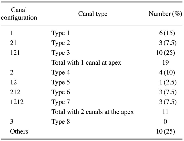
The sample size for the study was 40 teeth. The results aligned with the Vertucci classification in 75% (30/40) of the roots. The mesial roots exhibited canal configurations of type I (6, 15%), type II (3, 7.5%), type III (10, 25%), type IV (4, 10%), type V (1, 2.5%), type VI (3, 7.5%), and type VII (3, 7.5%) (Table 1). Representative examples are shown in Figure 1.
Table 1. Distribution of canal types according to the Vertucci classification among mesial roots of mandibular first molars.
Fig. 1. Representative examples of root canal configuration types that fit the Vertucci classification, based on data obtained using micro-computed tomography. A. Type I (1). B. Type II (2-1). C. Type III (1-2-1). D. Type IV (2). E. Type V (1-2). F. Type VI (2-1-2). G. Type VII (1-2-1-2).
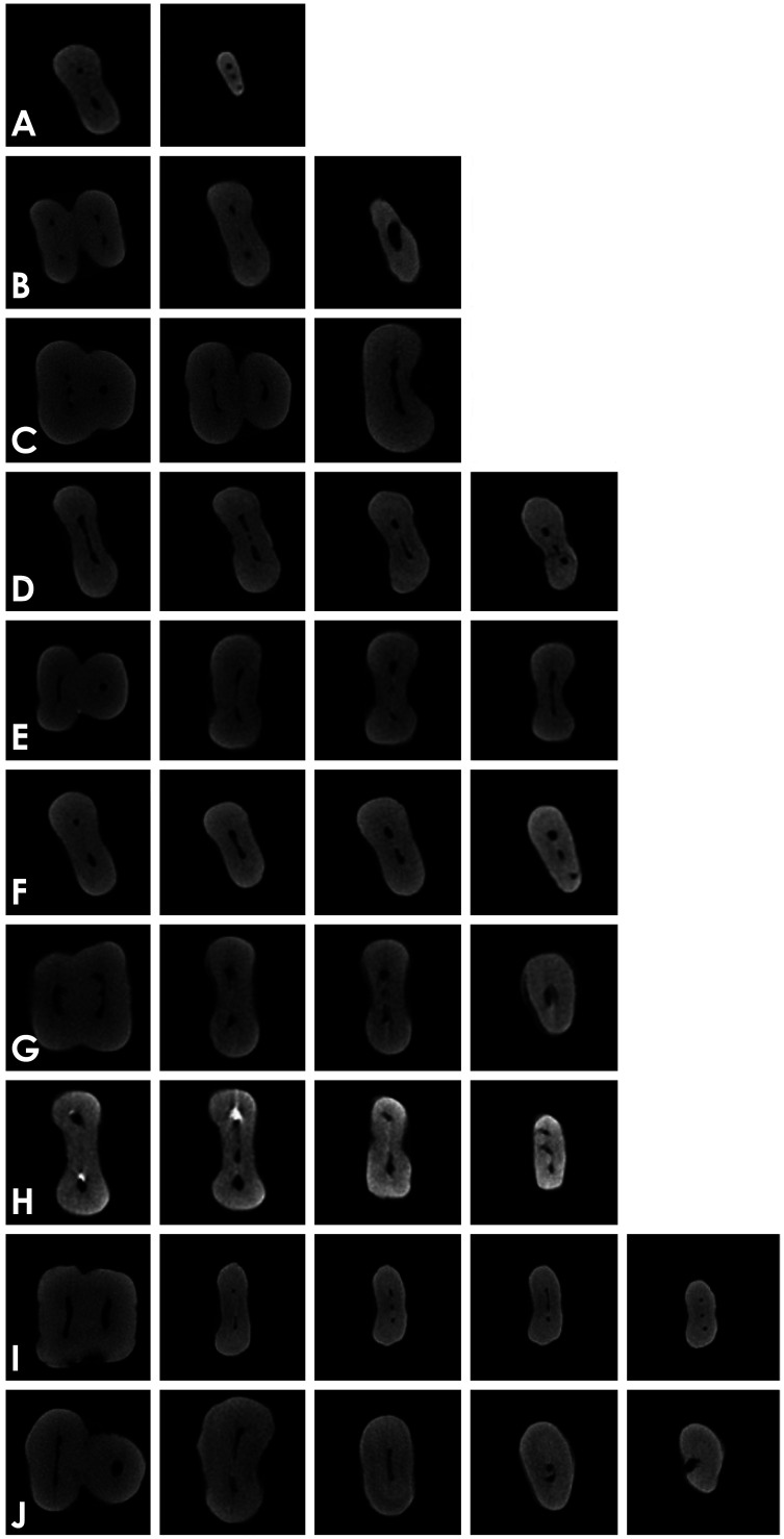
The examined images also revealed 10 configurations (25%) that did not fit into the Vertucci classification. These were types 2-3, 2-3-1, 3-2-1, 1-3-2-3, 1-2-3-1, 2-1-2-3, 1-2-3-1, 2-3-2-3, 1-2-3-2-3, 1-2-1-2-1). Among these, 8 configurations (3-2-1, 1-3-2-3, 1-2-3-1, 2-1-2-3, 1-2-3-1, 2-3-2-3, 1-2-3-2-3, 1-2-1-2-1) have not been previously reported in academic studies. Representative examples are shown in Figure 2.
Fig. 2. Representative examples of root canal configuration types that do not fit the Vertucci classification, based on data obtained from DataViewer on axial slices. A. Type (2-3). B. Type (2-3-1). C. Type (3-2-1). D. Type (1-3-2-3). E. Type (1-2-3-1). F. Type (2-1-2-3). G. Type (1-2-3-1). H. Type (2-3-2-3). I. Type (1-2-3-2-3). J. Type (1-2-1-2-1).
The differences in intra-observer agreement were not statistically significant (P>0.05). The inter-observer agreement value was 0.957, showing a high level of both intra- and inter-observer reliability.
Discussion
Many researchers have examined the root canal morphology of human permanent teeth using a number of methods, such as analyzing transparencies of previously stained samples, macroscopic sections, and radiographs of extracted teeth.22 The development of X-ray micro-CT imaging has gained increasing importance in the study of dental tissues.23 It has been used as a tool for the 3D reconstruction of internal and external tooth morphology due to its high-resolution capability, which is perfectly suited for root canal studies. Moreover, micro-CT provides quantitative and qualitative assessment of the root canal system under in vitro conditions.24
Ordinola-Zapata et al.5 examined the mesial roots of 32 mandibular first molars with multiple methods that included micro-CT. They stated that the evaluation method and the anatomy type are both important for accurately defining the canal configuration in the mesial root. They also concluded that the micro-CT method had the highest level of accuracy. The present study used micro-CT to identify different canal configurations of mesial roots of mandibular molars.
Weine25 reported a mandibular first molar with 3 canals in the mesial root. Vertucci11 reported that the type IV (2) canal was observed in 43% of mesial roots in their study, while the type II (2-1) canal forms were observed in 28%. Berna and Badanelli26 reported 2 cases in which the first molars had 3 canals in the mesial and distal roots.26 Calıskan et al.27 encountered type II (2-1) morphology in 37% of mesial roots and type IV in 44%. Gulabivala et al.8 reported that the most frequent canal configurations were type IV (n=53; 38.1%) and type II (n=40; 28.8%) in their study. Sert et al.28 reported that the most common findings were type II (1-2) (44%) and type IV (2) (43%). Pablo et al.29 stated that their most commonly observed canal types were type IV and type II. Harris et al.30 observed highly variable canal morphology in the mesial root, with the most common configuration being type V (22.7%). Gambarini et al.31 also assessed the canal configurations of mandibular first molar mesial roots and reported that 59% of cases were type IV, while 41% were type II. Marceliano-Alves et al.32 recorded the highest frequency as belonging to type IV (n=48; 46.2%), followed by type II (n=17; 16.3%). Type VIII was detected in 7.7% (n=8) of the samples.
In contrast to those previous studies, the most frequent canal configuration observed in this research was type III, followed by type I. This shows that different results may be obtained when analyzing canal configurations across different studies. Factors such as sex, ethnicity, and population type may play a role in forming the basis of these differences. It has also been stated that variations in the root canal morphology of mandibular molars are racially and genetically determined.18
In other studies, several canal configurations that do not fit within the Vertucci classification have been reported. These are type IX (1-3), type X (1-2-3-2), type XI (1-2-3-4), type XII (2-3-1), type XIII (1-2-1-3), type (4-2), type (3-2), type (1-3-1), type (2-3), type (2-1-2-1), type (3-1), type (4), type (4-1), and type (5-4).8,17,33 In this study, 2 types were observed to fit within these additional configurations: type (2-3) and type XII (2-3-1). In addition, 8 new configurations were found (3-2-1, 1-3-2-3, 1-2-3-1, 2-1-2-3, 1-2-3-1, 2-3-2-3, 1-2-3-2-3, 1-2-1-2-1), and this study marks the first time they were observed in mesial roots of mandibular first molars.
The canal configuration type plays an important role in predicting endodontic treatment success. It is envisaged that teeth with simple tubular configurations (types I, IV, and VIII) will be shaped and filled successfully; however, debridement and shaping of teeth with branched canal configurations (types III, VI, and VII) can prove difficult.8 Techniques can be divided into 2 categories: in vivo (performed directly in patients) and ex vivo (performed using extracted teeth).31 Although the current study shows that micro-CT is useful for the examination of root canal morphology, it should only be used when it is impossible to assess the root canal system accurately by conventional techniques, such as periapical radiography. The reason for this is that micro-CT is recommended for in vitro investigations of the internal and external morphology of dental structures, not in living persons. When there are abnormal findings, CBCT may be necessary for in vivo imaging.18
The present study has some limitations. The sample size was relatively small, and further studies are needed on larger cohorts regarding this issue.
In the current study, a high rate of variation was detected in the morphology of mesial roots of mandibular first molars. The high prevalence of additional configurations demonstrates the inherent complexity of the mesial root canal anatomy. Clinicians should take this complexity into consideration before commencing root canal treatment procedures to prevent the incidence of iatrogenic complications related to shaping and cleaning procedures. Furthermore, micro-CT is a highly suitable method to obtain 3D imagery that accurately reveals the intricacy of root canal morphology.
Footnotes
This work was supported by the Beykent University Scientific Research Projects Coordination Unit.
Conflicts of Interest: None
References
- 1.Wayman BE, Patten JA, Dazey SE. Relative frequency of teeth needing endodontic treatment in 3350 consecutive endodontic patients. J Endod. 1994;20:399–401. doi: 10.1016/S0099-2399(06)80299-2. [DOI] [PubMed] [Google Scholar]
- 2.Scavo R, Martinez Lalis R, Zmener O, Dipietro S, Grana D, Pameijer CH. Frequency and distribution of teeth requiring endodontic therapy in an Argentine population attending a specialty clinic in endodontics. Int Dent J. 2011;61:257–260. doi: 10.1111/j.1875-595X.2011.00069.x. [DOI] [PMC free article] [PubMed] [Google Scholar]
- 3.D'Arcangelo C, Varvara G, De Fazio P. Root canal treatment in mandibular canines with two roots: a report of two cases. Int Endod J. 2001;34:331–334. doi: 10.1046/j.1365-2591.2001.00376.x. [DOI] [PubMed] [Google Scholar]
- 4.Ahmed HM, Versiani MA, De-Deus G, Dummer PM. A new system for classifying root and root canal morphology. Int Endod J. 2017;50:761–770. doi: 10.1111/iej.12685. [DOI] [PubMed] [Google Scholar]
- 5.Ordinola-Zapata R, Bramante CM, Versiani MA, Moldauer BI, Topham G, Gutmann JL, et al. Comparative accuracy of the clearing technique, CBCT and micro-CT methods in studying the mesial root canal configuration of mandibular first molars. Int Endod J. 2017;50:90–96. doi: 10.1111/iej.12593. [DOI] [PubMed] [Google Scholar]
- 6.Villas-Bôas MH, Bernardineli N, Cavenago BC, Marciano M, Del Carpio-Perochena A, de Moraes IG, et al. Micro-computed tomography study of the internal anatomy of mesial root canals of mandibular molars. J Endod. 2011;37:1682–1686. doi: 10.1016/j.joen.2011.08.001. [DOI] [PubMed] [Google Scholar]
- 7.Keleş A, Keskin C. Apical root canal morphology of mesial roots of mandibular first molar teeth with Vertucci type II configuration by means of micro-computed tomography. J Endod. 2017;43:481–485. doi: 10.1016/j.joen.2016.10.045. [DOI] [PubMed] [Google Scholar]
- 8.Gulabivala K, Aung TH, Alavi A, Ng YL. Root and canal morphology of Burmese mandibular molars. Int Endod J. 2001;34:359–370. doi: 10.1046/j.1365-2591.2001.00399.x. [DOI] [PubMed] [Google Scholar]
- 9.Karabucak B, Bunes A, Chehoud C, Kohli MR, Setzer F. Prevalence of apical periodontitis in endodontically treated premolars and molars with untreated canal: a cone-beam computed tomography study. J Endod. 2016;42:538–541. doi: 10.1016/j.joen.2015.12.026. [DOI] [PubMed] [Google Scholar]
- 10.Costa FF, Pacheco-Yanes J, Siqueira JF, Jr, Oliveira AC, Gazzaneo I, Amorim CA, et al. Association between missed canals and apical periodontitis. Int Endod J. 2019;52:400–406. doi: 10.1111/iej.13022. [DOI] [PubMed] [Google Scholar]
- 11.Vertucci FJ. Root canal anatomy of the human permanent teeth. Oral Surg Oral Med Oral Pathol. 1984;58:589–599. doi: 10.1016/0030-4220(84)90085-9. [DOI] [PubMed] [Google Scholar]
- 12.Fan B, Ye W, Xie E, Wu H, Gutmann JL. Three-dimensional morphological analysis of C-shaped canals in mandibular first premolars in a Chinese population. Int Endod J. 2012;45:1035–1041. doi: 10.1111/j.1365-2591.2012.02070.x. [DOI] [PubMed] [Google Scholar]
- 13.Grande NM, Plotino G, Gambarini G, Testarelli L, D'Ambrosio F, Pecci R, et al. Present and future in the use of micro-CT scanner 3D analysis for the study of dental and root canal morphology. Ann Ist Super Sanita. 2012;48:26–34. doi: 10.4415/ANN_12_01_05. [DOI] [PubMed] [Google Scholar]
- 14.Kim Y, Perinpanayagam H, Lee JK, Yoo YJ, Oh S, Gu Y, et al. Comparison of mandibular first molar mesial root canal morphology using micro-computed tomography and clearing technique. Acta Odontol Scand. 2015;73:427–432. doi: 10.3109/00016357.2014.976263. [DOI] [PubMed] [Google Scholar]
- 15.Weine FS, Pasiewicz RA, Rice RT. Canal configuration of the mandibular second molar using a clinically oriented in vitro method. J Endod. 1988;14:207–213. doi: 10.1016/S0099-2399(88)80171-7. [DOI] [PubMed] [Google Scholar]
- 16.Bansal R, Hegde S, Astekar MS. Classification of root canal configurations: a review and a new proposal of nomenclature system for root canal configuration. J Clin Diagn Res. 2018;12:ZE01–ZE05. [Google Scholar]
- 17.Kartal N, Yanikoğlu FÇ. Root canal morphology of mandibular incisors. J Endod. 1992;18:562–564. doi: 10.1016/S0099-2399(06)81215-X. [DOI] [PubMed] [Google Scholar]
- 18.Zhang R, Wang H, Tian YY, Yu X, Hu T, Dummer PM. Use of cone-beam computed tomography to evaluate root and canal morphology of mandibular molars in Chinese individuals. Int Endod J. 2011;44:990–999. doi: 10.1111/j.1365-2591.2011.01904.x. [DOI] [PubMed] [Google Scholar]
- 19.Peiris R, Malwatte U, Abayakoon J, Wettasinghe A. Variations in the root form and root canal morphology of permanent mandibular first molars in a Sri Lankan population. Anat Res Int. 2015;2015:803671. doi: 10.1155/2015/803671. [DOI] [PMC free article] [PubMed] [Google Scholar]
- 20.Alkaabi W, AlShwaimi E, Farooq I, Goodis HE, Chogle SM. A micro-computed tomography study of the root canal morphology of mandibular first premolars in an Emirati population. Med Princ Pract. 2017;26:118–124. doi: 10.1159/000453039. [DOI] [PMC free article] [PubMed] [Google Scholar]
- 21.Walker RT. Root form and canal anatomy of mandibular first molars in a southern Chinese population. Endod Dent Traumatol. 1988;4:19–22. doi: 10.1111/j.1600-9657.1988.tb00287.x. [DOI] [PubMed] [Google Scholar]
- 22.Peiris R, Takahashi M, Sasaki K, Kanazawa E. Root and canal morphology of permanent mandibular molars in a Sri Lankan population. Odontology. 2007;95:16–23. doi: 10.1007/s10266-007-0074-8. [DOI] [PubMed] [Google Scholar]
- 23.Fumes AC, Sousa-Neto MD, Leoni GB, Versiani MA, da Silva LA, da Silva RA, et al. Root canal morphology of primary molars: a micro-computed tomography study. Eur Arch Paediatr Dent. 2014;15:317–326. doi: 10.1007/s40368-014-0117-0. [DOI] [PubMed] [Google Scholar]
- 24.Asgary S, Nikneshan S, Akbarzadeh-Bagheban A, Emadi N. Evaluation of diagnostic accuracy and dimensional measurements by using CBCT in mandibular first molars. J Clin Exp Dent. 2016;8:e1–e8. doi: 10.4317/jced.52570. [DOI] [PMC free article] [PubMed] [Google Scholar]
- 25.Weine FS. Case report: three canals in the mesial root of a mandibular first molar (?) J Endod. 1982;8:517–520. doi: 10.1016/S0099-2399(82)80080-0. [DOI] [PubMed] [Google Scholar]
- 26.Martínez-Berná A, Badanelli P. Mandibular first molars with six root canals. J Endod. 1985;11:348–352. doi: 10.1016/S0099-2399(85)80043-1. [DOI] [PubMed] [Google Scholar]
- 27.Çalişkan MK, Pehlivan Y, Sepetçioǧlu F, Türkün M, Tuncer SŞ. Root canal morphology of human permanent teeth in a Turkish population. J Endod. 1995;21:200–204. doi: 10.1016/S0099-2399(06)80566-2. [DOI] [PubMed] [Google Scholar]
- 28.Sert S, Aslanalp V, Tanalp J. Investigation of the root canal configurations of mandibular permanent teeth in the Turkish population. Int Endod J. 2004;37:494–499. doi: 10.1111/j.1365-2591.2004.00837.x. [DOI] [PubMed] [Google Scholar]
- 29.de Pablo OV, Estevez R, Péix Sánchez M, Heilborn C, Cohenca N. Root anatomy and canal configuration of the permanent mandibular first molar: a systematic review. J Endod. 2010;36:1919–1931. doi: 10.1016/j.joen.2010.08.055. [DOI] [PubMed] [Google Scholar]
- 30.Harris SP, Bowles WR, Fok A, McClanahan SB. An Anatomic Investigation of the Mandibular First Molar Using Micro-Computed Tomography. J Endod. 2013;39:1374–1378. doi: 10.1016/j.joen.2013.06.034. [DOI] [PubMed] [Google Scholar]
- 31.Gambarini G, Piasecki L, Ropini P, Miccoli G, Nardo DD, Testarelli L. Cone-beam computed tomographic analysis on root and canal morphology of mandibular first permanent molar among multiracial population in Western European population. Eur J Dent. 2018;12:434–438. doi: 10.4103/ejd.ejd_116_18. [DOI] [PMC free article] [PubMed] [Google Scholar]
- 32.Marceliano-Alves MF, Lima CO, Bastos LG, Bruno AM, Vidaurre F, Coutinho TM, et al. Mandibular mesial root canal morphology using micro-computed tomography in a Brazilian population. Aust Endod J. 2019;45:51–56. doi: 10.1111/aej.12265. [DOI] [PubMed] [Google Scholar]
- 33.Ng YL, Aung TH, Alavi A, Gulabivala K. Root and canal morphology of Burmese maxillary molars. Int Endod J. 2001;34:620–630. doi: 10.1046/j.1365-2591.2001.00438.x. [DOI] [PubMed] [Google Scholar]


