Abstract
Purpose
The pulp length to width (PL/W) ratio of the maxillary lateral incisor can be used as an age determination method. This study aimed to investigate the correlation between the PL/W ratio of the maxillary lateral incisor on panoramic radiographs and human chronological age in Indonesian subjects.
Materials and Methods
This study analyzed with 134 maxillary lateral incisors on 113 panoramic radiographs from patients who visited the Oral and Maxillofacial Radiology Unit of Dental Hospital Universitas Padjadjaran, Bandung, Jawa Barat, Indonesia, from 2013 to 2018 (age range: between 11 to 60 years). The pulp length was measured from the pulp chamber roof to the apical foramen, and the pulp width was measured on the cervical area of the cementoenamel junction in millimeters using Fiji ImageJ open-source software. Simple linear regression (in SPSS) was used to analyze the results. The reliability of the observers was evaluated.
Results
The PL/W ratio of the maxillary lateral incisor was significantly correlated with chronological age (P<0.01). No statistically significant difference was found in the PL/W ratio between the left and right maxillary lateral incisors (P=0.333). There was a very strong correlation (r=0.939) between the PL/W ratio of the maxillary lateral incisor and human chronological age, with the following formula: age= -3.057+1.875×PL/W ratio (R2=0.882, standard error of estimate: 4.659).
Conclusion
The PL/W ratio of the maxillary lateral incisor on panoramic radiograph can be used for age determination in Indonesian subjects.
Keywords: Age Determination by Teeth; Incisor; Dental Pulp; Radiography, Panoramic
Introduction
Indonesia is located on the 3 major tectonic plates: Eurasian, Indian-Australian, and Pacific. Due to its geographical conditions, Indonesia is prone to natural disasters, such as volcanic eruptions, tsunamis, earthquakes, and landslides.1,2 Natural disasters can lead to many deaths, requiring human identification techniques to obtain information on the identity of the victims.3 Age determination based on dental findings plays an essential role in identifying individuals in natural disasters, as well as for judicial purposes, in accident cases, and after terrorist attacks.4,5,6,7,8 However, methods of identifying individuals must be accurate, economical, and reliable, especially when determining the age of victims and suspects.
There are 5 methods of identifying an individual's age through the teeth: visual, morphological, biochemical, histological, and radiographic. The radiographic method has several advantages over other methods; for example, it is simpler and cost-effective, can be applied to living individuals or remains, and does not require tooth extraction. The radiographic method involves performing an assessment based on tooth maturity through radiographic images.9 Environmental, dietary, and endocrine factors have minimal effects on tooth maturity; therefore, this method is reliable for age determination.10 However, once all the permanent teeth have erupted, age determination can no longer be done by studying their development. Nonetheless, some changes in the tooth tissue and structure due to normal aging, such as secondary dentin, can be used for age determination.11,12 After root development finishes and the tooth can work functionally, secondary dentin apposition takes place. Secondary dentin continues to form throughout the lifespan and is correlated with age.8,12,13
Secondary dentin apposition occurs in the pulp cavity, resulting in a significant pulp size reduction with age.10,12,14 As has previously been reported by Solheim15 in Caucasians, Oscandar et al.16 in the Deutero-Malay subrace, and Rieuwpassa et al.17 in a Makassar population, the pulp is smaller in older than in younger individuals because of the dentinal matrix secretion of odontoblasts (physiological secondary odontogenesis). For this reason, pulp size can be used as an age determination method. Various non-invasive procedures have been developed for age determination by analyzing the pulp size; one such method involves using radiographic images. A previous study by Kvaal et al.18 reported that the pulp-to-tooth ratio was significantly correlated with chronological age using periapical radiographs in a Norwegian population, implying that this parameter could be used for age determination. Another study by Indira et al.19 using periapical radiographs noted that the ratio between the total pulp length and cervical pulpal width was correlated with chronological age. This method has many benefits because the pulp ratio measurement is not affected by attrition or caries, and using this parameter can reduce the need for radiograph magnification and angulation. However, an age determination study using the pulp length to width (PL/W) ratio method has not been carried out in Indonesian subjects.
Panoramic radiography is one of the most frequently used imaging methods in dentistry and is widely available in various countries.20 This modality has many advantages; panoramic radiography is a simple, fast, low-cost, and comfortable procedure that obviates the need for taking repeated radiographs, has a low radiation dose, and can show the entire dentition in a single radiographic image.21,22,23 Although the superimposition phenomenon of anterior teeth on panoramic radiographs is unavoidable, this phenomenon has been minimized as much as possible with the latest panoramic radiograph machines.21,24 In addition, measurements can be made on single panoramic radiographs, which can be useful when a measurement is made on both sides.25 In previous studies, panoramic radiographs were used for age determination based on the pulp size by Erbudak et al.14 in Turkish and Li et al.26 in Chinese populations, because panoramic radiography allowed measurements to be obtained easily. Those studies showed that anterior teeth had a closer correlation with chronological age than posterior teeth. In brief, pulp size can be measured on panoramic radiographs to determine human chronological age.
Maxillary teeth have a closer correlation with human chronological age than mandibular teeth because maxillary teeth have more regular and distinct growth layers.27,28 Previous studies by Paewinsky et al.29 in German, Cameriere et al.30 in Portuguese, Roh et al.31 in Korean, and Li et al.26 in Chinese populations found that the pulp size of the maxillary lateral incisor had the strongest correlation with chronological age compared to other teeth with a single root canal. Anatomically, the maxillary lateral incisor has a large pulp chamber and a single root canal that makes pulp measurement easier.32,33 To summarize, the pulp size of the maxillary lateral incisor can be used for age determination.
This study aimed to investigate the correlation between the PL/W ratio of the maxillary lateral incisor on panoramic radiographs and chronological age in Indonesian subjects.
Materials and Methods
The analytical study was done with a database of patients who visited the Oral and Maxillofacial Radiology Unit of Dental Hospital Universitas Padjadjaran, Bandung, Jawa Barat, Indonesia, from 2013 to 2018. The inclusion criteria for the sample were patients between 11 to 60 years of age (as confirmed from the medical records), complete closure of the apical foramen of the maxillary lateral incisor, and good quality of the panoramic radiograph in terms of anatomical coverage, density, contrast, and resolution. Maxillary lateral incisors with pathologic conditions (caries, periodontitis, periapical lesion, rotation, root resorption, attrition, abrasion, erosion, or impaction), 2 or more root canals, nonvital status, root and crown anomalies (peg-shaped lateral incisors, concrescence fusion, germination, dilaceration, supernumerary root, dens evaginatus, or dens invaginatus), an orthodontic appliance, a restoration, or a prosthesis were excluded.
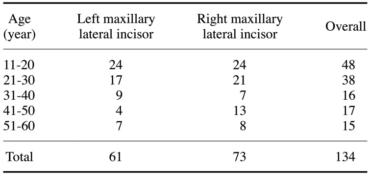
This study analyzed 113 panoramic radiographs with a total of 134 maxillary lateral incisors on both sides (Table 1). All the panoramic radiographs were taken with a Vatech Picasso Trio (Vatech, Suwon, Korea; scan parameters: 90 kVp, 10 mAs, and 12 cm×8.5 cm field of view) in JPG format, and the records also contained the patient's chronological age at the time of radiography exposure.
Table 1. Sample distribution according to patients' age.
The Ethics Committee of the Faculty of Medicine, Universitas Padjadjaran, Bandung, Jawa Barat, Indonesia, approved this study (registration number: 75/UN6.KEP/EC/2021) on January 28, 2021. This study was conducted at the Oral and Maxillofacial Radiology Unit of Dental Hospital Universitas Padjadjaran, Bandung, Jawa Barat, Indonesia, from January to March 2021.
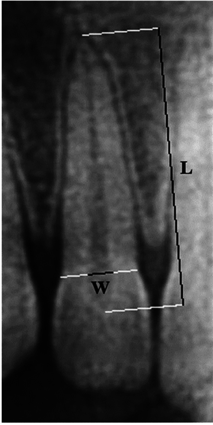
The Fiji ImageJ open-source software (ImageJ, 1.34n; National Institute of Health, Bethesda, MD, USA) was used to analyze the PL/W ratio of the maxillary lateral incisors. The PL/W ratio was calculated following the age determination method of Indira et al.19 as the pulp length measured from the pulp chamber roof to the apical foramen (L) divided by the pulp width measured on the cervical area of the cementoenamel junction (W) in millimeters (Fig. 1). The results were tabulated in Microsoft Excel (Microsoft Corp, Redmond, WA, USA).
Fig. 1. The pulp length (L) and width (W) measurements of a right maxillary lateral incisor in millimeters made using the Fiji ImageJ open-source software (ImageJ, 1.34n; National Institute of Health, Bethesda, MD, USA).
The observers were oral and maxillofacial radiologists, and analyses of intra- and inter-observer reliability were conducted. The first observer measured 25 samples with 1 repetition at a 3-week interval for an intra-observer analysis. Meanwhile, the first and second observers measured the same 25 samples at the same time to analyze inter-observer reliability. The Cronbach alpha coefficient was used to analyze both intra- and inter-observer reliability. The independent t-test was performed to evaluate the significance of differences between the left and right maxillary lateral incisors. The correlation coefficient (r), coefficient of determination (R2), and standard error of estimate (SEE) were determined with simple linear regression using SPSS version 23.0 (IBM, Armonk, NY, USA).
Results
Excellent results were found for intra- (r=0.995) and inter-observer (r=0.973) reliability, confirming measurement validity for both observers. The independent t-test showed no statistically significant difference between the average score of the left and right maxillary lateral incisors (P=0.333). The PL/W ratio of the maxillary lateral incisor had a statistically significant correlation with chronological age (P=0.000). The regression equation of the PL/W ratio of maxillary lateral incisor that best-fitted the data was as follows:
| age= -3.057+1.875×PL/W ratio |
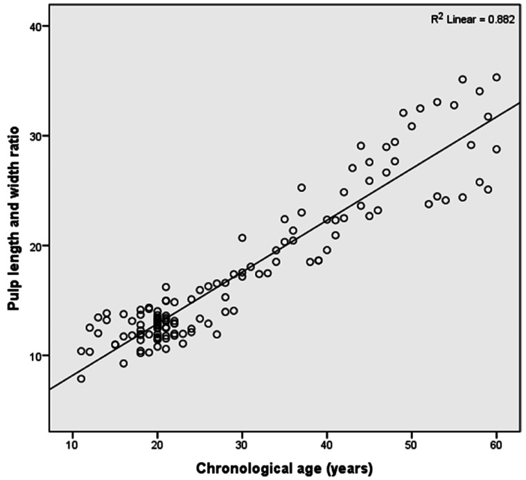
The equation represents age as the dependent variable and the PL/W ratio as the independent variable. A scatter plot showed that the PL/W ratio increased with chronological age (Fig. 2).
Fig. 2. Scatter plot showing the relationship between the pulp length and width ratio and chronological age.
The linear regression equation yielded the following results: r=0.939, R2=0.882, and SEE=4.659. This implies that the PL/W ratio explained 88.2% of variation in chronological age. The other 11.8% was influenced by other variables outside the scope of this study.
Discussion
There are various purposes for identifying living individuals or remains, such as legal status or age status relevant for the population administration system. Teeth are used to chew food and also have an aesthetic impact on the face. Since teeth are the strongest structure of the human body, they can provide information that can be used for individual identification, such as human chronological age. Pulp reduction due to secondary dentin apposition can be seen on radiographs and is used to estimate chronological age.
In the present study, the intra- and inter-observer reliability test showed that the observers made consistent measurements. This finding aligns with that of Karkhanis et al.10 in a West Australian population, wherein acceptable intra-observer reliability was reported when using the Fiji ImageJ open-source software (r>0.75). This present study also aligns with the previous study by Juneja et al.34 conducted in Karnataka population, which reported excellent intra- and inter-observer reliability when using the Fiji ImageJ open-source software (r=0.946 and r=0.882, respectively). The use of the Fiji ImageJ open-source software with its straight-line tool provides consistent results for the pulp size, minimizing observer-related variability. Nonetheless, Fiji ImageJ is a semiautomatic program that still requires observers to analyze the pulp size.
In the present study, there was no statistically significant pulp size difference between the left and right maxillary lateral incisors. Consistent findings were reported by Kvaal et al.18 in Norwegian, Zaher et al.27 in Egyptian, Penaloza et al.35 in Malaysian, and Li et al.26 in Chinese populations. The same regression equation can be used for the left and right maxillary lateral incisors, showing that secondary dentin apposition occurred simultaneously and evenly on both sides.
The maxillary lateral incisor was chosen because it was easy to measure. The authors preferred the maxillary lateral incisor over the maxillary central incisor because a dark air space was often found on the central incisor apex on panoramic radiographs.36 In the present study, the PL/W ratio of the maxillary lateral incisor showed a significant relationship with chronological age and yielded a linear regression model with a close fit (P=0.000). This study agrees with the previous study by Zaher et al.27 in an Egyptian population, which reported a significant correlation between the pulp-to-tooth-area ratio of maxillary lateral incisor and chronological age. In contrast, Li et al.26 in a Chinese population found that only the pulp-to-tooth-width ratio of the maxillary lateral incisor was significantly correlated with chronological age, and those authors suggested that the speed of secondary dentin formation might differ across people of the same ethnicity.
In the present study, the PL/W ratio of the maxillary lateral incisor (independent variable) was used to estimate age (dependent variable), and a positive correlation was found (Fig. 2). In contrast, a previous study by Indira et al.19 reported that the ratio between the total pulp length and the cervical pulpal width of the maxillary central incisor on periapical radiographs was negatively correlated with age in the range of 16 to 50 years. An explanation for this discrepancy is that the previous study measured pulp length and width manually. Software assistance with morphometric measurements is highly recommended to improve the accuracy. Moreover, the previous study also did not report the intra- and inter-observer reliability. The different population in the previous study may also have contributed to this discrepancy because there are differences among populations in dental maturity.
The authors found that the PL/W ratio of the maxillary lateral incisor had a very strong correlation with chronological age (r=0.939). The authors assumed that this strong correlation may have been due to secondary dentin apposition in Indonesian subjects. This assumption needs to be explored in further research. The findings of this study partially agree with those of Roh et al.,31 who reported that the sum of the pulp-to-tooth-width ratio value of the maxillary lateral incisor according to the Kvaal method had a higher correlation than other single-rooted teeth using digital panoramic radiographs of Koreans (r=0.60). That previous study noted that the equation derived from the width ratio was the most appropriate (i.e., without the length ratio) for age determination in the Korean population. However, in the present study, the correlation coefficient obtained using the PL/W ratio was higher in Indonesian subjects. In contrast, Zaher et al.27 reported a low correlation coefficient between the pulp-to-tooth-area ratio of the maxillary lateral incisor and chronological age (r=-0.2) in an Egyptian population using periapical radiographs, and they suggested that secondary dentin deposition was relatively slow in this population.
In the present study, there was a significant correlation between the PL/W ratio of the maxillary lateral incisor and chronological age (R2=0.882), and the regression formula had a high accuracy (SEE=4.659). An explanation for this may be that the variation in maxillary lateral incisor pulp length and width was small and the pulp size to be measured was large. This finding is similar to that of the previous study by Cameriere et al.,30 which reported that the R2 value of the pulp-to-tooth-area ratio of the maxillary lateral incisor in the Portuguese population was 0.816, but the accuracy of the regression formula (SEE=6.64) was lower than the present study. This difference in results might be explained by the larger sample size in the present study, which allowed more reliable results. The present study contrasts with another study by Ge et al.,37 which found that the R2 of maxillary lateral incisor pulp cavity volume was 0.311 (SEE=9.402) in a Chinese population using CBCT images. Another study that contrasts with the present study was conducted by Akay et al.38, which stated that the R2 value of the maxillary lateral incisor pulp-to-tooth volume ratio was 0.197 in a Turkish population with a mean square error of 6.72 using CBCT images. These results showed that pulp size measurements using 2-dimensional images could be more accurate than those made using 3-dimensional images. Further study is necessary to compare various pulp volume measurements of the maxillary lateral incisor. On the contrary, Hisham et al.39 noted that the R2 value of the pulp-to-tooth ratio was 0.076 (SEE=11.192) in a Malaysian population. The R2 values were lower and the SEE values were higher in previous studies than in the present study. Making measurements using the Fiji ImageJ open-source software requires skilled observers with expertise in oral and maxillofacial radiology. Although the number of samples used in their study was larger, the measurements in the present study were made by 2 oral and maxillofacial radiologists and were tested for intra- and inter-observer reliability. The R2 value was higher and the SEE was lower in the present study than in the previous studies that used different methods to analyze maxillary lateral incisors. A reason for this may be the fact that the authors did not measure the length and width of the tooth's outer surface, which is susceptible to attrition and caries. Dietary patterns and chewing habits can also be assumed to affect these parameters because they influence secondary dentin apposition, but these variables were not analyzed in the present study.26
The previous study by Indira et al.19 used periapical radiographs to analyze pulp length and width. Their findings contrast with those of the present study that used digital panoramic radiographs, which provide a better view of the dental anatomy, contrast, brightness, and density than conventional panoramic radiographs.40 The main disadvantage of using panoramic radiographs is that the areas of diagnostic interest may lie outside the focal trough, which causes the image to be blurred, enlarged, reduced, distorted, a ghost image, a double image, or a triple picture. The patient's dental arch must be placed in the focal trough to obtain a high-quality image even though the tooth inclination and skeletal base can limit the placement and allow some movement.36,41,42 However, panoramic radiographs are more commonly used in oral radiodiagnostics for age determination than periapical radiographs.
Digital software imaging analysis is a helpful technology for clinical use, especially in dentistry. This study analyzed the PL/W ratio of the maxillary lateral incisor using the Fiji ImageJ open-source software, which was also used for pulp analysis by Erbudak et al.14 in Turkish, Karkhanis et al.10 in Western Australian, Juneja et al.34 in Karnatakan, and Hisham et al.39 in Malaysian populations. On the contrary, the previous study by Indira et al.19 used digimatic vernier calipers to measure the pulp length and width. Another study by Cameriere et al.30 in the Portuguese population used Adobe Photoshop® CS4 (Adobe Systems Incorporated, San Jose, CA, USA) to analyze the pulp, but this software is not designed explicitly for clinical purposes. The Fiji ImageJ open-source software is used frequently for measurements in image manipulation and is preferable to other methods because it makes quantitative image analysis easier, supports image manipulation, and is faster. Determining a reference point in digital software requires skilled and experienced observers. The authors suggest reducing measurement limitations by zooming in on the image and using the smallest level of pixel in the image.
In the present study, the sample may not be representative of the entire Indonesian population because of the small sample size. The data did not have a fully even distribution because it was difficult to find older adults who met the criteria. However, the findings of the present study constitute an important scientific contribution for reference in oral and maxillofacial radiology, forensic odontology, and dentistry research in Indonesia.
The PL/W ratio of the maxillary lateral incisor on panoramic radiographs can be used as an alternative method to identify human age in either living or dead individuals. An advantage of this method is that the pulp is only slightly affected by external factors, such as attrition, trauma occlusion, and caries. Moreover, the Fiji ImageJ open-source software and the morphological condition of the maxillary lateral incisor facilitated accessible measurements of the PL/W ratio. The principal limitations of this study are that the PL/W ratio can not be applied to multi-rooted teeth and that the measurement depends on the radiographic quality. A challenge for future studies will be to incorporate in-depth analyses of sex, ethnicity, dietary patterns, and different populations to improve the accuracy.
In conclusion, based on our findings, the PL/W ratio of the maxillary lateral incisor on panoramic radiographs can be used for age determination in Indonesian subjects.
Acknowledgements
We would like to express special thanks to Dr. Marry Siti Mariam, drg., M.Kes., Dr. Endah Mardiati, drg., MS., Sp.Ort (K), and Nani Murniati, drg., M.Kes., who gave the advice in completing this study. We also acknowledge the oral and maxillofacial radiology and forensic odontology staff of Faculty of Dentistry, Universitas Padjadjaran, Bandung, Jawa Barat, Indonesia.
Footnotes
Conflicts of Interest: None
References
- 1.Anugrahadi A, Sumotarto U, Purwiyono TT. Geomorphological impact of Palu earthquake. J Phys Conf Ser. 2019;1402:033015 [Google Scholar]
- 2.Kusumayudha SB, Lestari P, Paripurno ET. Eruption characteristic of the sleeping volcano, Sinabung, North Sumatera, Indonesia, and SMS gateway for disaster early warning system. Indones J Geogr. 2018;50:70–77. [Google Scholar]
- 3.Zikir A, Mânica S. Forensic dentistry and disaster victim identification (DVI) in Indonesia. Aust J Forensic Sci. 2019;53:75–83. [Google Scholar]
- 4.Kanchan-Talreja P, Acharya AB, Naikmasur VG. An assessment of the versatility of Kvaal's method of adult dental age estimation in Indians. Arch Oral Biol. 2012;57:277–284. doi: 10.1016/j.archoralbio.2011.08.020. [DOI] [PubMed] [Google Scholar]
- 5.Divakar KP. Forensic odontology: the new dimension in dental analysis. Int J Biomed Sci. 2017;13:1–5. [PMC free article] [PubMed] [Google Scholar]
- 6.Kumar R, Athota A, Rastogi T, Karumuri SK. Forensic radiology: an emerging tool in identification. J Indian Acad Oral Med Radiol. 2015;27:416–422. [Google Scholar]
- 7.Prajapati G, Sarode SC, Sarode GS, Shelke P, Awan KH, Patil S. Role of forensic odontology in the identification of victims of major mass disasters across the world: a systematic review. PLoS One. 2018;13:e0199791. doi: 10.1371/journal.pone.0199791. [DOI] [PMC free article] [PubMed] [Google Scholar]
- 8.Krishan K, Kanchan T, Garg AK. Dental evidence in forensic identification - an overview, methodology and present status. Open Dent J. 2015;9:250–256. doi: 10.2174/1874210601509010250. [DOI] [PMC free article] [PubMed] [Google Scholar]
- 9.Panchbhai AS. Dental radiographic indicators, a key to age estimation. Dentomaxillofacial Radiol. 2011;40:199–212. doi: 10.1259/dmfr/19478385. [DOI] [PMC free article] [PubMed] [Google Scholar]
- 10.Karkhanis S, Mack P, Franklin D. Age estimation standards for a Western Australian population using the coronal pulp cavity index. Forensic Sci Int. 2013;231:412.e1–412.e6. doi: 10.1016/j.forsciint.2013.04.004. [DOI] [PubMed] [Google Scholar]
- 11.Uzuner FD, Kaygısız E, Darendeliler N. In: Post mortem examination and autopsy-current issues from death to laboratory analysis. Dogan KH, editor. London: IntechOpen; 2017. Defining dental age for chronological age determination; pp. 77–95. [Google Scholar]
- 12.Ellingham S, Adserias-Garriga J. In: Age estimation: a multidisciplinary approach. Adserias-Garriga J, editor. Cambridge: Academic Press; 2019. Complexities and considerations of human age estimation; pp. 1–15. [Google Scholar]
- 13.de las Heras SM. In: Age estimation: a multidisciplinary approach. Adserias-Garriga J, editor. Cambridge: Academic Press; 2019. Dental age estimation in adults; pp. 125–140. [Google Scholar]
- 14.Erbudak HÖ, Ozbek M, Uysal S, Karabulut E. Application of Kvaal et al.'s age estimation method to panoramic radiographs from Turkish individuals. Forensic Sci Int. 2012;219:141–146. doi: 10.1016/j.forsciint.2011.12.012. [DOI] [PubMed] [Google Scholar]
- 15.Solheim T. Amount of secondary dentin as an indicator of age. Scand J Dent Res. 1992;100:193–199. doi: 10.1111/j.1600-0722.1992.tb01740.x. [DOI] [PubMed] [Google Scholar]
- 16.Oscandar F, Rojiun ZA, Ibrahim N, Supian S, Tharmalingam D, Malinda Y. Preliminary research: correlation between pulp chamber volume of mandibular first molar and chronological age on deuteromalay subrace. Int J Med Toxicol Leg Med. 2018;21:34–36. [Google Scholar]
- 17.Rieuwpassa IE, Alfian AA, Hasyim R. Age estimation based on dental panoramic radiography overview with coronal pulp cavity index (CPCI) methods. Int J Adv Sci Technol. 2020;29:8872–8879. [Google Scholar]
- 18.Kvaal SI, Kolltveit KM, Thomsen IO, Solheim T. Age estimation of adults from dental radiographs. Forensic Sci Int. 1995;74:175–185. doi: 10.1016/0379-0738(95)01760-g. [DOI] [PubMed] [Google Scholar]
- 19.Indira AP, Shashikala R, Nagaraj T, Santosh HN. Age estimation of adults using dental pulp: a cross-sectional radiographic study. J Adv Clin Res Insights. 2015;2:131–134. [Google Scholar]
- 20.Yeung AW, Mozos I. The innovative and sustainable use of dental panoramic radiographs for the detection of osteoporosis. Int J Environ Res Public Health. 2020;17:2449. doi: 10.3390/ijerph17072449. [DOI] [PMC free article] [PubMed] [Google Scholar]
- 21.White SC, Pharoah MJ. Oral radiology: principles and interpretation. 7th ed. St. Louis: Elsevier; 2014. [Google Scholar]
- 22.Ramos BC, da Silva Izar BR, Pereira JLC, Souza PS, Valerio CS, Tuji FM, et al. Formation of ghost images due to metal objects on the surface of the patient's face: a pictorial essay. Imaging Sci Dent. 2016;46:63–68. doi: 10.5624/isd.2016.46.1.63. [DOI] [PMC free article] [PubMed] [Google Scholar]
- 23.Suomalainen A, Pakbaznejad Esmaeili E, Robinson S. Dentomaxillofacial imaging with panoramic views and cone beam CT. Insights Imaging. 2015;6:1–16. doi: 10.1007/s13244-014-0379-4. [DOI] [PMC free article] [PubMed] [Google Scholar]
- 24.Kayal RA. Distortion of digital panoramic radiographs used for implant site assessment. J Orthod Sci. 2016;5:117–120. doi: 10.4103/2278-0203.192113. [DOI] [PMC free article] [PubMed] [Google Scholar]
- 25.Limdiwala PG, Shah JS. Age estimation by using dental radiographs. J Forensic Dent Sci. 2013;5:118–122. doi: 10.4103/0975-1475.119778. [DOI] [PMC free article] [PubMed] [Google Scholar]
- 26.Li MJ, Chu G, Han MQ, Chen T, Zhou H, Guo YC. Application of the Kvaal method for age estimation using digital panoramic radiography of Chinese individuals. Forensic Sci Int. 2019;301:76–81. doi: 10.1016/j.forsciint.2019.05.015. [DOI] [PubMed] [Google Scholar]
- 27.Zaher JF, Fawzy IA, Habib SR, Ali MM. Age estimation from pulp/tooth area ratio in maxillary incisors among Egyptians using dental radiographic images. J Forensic Leg Med. 2011;18:62–65. doi: 10.1016/j.jflm.2010.12.004. [DOI] [PubMed] [Google Scholar]
- 28.Fancy SG. Preparation of mammalian teeth for age determination by cementum layers: a review. Wildl Soc Bull. 1980;8:242–248. [Google Scholar]
- 29.Paewinsky E, Pfeiffer H, Brinkmann B. Quantification of secondary dentine formation from orthopantomograms - a contribution to forensic age estimation methods in adults. Int J Legal Med. 2005;119:27–30. doi: 10.1007/s00414-004-0492-x. [DOI] [PubMed] [Google Scholar]
- 30.Cameriere R, Cunha E, Wasterlain SN, De Luca S, Sassaroli E, Pagliara F, et al. Age estimation by pulp/tooth ratio in lateral and central incisors by peri-apical X-ray. J Forensic Leg Med. 2013;20:530–536. doi: 10.1016/j.jflm.2013.02.012. [DOI] [PubMed] [Google Scholar]
- 31.Roh BY, Lee WJ, Ryu JW, Ahn JM, Yoon CL, Lee SS. The application of the Kvaal method to estimate the age of live Korean subjects using digital panoramic radiographs. Int J Legal Med. 2018;132:1161–1166. doi: 10.1007/s00414-017-1762-8. [DOI] [PubMed] [Google Scholar]
- 32.Saini A, Garg A. Age estimation of an individual by pulp/tooth ratio by maxillary lateral incisor using periapical radiographs (RVG) Int J Forensic Odontol. 2019;4:27–33. [Google Scholar]
- 33.Altunsoy M, Ok E, Nur BG, Aglarci OS, Gungor E, Colak M. A cone-beam computed tomography study of the root canal morphology of anterior teeth in a Turkish population. Eur J Dent. 2014;8:302–306. doi: 10.4103/1305-7456.137630. [DOI] [PMC free article] [PubMed] [Google Scholar]
- 34.Juneja M, Devi YB, Rakesh N, Juneja S. Age estimation using pulp/tooth area ratio in maxillary canines - a digital image analysis. J Forensic Dent Sci. 2014;6:160–165. doi: 10.4103/0975-1475.137047. [DOI] [PMC free article] [PubMed] [Google Scholar]
- 35.Penaloza TYM, Karkhanis S, Kvaal SI, Nurul F, Kanagasingam S, Franklin D, et al. Application of the Kvaal method for adult dental age estimation using cone beam computed tomography (CBCT) J Forensic Leg Med. 2016;44:178–182. doi: 10.1016/j.jflm.2016.10.013. [DOI] [PubMed] [Google Scholar]
- 36.Rondon RH, Pereira YC, do Nascimento GC. Common positioning errors in panoramic radiography: a review. Imaging Sci Dent. 2014;44:1–6. doi: 10.5624/isd.2014.44.1.1. [DOI] [PMC free article] [PubMed] [Google Scholar]
- 37.Ge ZP, Yang P, Li G, Zhang JZ, Ma XC. Age estimation based on pulp cavity/chamber volume of 13 types of tooth from cone beam computed tomography images. Int J Legal Med. 2016;130:1159–1167. doi: 10.1007/s00414-016-1384-6. [DOI] [PubMed] [Google Scholar]
- 38.Akay G, Gungor K, Gurcan S. The applicability of Kvaal methods and pulp/tooth volume ratio for age estimation of the Turkish adult population on cone beam computed tomography images. Aust J Forensic Sci. 2019;51:251–265. [Google Scholar]
- 39.Hisham S, Abdullah N, Noor MH, Franklin D. Quantification of secondary dentin formation using dental orthopantomographs in a contemporary Malaysian population. Aust J Forensic Sci. 2019;51:S180–S183. [Google Scholar]
- 40.Villoria EM, Rodrigues RC, do Nascimento Pereira CH, Conceição GS, Soares RV. The importance of digital radiographic systems in dental schools and oral radiology centers as part of reopening during the COVID-19 pandemic. Imaging Sci Dent. 2021;51:91–92. doi: 10.5624/isd.20200216. [DOI] [PMC free article] [PubMed] [Google Scholar]
- 41.Park JW, Huh KH, Yi WJ, Heo MS, Lee SS, Choi SC. Comparison of the reproducibility of panoramic radiographs between dentulous and edentulous patients. Imaging Sci Dent. 2014;44:95–102. doi: 10.5624/isd.2014.44.2.95. [DOI] [PMC free article] [PubMed] [Google Scholar]
- 42.Nikneshan S, Sharafi M, Emadi N. Evaluation of the accuracy of linear and angular measurements on panoramic radiographs taken at different positions. Imaging Sci Dent. 2013;43:191–196. doi: 10.5624/isd.2013.43.3.191. [DOI] [PMC free article] [PubMed] [Google Scholar]


