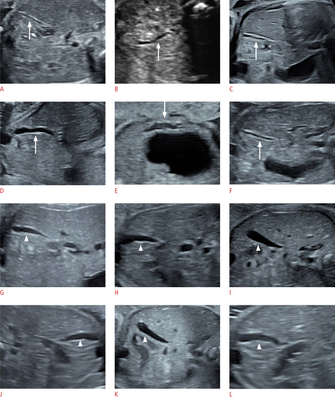
Fig. 2. Longitudinal ultrasound images of gallbladders (GBs) in cystic biliary atresia (CBA) and choledochal cyst (CC) fetuses.
A-F. In the CBA group, the GBs (arrow) present with abnormal morphology, either with a thick and rigid GB wall or with a tortuous tubular shape. G-L. In the CC group, the GBs (arrowhead) are well-filled, with smooth pear-shaped structures.

