Abstract
Background:
Tarsometatarsal (TMT) arthrodesis is commonly performed in the management of midfoot arthritis, trauma, or deformity. The purpose of this study was to collect aggregate data (demographic, surgical, and perioperative outcomes) on patients who previously had a TMT fusion with BME compression staples.
Methods:
Sixty-six patients underwent TMT fusion with BME compression staples. Outcomes included demographics, surgical information, the Veterans Rand VR-12 Health Survey, Foot and Ankle Ability Measure (FAAM), visual analog scale (VAS), Revised-Foot Function Index (FFI-R), Ankle Osteoarthritis Scale (AOS), patient satisfaction survey scores, radiographic fusion rate, level of pain reduction, and complications. Sixty-six patients (68 feet) were analyzed (59 females) with an average age of 64 years (range, 18-83). The mean latest follow-up was 35.9 (range, 6-56.6 months).
Results:
The average surgical time was 38.1±14.3 minutes (range, 11-75). All outcomes improved significantly (P < .001) from preoperative to latest follow-up except for the VR-12 Mental and Physical score. The average time to fusion determined by radiographs was 8.4 weeks (range, 6.1-46.1 weeks). Wound complications were not seen. Indications for subsequent surgeries (26.5%, 18/68 feet) in this current study included pain (n = 14), broken staples, and nonunion (n = 3).
Conclusions:
The fusion rate in this study, 89.7%, was similar to values reported in the literature. The patient satisfaction score of 81.9 at latest follow-up is consistent with patient satisfaction for other methods of fusion.
Level of Evidence:
Level IV, retrospective case series.
Keywords: tarsometatarsal fusion, staples
Introduction
Tarsometatarsal (TMT) arthrodesis is commonly performed in the management of midfoot arthritis, trauma, or deformity. 25 The vast majority of patients experience substantial improvement in both pain and function with a successful fusion. 11 A variety of conventional fixation devices have been used in TMT fusion, including Kirschner wires, lag screws, staples, compression plate devices, external fixators, and their combinations. Conventional screw placement across the TMT joints is difficult because of the acute angle of screw insertion. Recently, there has been an increase in popularity in the use of shape memory compression staples in orthopedic surgical procedures, including TMT fusions. 1,18 Purported benefits of staple fixation include ease of insertion, faster time to union, low-profile design, and maximization of joint coaptation. Older-generation nitinol staples required refrigeration prior to implantation and subsequent heating after implantation to achieve their dynamic compression state. 14 In contrast, new generation of nitinol staples, including BME ELITE (Synthes GmbH, Oberdorf, Switzerland), have the ability to elastically recover from deformations, which may occur in vivo, imparting a dynamic compressive capability not possible in conventional fusion methods. This has been demonstrated in numerous in vitro biomechanical studies. 1,9,22 This feature of recovering a prior shape, enables the specific staple used in this series to be implanted after reduction and alignment of the fusion site with the expectation that the staple would begin to impart compression at the intended fusion site after it was released from its “inserter.” It is reported that compression staples generate a significantly greater compression force across a stimulated osteotomy compared to mechanical staples, and resist permanent deformation, fully recovering their shape following loading. 1 -3,5,9,14
Numerous studies have demonstrated the safety and efficacy of the use of nitinol compression staples in TMT fusion 3,9,13,14,16,23 and generally have fusion rates near 90%, comparable to literature rated for conventional fixation. 19 Although some clinical data exist regarding union rate using BME ELITE compression staples in TMT fusion, the data lack objective preoperative and postoperative data and analysis. In this report, validated clinical outcome scores were used to assess the subjective efficacy of midfoot arthrodesis using the new generation of nitinol staples. The purpose of this study was to collect aggregate data (demographic, surgical, and perioperative outcomes) on patients who previously had a TMT fusion with BME compression staples. Primary endpoints include arthrodesis rate and level of pain reduction. Whereas secondary endpoints include Veterans Rand 12-Item Health Survey (VR-12), Foot and Ankle Ability Measure (FAAM), visual analog scale (VAS) for pain, Revised-Foot Function Index (FFI-R), Ankle Osteoarthritis Scale (AOS), and patient satisfaction.
Methods
After institutional review board approval was obtained, a retrospective chart review of prospectively collected outcome data was conducted to investigate the surgical and perioperative outcomes on TMT fusion with BME compression staples (Synthes USA, LLC, Monument, CO; or BioMedical Enterprises, Inc, San Antonio, TX). All procedures were performed by a single, fellowship-trained foot and ankle surgeon (J.C.C.) between March 2014 and April 2018.
Inclusion criteria were patients between the age of 18 and 85 years, a prior single or multi-TMT joint primary or revision (previously failed TMT fusion procedure) fusion with the use of BME ELITE compression staples (first, second, or third TMT joint, with or without naviculocuneiform joint); and willingness to participate in external research via their clinic admitting form. Patients were excluded if they were younger than 18 years, had diabetes, less than 6 months of follow-up, and/or had an associated talonavicular or calcaneocuboid fusion.
Data collection included patient demographics; medical and surgical history; complications; and pre- and postoperative patient-reported outcomes. Outcome scores included the VR-12, 12 AOS, 8 VAS, 26 FAAM, 17 and a patient satisfaction survey. The VR-12 evaluated 8 domains, the scores are tabulated into a summary physical score (PCS) and a summary mental score (MCS) and it follows patient-reported changes in physical and emotional health over time. 12 AOS 8 is a validated and reliable outcome measure derived from the Foot Function Index. The patient satisfaction survey consists of 6 questions: 5 multiple-choice questions ask the patient to describe their pain relief, ability to perform daily tasks, ability to perform heavy work or recreational activities, meeting expectations (answers range from excellent to poor), and if they would have the operation again. The last question on the patient satisfaction asked, “How satisfied are you with your medical care?” and is a standard 0-100 numeric rating scale, with 0 denoting “least satisfied” and 100 being “most satisfied.”
Eighty-four patients were originally screened, 18 patients were excluded. Sixty-six patients (68 feet) were analyzed (59 women), with an average age of 64 years (range, 18-83). The mean follow-up was 35.9 months (range, 6-56.6). The majority (48/66) of patients were nonsmokers whereas 25.8% (17/66) of patients were former smokers. The average body mass index was 29.6 (range, 20.7-44.3). Primary TMT fusions accounted for 92.4% of the cohort whereas 5 patients/5 feet had a previously failed TMT fusion procedure. The fusions performed included 32 single-TMT fusions, 27 multiple-TMT fusions, 4 naviculocuneiform and single-TMT fusions, and 5 naviculocuneiform and multiple TMT fusions (Table 1). The average surgical time was 38.1±14.4 minutes (range, 11-75).
Table 1.
Fusion Procedures.
| First TMT | Second TMT | Third TMT | |
|---|---|---|---|
| Single TMT | 4 | 25 | 3 |
| Multiple TMT | 2 | 27 | 25 |
| NC and single TMT | 0 | 4 | 0 |
| NC and multiple TMT | 0 | 5 | 5 |
Abbreviations: NC, naviculocuneiform; TMT, tarsometatarsal.
Surgical Procedure
Surgery was performed as an outpatient procedure. The decision to use 1 or 2 incisions was dependent on the number of TMT joints involved in the injury or arthritic process. Second- and third-TMT joint fusions were performed using 1 incision; however, if the first TMT was also involved, 2 dorsal longitudinal incision were made.
The first incision was made over the first TMT joint, just lateral to the extensor hallucis longus. This allowed access to the first and most of the second TMT joints. Pathology involving only the medial 2 TMT joints could be corrected with this single incision. Accessing the entire second TMT joint through this incision carried a risk of injury to the dorsalis pedis artery and deep peroneal nerve. If there was any concern about the reduction accuracy of the second TMT joint or if a surgery included a third TMT joint fusion, a second more lateral incision was used to facilitate exposure and visualization of the second and third TMT joint. The second, lateral incision was in line with the third dorsal webspace, which was further lateral than what is typically appreciated.
Dorsal bone spurs were removed to expose the joints. If the joints were well aligned, small osteotomes and curettes were used to remove articular cartilage remnants and expose subchondral bone. A saw was used only when significant angular correction was needed to correct the alignment. The opposing surfaces of the joints were perforated with a 2 mm diameter drill to enter subchondral bone. A small curved osteotome was used to microfracture the subchondral bone. If there are small gaps it was filled with local bone graft. As a general rule, the second and third TMT joints are immobilized with a single staple. With the firstt TMT joint, there is enough room to use two staples for added stability and strength. It can either be placed next to each other over the dorsum, or one over the dorsum and a second medial, or dorso-medial.
The patients were immobilized in a short leg cast splint for 2 weeks, followed by a controlled ankle movement boot for 4 weeks. During that time, patients were encouraged to do active range of motion and did not have to sleep with the boot. Patients were advised to be heel touch weightbearing for the first 6 weeks and could then progress to weightbearing as tolerated if the radiographs looked fine.
Statistical Analysis
Paired-sample t tests were used to determine significant differences in outcome variables from preoperative to latest follow-up. Statistical analyses were performed with SPSS, version 24.0 (IBM Corp, Armonk, NY), and significance was set at P < .05.
Results
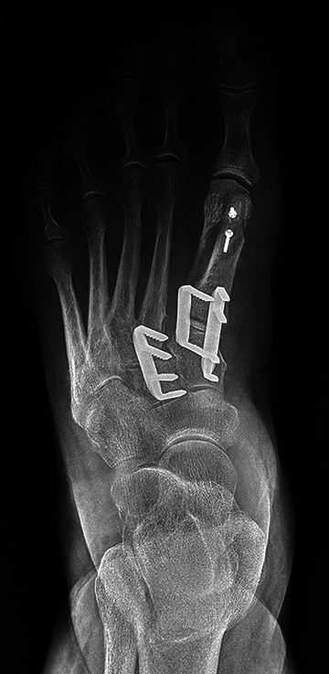
Implant size selections were made by the surgeon. Of the 6 first-TMT fusions, 4 two-prong staples and 1 four-prong staples were used. One patient had both two-prong and four-prong staples implanted in the first TMT (Figure 1). For the second TMT, 41 two-prong staples and 20 four-prong staples were used. For the third TMT, 32 two-prong staples and 1 four-prong staple were used.
Figure 1.

Anteroposterior view of a patient who had first-TMT and second-TMT fusions using BME ELITE staples. The first TMT shows both a 2-prong and 4-prong staples implanted. TMT, tarsometatarsal.
Clinical outcome scores are summarized in Table 2. Good to excellent relief of their pain after surgery was reported by 63.6% of patients whereas 59.1% patients reported that the surgery met their expectations. Eighty-six percent of the patients stated they would definitely or probably have the operation again.
Table 2.
Clinical Outcome Scores (Mean ± SD).
| Total Patient Population | |||
|---|---|---|---|
| Clinical Outcome Measure | Preoperative | Latest Follow-up | P Value |
| VR-12 Physical | 35.9 ± 10.1 | 40.1 ± 12.8 | .007 |
| VR-12 Mental | 54.8 ± 10.1 | 55.0 ± 9.7 | .881 |
| AOS Pain | 55.4 ± 22.6 | 27.6 ± 23.5 | <.001 |
| AOS Disability | 59.8 ± 23.5 | 34.2 ± 26.6 | <.001 |
| FFI-R | 69.7 ± 21.3 | 50.3 ± 19.0 | <.001 |
| FAAM ADL | 53.3 ± 18.6 | 81.3 ± 19.9 | <.001 |
| FAAM Sports | 33.4 ± 26.5 | 63.4 ± 33.1 | <.001 |
| VAS | 6.0 ± 2.2 | 2.5 ± 2.3 | <.001 |
| Patient satisfaction | 81.9 ± 22.3 | ||
Abbreviations: ADL, Activities of Daily Living; AOS, Ankle Osteoarthritis Scale; FFI-R, Revised-Foot Function Index; FAAM, Foot and Ankle Ability Measure; VAS, visual analog scale for pain; VR-12, Veterans Rand 12-Item Health Survey.
To determine time to fusion and fusion rate, a single orthopedic surgeon (K.L.F.) not involved in the surgical or clinical care of the patient reviewed sequential postoperative radiographs. Fusion rate was reported for each joint fused as well as the presence of any hardware complications. The radiographic end point fusion was defined as at least 50% osseous bridging across each joint. 19 The average time to fusion was 8.4 weeks (range, 6.1-46.1 weeks), with the longest time of 12.7 weeks for the first TMT compared to the second (7.0 weeks) and third TMT (8.3 weeks) joints, respectively.
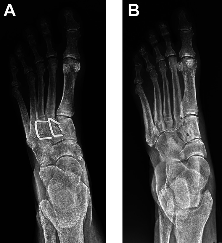
Indications for subsequent surgeries (26.5%, 18/68 feet) in this current study included discomfort over the hardware (n = 14), shortening osteotomies (n = 1), and revision surgery for nonunion of the joint (n = 3). The average time to subsequent surgery was 16.8 months (6-48 months). Of the 14 feet with discomfort over the hardware, 5 surgeries were performed because of pain over a single staple and 9 because of pain over multiple staples. Four patients had broken staples; 3 were broken 2-prong staples of the third TMT joint (Figure 2A) and occurred at 6, 15, and 48 months postoperatively. One patient had broken staples of both the second (4-prong) and third (2-prong) TMT joints at 14 months postoperatively. Hardware removal performed because of pain resulted in resolution of symptoms in all patients (Figure 2B). One other surgery included distal metatarsal shortening osteotomies due to overload. There were no wound complications.
Figure 2.

(A) Patient who had second TMT and third TMT fusions using 2-prong BME staples. The staple is broken across the third TMT joint in this AP view. (B) Oblique radiograph view of a surgery to remove the broken staple across the third TMT joint. AP, anteroposterior; TMT, tarsometatarsal.
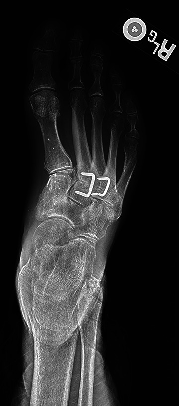
There were 8 nonunions. Nonunions were defined as no radiologic sign of healing on plain radiographs or computed tomographic (CT) scan. Five of the 8 nonunions were obvious on plain radiographs, whereas 3 were questionable and then confirmed with CT scan. Four of the 8 nonunions had broken hardware, whereas in the other 4 the staples were intact. Four patients had a single first-TMT joint nonunion whereas the other 4 patients were multi-TMT joint. Of the single-joint nonunions, 1 presented in the first TMT, 1 in the second TMT joint, and 2 in the third TMT joint. Four patients had nonunions of both the second- and third-TMT joints. Three of the 8 nonunions had a subsequent revision surgery. The average time to the revision surgery was 20.6 months (range, 6-48). In the 3 patients who underwent nonunion revision surgery, BME staples were removed (1 patient had a broken staple; Figure 3), and different fixation methods were employed (Infuse, fusion with 2 compression screws; CrossRoads, Memphis, TN).
Figure 3.

Patient presenting with a nonunion and a broken staple across the third TMT joint. TMT, tarsometatarsal.
Discussion
Most contemporary studies on TMT fusions report fusion rates of greater than 90% and satisfaction scores surpassing 85%. 20 The occasional discord between lower patient satisfaction despite successful fusion is typically the result of mild residual pain, sesamoid discomfort, or persistent functional limitation. 20 The arthrodesis rate, 89.7% (61/68 feet), and the overall patient satisfaction score, 81.9, in this study are in accordance with literature values.
Smoking has been found to significantly increase the nonunion rate in patients undergoing conventional TMT fusions, with rates ranging from 18.6% to 27%. 10,15 In the current study, subsequent surgeries in the population of former smokers and current smokers consisted of 5.9% of this cohort (4/68). None of these were nonunions. The current smoker had hardware related pain and removal of both staples at 10.8 months. The other 3 patients needing surgery were former smokers who also had continued pain; in 1 patient, the hardware was removed and shortening osteotomies to unload the metatarsals was performed, and for the other 2 patients’ painful hardware was removed.
Symptomatic hardware with conventional methods is common and has been reported to require subsequent hardware removal in 9% to 25% of patients; fortunately, patients predictably experience pain relief with hardware removal. 11,20 In the current study, subsequent surgeries due to symptomatic hardware was 20.6% (14/68). Eight patients reported improvement in pain and less swelling, but 6 had ongoing pain issues without any identifiable reason for pain.
Recent studies have found nonunion rates with conventional fixation methods to range between 0% and 10% for isolated TMT fusions, 4,6,7,16,20,22 with the naviculocuneiform and talonavicular joints generally having the highest rates of nonunion. 20,24 In this current study, the focus was on the tarsometatarsal joints only. With time, this can be expanded to the larger joints as well.
One of the concerns of using nitinol staples was that the hardware failure rate would be unacceptably high. This did not prove to be the case in this study.
Reduction and fixation can be challenging with midfoot fusion, especially the second- and third-TMT joints. Staple fixation appeared to be simple and predictable in this patient cohort.
This study is not without limitations. There was no control group or comparative intervention cohort in this study because of the number of surgical options (plates, screws, other staples) for this pathology. Also, because of the retrospective case-series design of the study, a nonresponder bias exists because of incomplete patient data or inability to contact patients for follow-up for outcome data and complication variables. Furthermore, CT scans would have been more reliable assessing the extent and accuracy of fusion.
Conclusion
Numerous studies have demonstrated the safety and efficacy of nitinol compression staples used for in TMT fusion procedures. 3,9,14,15,21,23 TMT fusions performed using nitinol compression staples have fusion rates near 90%, which is comparable to the reported values for conventional fixation. 18 The fusion rate in this study, 89.7% is in agreement with the current available evidence. 26.5% of patients in this study underwent subsequent surgery. The patient satisfaction score of 81.9 at latest follow-up is consistent with the reported patient satisfaction for conventional methods of fusion. 20
Supplemental Material
Supplemental Material, FAO944904-ICMJE for Outcomes of Nitinol Compression Staples in Tarsometatarsal Fusion by Carissa C. Dock, Katie L. Freeman, J. Chris Coetzee, Rebecca Stone McGaver and M. Russell Giveans in Foot & Ankle Orthopaedics
Footnotes
Ethical Approval: Ethical approval for this study was obtained from IntegReview IRB (approved 11062017).
Declaration of Conflicting Interests: The author(s) declared the following potential conflicts of interest with respect to the research, authorship, and/or publication of this article: J. Chris Coetzee, MD, reports other from DePuy, during the conduct of the study. ICMJE forms for all authors are available online.
Funding: The author(s) disclosed receipt of the following financial support for the research, authorship, and/or publication of this article: The study was sponsored by DePuy Synthes Products, Inc.
ORCID iD: J. Chris Coetzee, MD,  https://orcid.org/0000-0001-6822-9512
References
- 1. Aiyer A, Russell NA, Pelletier MH, Myerson M, Walsh WR. The impact of nitinol staples on the compressive forces, contact area, and mechanical properties in comparison to a claw plate and crossed screws for the first tarsometatarsal arthrodesis. Foot Ankle Spec. 2016;9(3):232–240. [DOI] [PubMed] [Google Scholar]
- 2. Blitz NM, Lee T, Williams K, Barkan H, DiDimenico LA. Early weight bearing after modified Lapidus arthrodesis: a multicenter review of 80 cases. J Foot Ankle Surg. 2010;49(4):357–362. [DOI] [PubMed] [Google Scholar]
- 3. Choudhary RK, Theruvil B, Taylor GR. First metatarsophalangeal joint arthrodesis: a new technique of internal fixation by using memory compression staples. J Foot Ankle Surg. 2004;43(5):312–317. [DOI] [PubMed] [Google Scholar]
- 4. Coetzee JC, Wickum D. The Lapidus procedure: a prospective cohort outcome study. Foot Ankle Int. 2004;25(8):526–531. [DOI] [PubMed] [Google Scholar]
- 5. Dai KR, Hou XK, Sun YH, Tang RG, Qiu SJ, Ni C. Treatment of intra-articular fractures with shape memory compression staples. Injury. 1993;24(10):651–655. [DOI] [PubMed] [Google Scholar]
- 6. Dening J, van Erve RHGP. Arthrodesis of the first metatarsophalangeal joint: a retrospective analysis of plate versus screw fixation. J Foot Ankle Surg. 2012;51(2):172–175. [DOI] [PubMed] [Google Scholar]
- 7. DeVries JG, Granata JD, Hyer CF. Fixation of first tarsometatarsal arthrodesis: a retrospective comparative cohort of two techniques. Foot Ankle Int. 2011;32(2):158–162. [DOI] [PubMed] [Google Scholar]
- 8. Domsic RT, Saltzman CL. Ankle osteoarthritis scale. Foot Ankle Int. 1998;19(7):466–471. [DOI] [PubMed] [Google Scholar]
- 9. Hoon QJ, Pelletier MH, Christou C, Johnson KA, Walsh WR. Biomechanical evaluation of shape-memory alloy staples for internal fixation—an in vitro study. J Exp Orthop. 2016;3(1). [DOI] [PMC free article] [PubMed] [Google Scholar]
- 10. Ishikawa SN, Murphy GA, Richardson EG. The effect of cigarette smoking on hindfoot fusions. Foot Ankle Int. 2002;23(11):996–998. [DOI] [PubMed] [Google Scholar]
- 11. Jung HG, Myerson MS, Schon LC. Spectrum of operative treatments and clinical outcomes for atraumatic osteoarthritis of the tarsometatarsal joints. Foot Ankle Int. 2007;28(4):482–489. [DOI] [PubMed] [Google Scholar]
- 12. Kazis LE, Miller DR, Clark J, et al. Health-related quality of life in patients served by the department of veterans affairs: Results from the health study. Arch Intern Med. 1998;158(6):626–632. [DOI] [PubMed] [Google Scholar]
- 13. MacMahon A, Karbassi J, Burket JC, et al. Return to sports and physical activities after the modified Lapidus procedure for hallux valgus in young patients. Foot Ankle Int. 2016;37(4):378–385. [DOI] [PubMed] [Google Scholar]
- 14. Malal JJG, Hegde G, Ferdinand RD. Tarsal joint fusion using memory compression staples—a study of 10 cases. J Foot Ankle Surg. 2006;45(2):113–117. [DOI] [PubMed] [Google Scholar]
- 15. Mallette JP, Glenn CL, Glod DJ. The incidence of nonunion after Lapidus arthrodesis using staple fixation. J Foot Ankle Surg. 2014;53(3):303–306. [DOI] [PubMed] [Google Scholar]
- 16. Mani SB, Lloyd EW, MacMahon A, Roberts MM, Levine DS, Ellis SJ. Modified lapidus procedure with joint compression, meticulous surface preparation, and shear-strain-relieved bone graft yields low nonunion rate. HSS J. 2015;11(3):243–248. [DOI] [PMC free article] [PubMed] [Google Scholar]
- 17. Martin RRL, Irrgang JJ, Burdett RG, Conti SF, Van Swearingen JM. Evidence of validity for the Foot and Ankle Ability Measure (FAAM). Foot Ankle Int. 2005;26(11):968–983. [DOI] [PubMed] [Google Scholar]
- 18. Mereau TM, Ford TC. Nitinol compression staples for bone fixation in foot surgery. J Am Podiatr Med Assoc. 2006; 96(2):102–106. [DOI] [PubMed] [Google Scholar]
- 19. Myerson CL, Myerson MS, Coetzee JC, Stone McGaver R, Giveans MR. Multi-center, randomized, controlled study of subtalar arthrodesis using AlloStem versus autologous bone graft. J Bone Joint Surg Am. Published online September 20, 2019. doi: 10.2106/JBJS.18.01300. [DOI] [PubMed] [Google Scholar]
- 20. Nemec SA, Habbu RA, Anderson JG, Bohay DR. Outcomes following midfoot arthrodesis for primary arthritis. Foot Ankle Int. 2011;32(4):355–361. [DOI] [PubMed] [Google Scholar]
- 21. Rethnam U, Kuiper J, Makwana N. Mechanical characteristics of three staples commonly used in foot surgery. J Foot Ankle Res. 2009;2(1):1–5. [DOI] [PMC free article] [PubMed] [Google Scholar]
- 22. Saffo G, Wooster MF, Stevens M, Desnoyers R, Catanzariti AR. First metatarsocuneiform joint arthrodesis: A five-year retrospective analysis. J Foot Surg. 1989;28(5):459–465. [PubMed] [Google Scholar]
- 23. Schipper ON, Ellington JK. Nitinol compression staples in foot and ankle surgery. Orthop Clin North Am. 2019;50(3):391–399. [DOI] [PubMed] [Google Scholar]
- 24. Schipper ON, Ford SE, Moody PW, Van Doren B, Ellington JK. Radiographic results of nitinol compression staples for hindfoot and midfoot arthrodeses. Foot Ankle Int. 2018;39(2):172–179. [DOI] [PubMed] [Google Scholar]
- 25. Telleria JJM, Sangeorzan B. Tarsometatarsal Arthrodesis. In: Chiodo CP, Smith JT. editors. Foot and Ankle Fusions: Indications and Surgical Techniques. Cham, Switzerland: Springer; 2018. Chapter 6;81–100 [Google Scholar]
- 26. Wewers ME, Lowe NK. A critical review of visual analogue scales in the measurement of clinical phenomena. Res Nurs Health. 1990;3(4):227–236. [DOI] [PubMed] [Google Scholar]
Associated Data
This section collects any data citations, data availability statements, or supplementary materials included in this article.
Supplementary Materials
Supplemental Material, FAO944904-ICMJE for Outcomes of Nitinol Compression Staples in Tarsometatarsal Fusion by Carissa C. Dock, Katie L. Freeman, J. Chris Coetzee, Rebecca Stone McGaver and M. Russell Giveans in Foot & Ankle Orthopaedics


