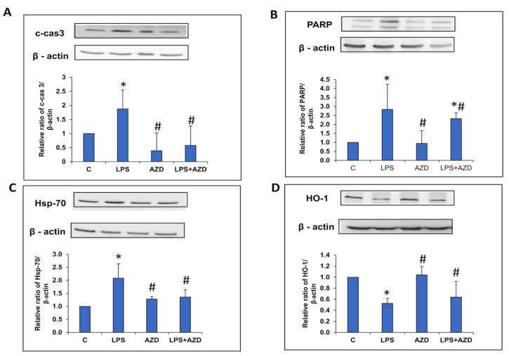
Figure 4.
LPS-induced expression of oxidative stress markers. Protein extracts from Rin-5F cells treated with LPS with/without AZD were separated on 12% SDS-PAGE and transferred on to nitrocellulose paper by Western blotting and protein bands detected using the appropriate primary antibodies. Specific antibodies against cleaved caspase 3 (A), PARP (B), Hsp-70 (C), and HO-1 (D) were used to detect the respective proteins and visualized by enhanced chemiluminescence using the Sapphire Biomolecular Imager (Azure biosystems, Dublin U.S.A) or using X-ray films. Beta actin was used as a loading control. Histograms represent the relative ratios of the quantitated proteins normalized against the loading control. The figures are a representative of at least three individual repetitive experiments. Asterisks indicate significant differences fixed at p ≤ 0.05 (* indicates significant difference relative to control untreated cells, whereas # indicates significant difference relative to LPS-treated cells).

