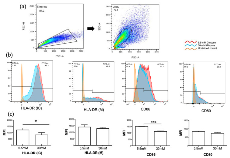
Figure 3.
Detection of MHC class II (HLA-DR) and co-stimulatory molecules (CD86, CD80) in 5.5 and 30mM of glucose. Monocyte derived macrophages (MDMs) were gated for singlets on a forward scatter height/forward scatter area density plot (a, left). MDMs were then gated on a side scatter area/forward scatter area density plot (a, right) for detection of intracellular HLA-DR (HLA-DR IC), membrane HLA-DR (HLA-DR M), CD86, and CD80. Histograms of each marker of a representative experiment are shown (b). (c) MFI of the indicated markers were measured; mean ± SD three independent experiments are shown. (d) Percentage of cells expressing each marker. Data were analyzed using unpaired t test. Differences were considered significant when p < 0.05 with asterisks as follows: * p < 0.01, ** p < 0.003 and *** p < 0.0003. MFI, mean fluorescence intensity.


