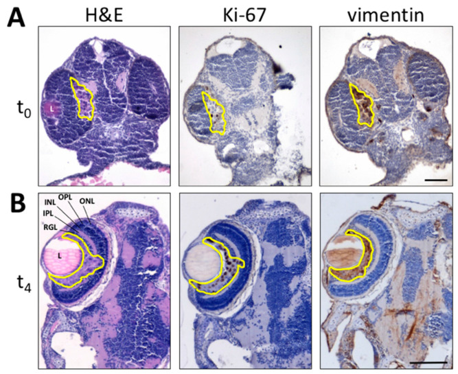
Figure 2.
Histological analysis of melanoma B16-BL6-DsRed+ xenografts. Paraffin sections of B16-BL6-DsRed+ cells grafted into zebrafish embryo eyes obtained at 1 h (t0) (A) or 4 days (t4) post implantation (B) are stained by H&E (left panel) whereas Ki-67 (central panel) and vimentin (right panel) immunoreactivity is shown in brown. Tumor area is highlighted in yellow. L, lens; INL, inner nuclear layer; IPL, inner plexiform layer; ONL, outer nuclear layer; OPL, outer plexiform layer; RGL, retinal ganglion cell layer. Scale bars: 50 µm.

