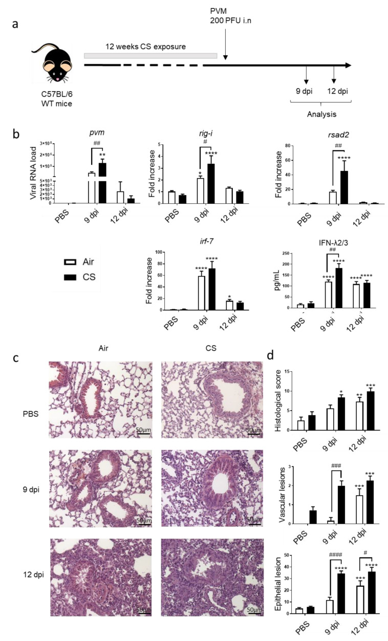
Figure 1.
PVM infection exacerbates COPD. (a) Experimental design of the study: the day of the infection was defined as day 0, allowing the association of this time point with the end of cigarette smoke (CS) protocol, consisting of mouse exposure to ambient air (Air) or CS (5 cigarettes/day, 5 days week) for 12 weeks. Analysis was performed at 9 and 12 days post infection (dpi). (b) Viral load and antiviral response, including mRNA expression rig-i, rsad-2 and irf-7, was evaluated by RT-qPCR in lung tissues. Results were expressed as fold increase compared to Air mice exposed to PBS using hprt1 expression as a house keeping gene. IFN-λ2/3 was evaluated by ELISA (pg/mL). (c) Histological changes were evaluated at 9 and 12 dpi. Representative slides are shown after HE staining. Scale bar = 50 µm. (d) Histological score analysis including vascular lesion and epithelial damages is expressed as mean ± SEM. Air mice (white bars) and CS mice (Black bars). * p < 0.05, ** p < 0.01, *** p < 0.001 and **** p < 0.0001 correspond to virus effect (PVM vs. PBS). # p < 0.05, ## p < 0.01, ### p < 0.001 and #### p < 0.0001 correspond to CS effect (CS vs. Air). Three independent experiments have been performed with 3–5 mice in each group per experiment.

