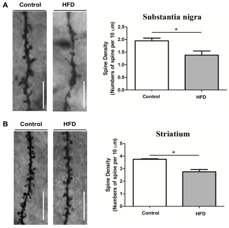
Figure 2.
Representative Golgi staining images of dopaminergic neurons in substantia nigra and striatum [90] (reused as per the International Journal of molecular Sciences copyright permission policy). (A) Represents decreased dendritic spine density in substantia nigra of high fat diet (HFD) mice compared to control. (B) Represents decreased dendritic spine density in striatum of high fat diet (HFD) mice compared to control. * indicates the significant difference with respect to control.

