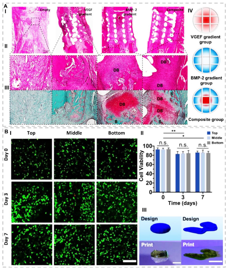
Figure 2.
(A). Investigation of impact of scaffolds containing BMP-2, VEGF, and their composition on defect regeneration after 2 weeks. (I) H&E stain with 500 µm and (II) with 100 µm scalebar. (III) Safranin O-stained DB points illustrate that cartilage develops to bone by undergoing endochondral ossification, and (B) points illustrate new bone tissues. (IV) Schematic of 3D-printed experimental groups, including key features of developed bioinks and segmental defect procedure. Construct design (4 mm in diameter, 5 mm in height)(reproduced content is open access) [48]. (B). (I) Representative Live/Dead images with 200 μm scale bar for cell viability and distribution in printed constructs for three layers (top, middle, and bottom) of scaffolds (II) quantification of cell viability, for top, middle, and bottom of printed discs after 0, 3, and 7 days of culture. n ≥ 3, * p < 0.05, ** p < 0.01, n.s. = not significant. (III) CAD design in comparison to representative image of a printed construct for designs of (right) a model femoral condyle and (left) a disc (~1.5 mm thickness, ~6.5 mm diameter). Scale bars = 1 cm (right) and 5 mm (left)(reproduced content is open access) [65].

