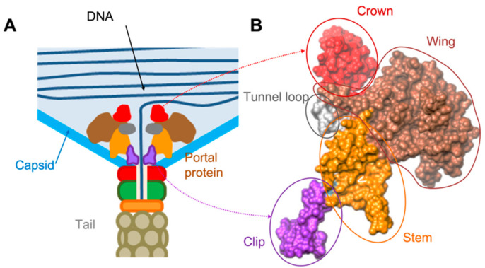
Figure 1.
Example of the of the portal protein location within the phage capsid and its domain organisation. (A) Overall diagram of the PP position. (B) One subunit from the portal protein complex gp6 of the SPP1 phage (pdb 2JES, [10]). Domains are indicated by colouring. Contacts with the capsid are made via wing and stem domains. Clip domains make contacts with accessory proteins of the tail.

