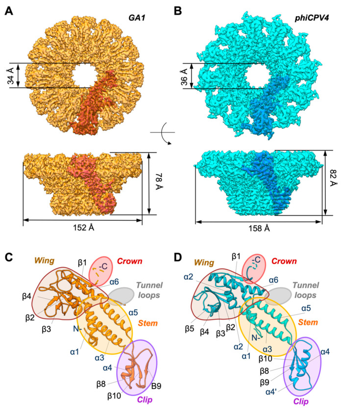
Figure 3.
Structures of the GA1 and phiCPV4 portal proteins. Sizes are as indicated; indicative monomer subunits are shown in deeper shade of orange on light orange and blue on cyan, respectively. (A) Top and side views of the GA1 PP. (B) Top and side views of the phiCPV4 PP. (C) Atomic model of the GA1 PP. (D) Atomic model of the phiCPV4 PP. Crown domains and DNA loops were not resolved (shown in pink and grey ovals correspondingly). The clip domain of the phiCPV4 PP is slightly bigger due to the presence of the additional short α4′ helix.

