Figure 4.
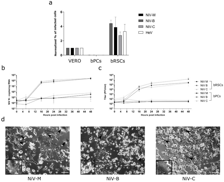
Susceptibility of bat reprogrammed cells (bRSCs) to henipaviruses. (a) Evaluation of entry of VSVΔG-RFP pseudotyped with Henipavirus glycoproteins into bPCs, bRSCs, and Vero cells. The infections of bPCs, bRSCs, and Vero cells were performed at an MOI of 0.01 and the percentages of infected cells were evaluated 6 h post-infection by measuring RFP by flow cytometry. (b–d) bPCs and bRSCs were infected with NiV-M, NiV-B and NiV-C isolate at an MOI of 0.1. The nucleocapsid gene transcription was quantified by RT-qPCR (b) and the release of virions into the supernatant was quantified by Vero plaque assay (c). (d) Cell cytopathic effects were observed under a light microscope at 24 h post-infection. Arrowheads show syncytia. Scale bar, 25 µm. Results labeled “bRSCs” correspond to bRSCs “EPI”. The same results were obtained with bRSCs “ESM2”.

