Abstract
B-cell maturation protein (BCMA) is a member of the tumor necrosis factor (TNF) receptor family and is expressed in B lymphocytes. BCMA binds two TNF family members, BAFF and APRIL, that stimulate cellular proliferation. BAFF in particular has been shown to influence B-cell survival and activation, and transgenic mice overexpressing BAFF have a lupus-like autoimmune disorder. We have inactivated BCMA in the mouse germ line. BCMA−/− mice have normal B-cell development, and the life span of mutant B lymphocytes is comparable to that of wild-type B cells. The humoral immune responses of BCMA−/− mice to T-cell-independent antigens as well as high and low doses of T-cell-dependent antigens are also intact. In addition, mutant mice have normal splenic architecture, and germinal centers are formed during an ongoing immune response. These data suggest a functional redundancy of BCMA in B-cell physiology that is probably due to the presence of TACI, another TNF receptor family member that is expressed on B cells and that can also bind BAFF and APRIL.
Members of the tumor necrosis factor (TNF) superfamily regulate a variety of cellular functions that include proliferation, differentiation, and apoptosis. In particular, several well-characterized members of the family such as TNF, lymphotoxins α and β, CD27 ligand (CD27L), CD30L, CD40L, OX40L, and FasL are known to be critical regulators of the immune system and are essential for lymphoid cell development and selection, immune tolerance, and cell death as well as immune responses against exogenous antigens (5, 7). Most TNF family members are synthesized as type II transmembrane precursors, and their extracellular domains can be cleaved to form soluble cytokines. However, both the soluble and the membrane-bound forms of the TNF ligand can bind to type I transmembrane receptors that contain one or more characteristic cysteine-rich motifs and belong to the TNF receptor family (7, 26, 29).
Recently, a new member of the TNF superfamily has been identified and termed BAFF (B-cell-activating factor belonging to the TNF family), BLyS (B-lymphocyte stimulator), TALL-1 (TNF and apoptosis ligand-related leukocyte-expressed ligand 1), THANK (TNF homologue that activates apoptosis, NF-κB, and c-Jun NH2-terminal kinase [JNK]), or zTNF4 (8, 19, 20, 23, 24). BAFF is expressed by monocytes and macrophages (21) as well as by T cells and dendritic cells (23). It has been shown specifically to bind to B cells (19, 23), suggesting that its receptor is expressed on this cell type. BAFF is known to stimulate B-cell proliferation and immunoglobulin secretion (19, 23) as well as modulate the survival of peripheral B cells (1, 3, 15, 27). Consistent with its role in regulating B-cell physiology, transgenic mice overexpressing BAFF develop a lupus-like autoimmune disorder (8, 12, 16), and human with systemic lupus erythematosus have elevated levels of BAFF in their blood (37).
The receptors for BAFF were identified as BCMA (B-cell maturation protein) and TACI (transmembrane activator and calcium modulator and cyclophilin ligand), two orphan members of the TNF receptor family (18, 25, 27, 32, 33, 35, 36). These two receptors are expressed on resting and activated B cells (6, 14, 17, 19, 23). Engagement of BCMA activates JNK, p38 mitogen activated protein kinase (MAPK) and the transcription factors NF-κB and Elk-1 (10), whereas cross-linking of TACI activates the transcription factors NF-κB and NF-AT (28). The physiological relevance of these two receptors was demonstrated by injecting soluble forms of either BCMA or TACI into mice. These decoy receptors disrupted immune responses and splenic architecture and prevented the accumulation of peripheral B cells (8, 27, 35, 36). In addition, they could even alleviate the autoimmune syndrome of lupus-prone mouse strains (8, 31).
Interestingly, both BCMA and TACI also bind APRIL (a proliferation-inducing ligand), another member of the TNF family that is closely related to BAFF (8, 11, 18, 22, 32, 33, 30, 36). APRIL has been shown to stimulate the proliferation of tumors (9) and, recently, B cells (36). The administration of recombinant APRIL to mice also led to an accumulation of B cells in vivo (36), similar to the effect of the administration of BAFF (19). Both APRIL and BAFF bind BCMA or TACI with equivalent affinity (8, 18, 22, 32, 33, 36), and it was not clear why there would be cross-interaction among the two ligands and two receptors.
Given the existence of two TNF ligands, APRIL and BAFF, that can bind independently to two TNF receptors, BCMA and TACI, it is difficult to deduce the relative contribution of each individual component of this dual receptor-dual ligand system to the regulation of B-cell physiology and humoral immune responses in vivo. Indeed, it is not known if one specific pair of ligand and receptor would play a more important role physiologically. We therefore undertook to dissect the system by selectively inactivating BCMA or/and TACI in the mouse germ line. In this report, we document the generation and characterization of mutant mice lacking BCMA.
MATERIALS AND METHODS
Generation of BCMA-deficient mice.
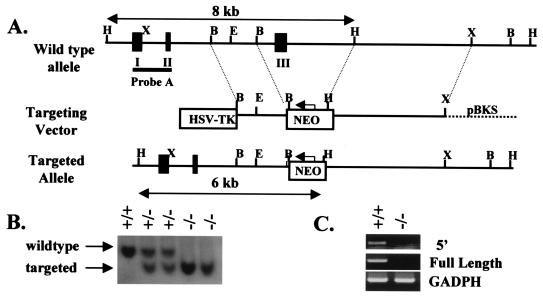
The cDNA for BCMA was obtained by reverse transcription-PCR (RT-PCR) of RNA isolated from mouse spleens, using the primers 5′-TCTTTCAGTGATCCAGTCCC-3′ and 5′-TCTCCTGACAGAAGGTTCTC-3′, and verified by sequencing. This cDNA is used to probe a mouse 129 genomic DNA library. Restriction enzyme digestion, Southern blotting, and DNA sequencing were used to map the genomic clone of BCMA. A targeting vector was constructed to replace the third and final exon of BCMA with a neo gene. A 4-kb HindIII-XhoI fragment 3′ and a 1.6-kb BamHI-BamHI fragment 5′ of the deleted exon were used as the long and short arms of homology, respectively. The targeting vector was linearized and electroporated into E14.1 embryonic stem (ES) cells, which were subsequently selected with 300 μg of G418 (Gibco) per ml and 2 μM ganciclovir. DNA was prepared from drug-resistant ES cell clones and digested with HindIII. Homologous recombinants were identified by Southern blotting using probe A (Fig. 1). The frequency of targeting was 1:50. Two targeted ES cell clones were injected into C57BL/6 blastocysts to generate chimeric mice for germ line transmission of the mutant allele. BCMA−/− mice were analyzed at 6 to 10 weeks of age and were of mixed 129-C57BL/6 background.
FIG. 1.
Gene inactivation of BCMA. (A) Partial restriction endonuclease map of the wild-type allele, the targeting vector, and the inactivated allele of BCMA (B, BamHI; E, EcoRI; H, HindIII; X, XhoI; pBKS, pBluescript KS). The black boxes represent exons. HindIII digestion of the genomic DNA will yield fragments of 8 and 6 kb for the wild-type and targeted alleles, respectively, as revealed by the external probe A. (B) Southern blot analysis of HindIII-digested tail DNA obtained from wild-type, BCMA+/− and BCMA−/− mice. (C) RT-PCR of splenic RNA samples obtained from wild-type and BCMA−/− mice. The 5′ RT-PCR identified the region corresponding to exon I of the gene. The housekeeping gene GADPH is included as control.
Examination of BCMA and TACI expression by RT-PCR.
Total RNA was extracted from splenocytes of wild-type and BCMA−/− mice by using TRIZOL reagent (GIBCO BRL) Five micrograms of the total RNA was reverse transcribed into cDNA, using the Superscript preamplification system (GIBCO BRL) in a reaction volume of 20 μl. The following primers were used to detect the presence of full-length BCMA cDNA (545 bp, primers 1 and 2) or the portion corresponding to exon I (278 bp, primers 1 and 3) of the BCMA cDNA: primer 1, 5′-TCTTTCAGTGATCCAGTCCC-3′; primer 2, 5′-CACTTTGCAAAGCAGTTGGC-3′; and primer 3, 5′-TTTGAGGCTCGTCCTTCAGG-3′. In addition, the expression of TACI was examined in a semiquantitative manner using the primer 5′-ATGGCTATGGCATTC-3′ and 5′-TCAGATCCCTGGTGCCTTCC-3′ in RT-PCR with various numbers of amplification cycles. Glyceraldehyde 3-phosphate dehydrogenase (GADPH) was also amplified as a control for the amount of template used in the RT-PCR.
Antibodies.
The antibodies used in flow cytometry and immunohistochemistry, all purchased from PharMingen (San Diego, Calif.), were anti-B220 (RA3-6B2), anti-immunoglobulin M (IgM) (R6-60.2), anti-CD5 (53-7.3), anti-CD21/35 (7G6), anti-CD23 (B3B4), and anti-Syndecan-1.
Flow cytometry analyses.
Single-cell suspensions were obtained from the spleen and lymph nodes by dissociation of the tissues with plastic mesh and rubber stoppers from 5-ml syringes. Peritoneal cavity and bone marrow cells were obtained by injecting staining medium (phosphate-buffered saline [PBS] containing 3% fetal bovine serum and 0.1% sodium azide) into the peritoneal cavity and into the femur and tibia, respectively, of each mouse. All cells were treated with red blood cell lysing solution (0.15 M NH4Cl, 1 mM KHCO3, 0.1 mM Na2EDTA) for 5 min at 4°C to eliminate erythrocytes. For flow cytometric analyses, cells were stained with optimal amounts of fluorescein isothiocyanate (FITC)-, phycoerythrin, and biotin-conjugated antibodies for 10 min at 4°C and subsequently washed twice with staining medium. Biotin-conjugated antibodies were revealed by second step staining with streptavidin-CyChrome. Flow cytometry analyses were performed on a FACScan (Becton Dickinson, Mountain View, Calif.) using CellQuest software.
BrdU incorporation studies.
Wild-type and mutant mice were fed continuously with water containing 1 mg of 5-bromo-2-deoxyuridinc (BrdU) per ml over a period of 1 to 3 weeks. Peritoneal cavity and splenic cells were harvested from the animals and surface stained with anti-B220 and anti-IgM antibodies to identify B cells. The cells were fixed for 30 min in 1 ml of ice-cold 70% ethanol and overnight in PBS containing 0.1% Tween 20 and 1% paraformaldehyde. Subsequently, the cells were washed once in PBS and incubated for 10 min at room temperature in a solution containing 150 mM NaCl, 5 mM MgCl2, 10 μM HCl, and 300 μg of DNase I per ml. Finally, the cells were stained intracellularly with the anti-BrdU antibody (Becton Dickinson) for 20 min at room temperature, washed twice in PBS, and analyzed on a FACScan.
Basal serum immunoglobulin levels.
To measure basal serum immunoglobulin levels, enzyme-linked immunosorbent assay (ELISA) plates were coated with rat anti-mouse Igκ antibodies (5 μg/ml). Sera obtained from wild-type and mutant mice were serially diluted and added into the wells. After the washing and blocking steps, horseradish peroxidase-coupled rat anti-mouse IgM, IgA, IgE, IgG1, IgG2a, IgG2b, or IgG3 antibodies were added; this was followed by addition of the ELISA substrate tetramethylbenzidine (Pierce). Levels of the various serum immunoglobulins were quantified against known standards that were also included in the assays.
Immunizations of BCMA-deficient mice with antigens.
The ability of BCMA−/− mice to mount a humoral immune response was assessed by immunizing the animals with the hapten 4-hydroxy-3-nitrophenyl acetyl (NP) as described previously (34). Wild-type and mutant mice were injected intraperitoneally with 10 μg of NP25-Ficoll (NP-Ficoll) in PBS to examine their immune responses to a T-cell-independent antigen. For the immune response to a T-cell-dependent antigen, mice were immunized intraperitoneally with 200 or 5 μg of alum-precipitated NP17-chicken globulin (NP-CG) in a high- or low-dose vaccination protocol, respectively. For the secondary immune response, all animals were challenged with 5 μg of alum-precipitated NP-CG. Sera were collected from the mice at various time points after immunizations to detect the presence of NP-specific antibodies in an ELISA. To detect NP-specific antibodies, the ELISA plates were coated with NP-bovine serum albumin (5 μg/ml; 50 μl/well) at 4°C overnight and subsequently blocked with (200 μl/well) 3% bovine serum albumin at room temperature for 2 h. Preimmune and immune sera were added at various dilutions to the wells of the ELISA plates at room temperature for 2 h; this was followed sequentially by addition of horseradish peroxidase-coupled rat anti-mouse antibodies and the ELISA substrate tetramethylbenzidine (Pierce). The wells of the ELISA plates were washed with PBS containing 0.02% Tween 20 between each incubation step. Specific antibodies of classes IgM and IgG3 and those of classes IgM and IgG1 were quantified for T-cell-independent and T-cell-dependent immune responses, respectively.
Immunohistochemistry.
Spleens from wild-type and BCMA−/− mice were snap-frozen in Tissue-Tek solution (Sakura, Finetek). Cryostat sections 8 to 10 μm thick were prepared on gelatin-coated slides and fixed in ice-cold acetone. The samples were rehydrated, blocked for 1 h in staining buffer (0.1% Triton X-100 in PBS) containing 5% heat-inactivated rat serum, and subsequently stained with FITC-and biotin-conjugated antibodies. The biotin-conjugated antibodies were revealed with streptavidin-Texas red (PharMingen). After the final washes, the slides were mounted in Gel Mount (Biomeda, Foster City, Calif.) and examined by laser confocal microscopy (MRC 1024; Bio-Rad).
RESULTS
Generation of BCMA-deficient mice.
BCMA was recently identified as a receptor for BAFF and APRIL, two TNF family members that play an important role in B-cell activation (8, 18, 25). To determine if BCMA is required for the in vivo development and activation of B lymphocytes, we have inactivated the receptor in the mouse. BCMA-deficient mice were generated by deleting the third exon of the bcma gene, which codes for amino acids 92 to 185 of the protein (Fig. 1A). This targeting strategy leads to the removal of most of the intracellular domain of BCMA which includes the region (amino acids 119 to 143) that is required for the binding of TNF receptor-associated factors, the activation of NF-κB, JNK, and p38 MAPK, and the induction of Elk-1 (10).
The deletion of exon III in mutant mice was verified by Southern blotting (Fig. 1B). To ensure that BCMA is indeed inactivated, RT-PCR was performed on RNA isolated from the spleens of mutant mice, using primers that correspond to exon I and to the full-length cDNA of BCMA. As shown in Fig. 1C, neither the full-length BCMA message nor a truncated version that would be encoded by exons I and II was detected in samples obtained from the mutant mice. This suggests that the bcma loci have been successfully disrupted in the mutant mice and that no aberrant or truncated protein is likely to be generated.
Normal B-cell development in BCMA-deficient mice.
BCMA is expressed only in B lymphocytes (6, 14) and thus may play an important role in B-cell development and/or activation. To determine the effect of BCMA inactivation in early B-cell development, we analyzed the bone marrow cells of mutant and wild-type mice by flow cytometry. As shown in Fig. 2A, B220+ IgM− pre-B cells as well as B220+ IgM+ immature B cells are found in equivalent proportions in BCMA−/− and wild-type mice. In addition, the populations of mature recirculating B220high IgM+ cells are also comparable in the wild-type and mutant animals. Thus, the data indicate that BCMA is not required for the development of early B cells. This is not surprising, as BCMA is expressed only at the mature stage of B-cell differentiation (6, 14).
FIG. 2.
B-cell populations in BCMA−/− mice, determined by flow cytometry analyses of B-cell populations found in bone marrow (A), spleens (B and C), and peritoneal cavities (D) of wild-type and BCMA−/− mice. Only IgM+ B cells are shown in panel C. Numbers indicate the percentages of cells within the lymphocyte forward and side scatter gates (A, B, and D) and percentages of IgM+ B cells for (D). The data shown are representative of more than five analyses.
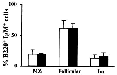
Since only B cells in the peripheral lymphoid organs express BCMA and its ligand BAFF is expressed by T cells and dendritic cells (23), which are known to interact with B cells, we next examine if the compositions of the various B-cell subpopulations found in the spleens of mice are altered in the absence of this protein. It has been reported that the treatment of mice with soluble BAFF expands certain B-cell subpopulations such as the marginal zone B cells in the spleen (1, 27). As shown in Fig. 2B, 2C, and 3, splenic B cells found in the mutant mice are indistinguishable with respect to phenotype or subpopulation distribution from those in wild-type animals. The B220+ IgM+ B cells are present within normal range in BCMA−/− mice compared to wild-type mice (Fig. 2B). In addition, the different subpopulations of immature, follicular, and marginal zone B cells, defined by their relative expression of the CD21/35 and CD23 antigens, are also unchanged (Fig. 2C and 3).
FIG. 3.
Distribution of various B-cell populations in spleens of BCMA−/− mice. The distribution of marginal zone (MZ), immature (Im), and follicular B cells as defined in Fig. 2C was examined in four wild-type (white boxes) and five mutant (black boxes) mice and expressed as percentage of total splenic B cells.
Next, we examine if the absence of BCMA would affect the development of the CD5+ B cells that reside predominantly in the peritoneal cavity of mice, given that its ligand BAFF is also produced and secreted by monocytes and myeloid cells (21) that are found in sizeable numbers at this location. CD5+ B cells can be distinguished from normal B cells by their expression of the CD5 antigen. As seen in Fig. 2D, CD5+ B cells are also found in the peritoneal cavities of BCMA−/− mice, suggesting that this B-cell subset is not perturbed by the inactivation of BCMA.
Finally, enumeration of the B cells in the bone marrow, spleens, lymph nodes, and peritoneal cavities of BCMA−/− mice indicates that their numbers are comparable to those of wild-type animals (data not shown). This suggests that the absence of BCMA has no effect on the size of the peripheral B-cell populations. Taken together, the data indicate that a BCMA deficiency does not alter the maturation of B cells or the distribution of the various subsets of peripheral B cells.
BCMA-deficient and wild-type B cells have comparable life spans.
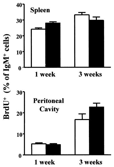
Recently, it has been reported that BAFF mediates the survival of peripheral B lymphocytes (1, 27). Therefore, we performed BrdU-labeling studies in vivo to determine if the life span of B cells is altered in the absence of BCMA. The life span of B cells is reflected by the turnover of the B-cell population in the periphery, which is measured by a change in the fraction of B cells that have incorporated BrdU over a period of time (4). An increased in the fraction of BrdU+ B cells would suggest a shortened life span, and vice versa. Mice were continuously fed with BrdU-containing water for a period of 1 or 3 weeks. As shown in Fig. 4, the fraction of IgM+ B cells in the spleen and peritoneal cavity that have incorporated BrdU is comparable between mutant and wild-type animals over a 1-week period. There seems to be a slightly higher turnover of the B-cell population in the peritoneal cavity of BCMA−/− mice compared to wild-type mice over a 3-week labeling period; however, this difference is not detected in the splenic B-cell population. Taken together, the data suggest that the average life span of B lymphocytes is not significantly altered in the absence of BCMA.
FIG. 4.
Turnover of wild-type and BCMA-deficient B cells. Mice were continuously fed with BrdU in drinking water for a period of 1 or 3 weeks. B220+ IgM+ splenic and peritoneal cavity B cells were stained for intracellular BrdU content. Groups of four wild-type (white box) and five mutant (black box) mice were analyzed for each time point.
BCMA-deficient mice have normal humoral immune responses.
Treatment of normal mice with soluble BAFF leads to elevated levels of serum immunoglobulins (19), and transgenic mice overexpressing BAFF develop a lupus-like syndrome characterized by the presence of autoantibodies (8, 12, 16). In addition, the administration of a soluble form of BCMA to mice inhibits (36), whereas treatment of mice with recombinant BAFF enhances (3), humoral immune responses. These findings together suggest that both BCMA and its ligand BAFF are involved in B-cell responses to antigens. To assess if the B cells in BCMA-deficient mice are functionally normal, we first examine the basal immunoglobulin levels in the sera of mutant mice. As shown in Fig. 5, the serum concentrations of the various classes of immunoglobulins such as IgM, IgG1, IgG2a, IgG2b, IgG3, and IgA as well as those of IgE (data not shown) are within the normal range, suggesting that there is no overt general activation or anergy of B lymphocytes in BCMA−/− mice.
FIG. 5.
Basal serum immunoglobulin levels in BCMA−/− mice. The concentrations of various serum immunoglobulin isotypes were measured by ELISA, and the value for each wild-type (open circles) and mutant (filled circles) mouse was plotted.
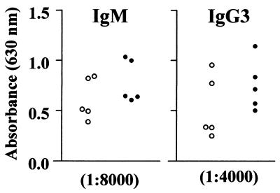
Antigens that elicit an antibody response from B cells can be classified as T cell independent or T cell dependent according to their dependency on CD4+ T-cell help. To determine if BCMA−/− mice could mount efficient immune responses against exogenous antigens, we first immunized the mice with a T-cell-independent antigen, NP-Ficoll. The primary antibody response to NP-Ficoll is mainly of the IgM and IgG3 class. As shown in Fig. 6, BCMA−/− mice are able to respond to T-cell-independent antigen, and the mutant B cells secrete equivalent if not slightly higher IgM and IgG3 antigen-specific antibodies compared to wild-type animals a week after the initial challenge with NP-Ficoll.
FIG. 6.
BCMA−/− mice have normal T-cell-independent immune responses. Wild-type (open circles) and mutant (filled circles) mice were immunized with 10 μg of the T-cell-independent antigen NP-Ficoll. The amount of antigen-specific antibodies of the IgM and IgG3 class was measured in an ELISA 8 days after the challenge. The value for each mouse was plotted. Preimmune sera were negative for the antigen-specific antibodies and are not shown.
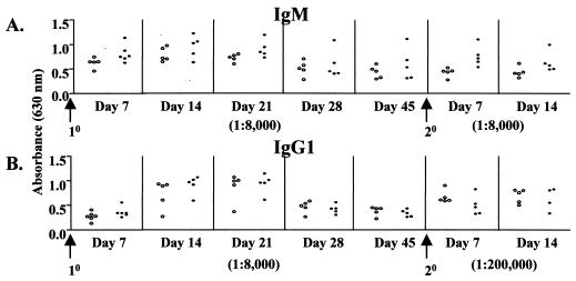
For the T-cell-dependent immune response, we immunized the mice with either a high or a low dose of the antigen NP-CG for the primary response and rechallenged the mice 45 days later with a low dose of the same antigen for the secondary response. The antibody response to NP-CG is mostly of the IgM and IgG1 class. In the high-dose immunization regimen (Fig. 7), the primary and secondary immune responses of BCMA-deficient mice are comparable to those of the wild-type mice across the various time points examined, although there seems to be a slightly higher antigen-specific IgM titer in the mutant animals within the first week of the primary and secondary challenge (Fig. 7A). The antigen-specific IgG titer is, however, comparable between the wild-type and mutant mice (Fig. 7B).
FIG. 7.
Primary and secondary immune responses of BCMA-deficient mice to a high dose of T-cell-dependent antigen. Wild-type (open circles) and mutant (filled circles) mice were immunized with 200 μg of the alum-precipitated T-cell-dependent antigen NP-CG for the primary (10) response and reboosted at day 45 with 5 μg of the antigen for the secondary (20) immune response. Sera were collected from the mice at various time points after primary and secondary immunizations and quantified for the presence of NP-specific antibodies of the IgM and IgG1 classes. The immune sera were diluted as indicated. The value for each mouse was plotted. Preimmune sera were negative for the antigen-specific antibodies and are not shown.
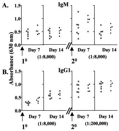
Similarly, in the low-dose immunization regimen (Fig. 8), BCMA−/− and wild-type mice do not differ significantly in their primary and secondary immune responses with the exception that there is again a slightly higher IgM titer immediately following a reboost.
FIG. 8.
Primary and secondary immune responses of BCMA-deficient mice to a low dose of T-cell-dependent antigen. Wild-type (open circles) and mutant (filled circles) mice were immunized with 5 μg of alum-precipitated NP-CG and reboosted at day 45 with the same amount of the antigen. The ELISA was performed as for Fig. 7.
Overall, the serological data indicate that BCMA−/− mice are able to mount normal humoral immune responses to T-cell-independent and T-cell-dependent antigens. Indeed, consistent with the serological data on the basal and antigen-specific antibody titers, our flow cytometry analysis indicates that plasma cells, as identified by their expression of the Syndecan-1 marker, are found in equivalent proportions in wild-type and BCMA−/− mice (Fig. 9).
FIG. 9.
Distribution of Syndecan-1-expressing B cells in BCMA−/− mice, determined by flow cytometry analyses of Syndecan-1-expressing B cells found in the spleens of naive mice (A) and wild-type and BCMA−/− mice 10 days post-NP-CG challenge (B). Numbers indicate percentages of total B cells present.
BCMA-deficient mice have normal splenic architecture.
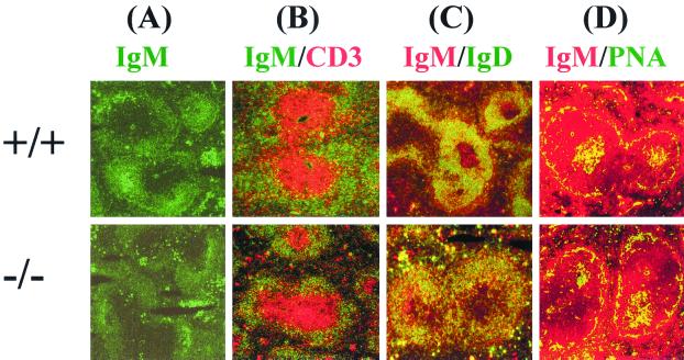
The various immune system cells are organized into discrete zones in the peripheral lymphoid organs (5), and certain members of the TNF/TNF receptor family, such as TNF receptor I, have been shown to be essential for this partitioning process (7). In addition, treatment of mice with a soluble form of BCMA can lead to disrupted splenic architecture (36). We thus examined whether the disruption of BCMA would affect the morphology of the splenic architecture by staining cryosections with fluorescence-labeled antibodies that recognize B and T cells. The primary follicle comprising the B- and T-cell zones appears to be normal, as judged by the anti-IgM and anti-CD3 staining that recognize separately B and T cells (Fig. 10A and B). In addition, the IgMhigh marginal zone B cells are present, as revealed by anti-IgM and anti-IgD staining (Fig. 10C), consistent with the flow cytometry data in Fig. 2C indicating the presence of this B-cell subpopulation. During an ongoing immune response, germinal centers comprising antigen-specific B cells are formed, and these structures are readily identified with peanut agglutinin staining. As shown in Fig. 10D, germinal centers are observed in both wild-type and mutant mice following immunizations with NP-CG. Thus, in summary, no obvious differences were observed in the splenic architecture of BCMA−/− mice.
FIG. 10.
Structures of primary follicles and germinal centers in spleens of BCMA-deficient mice. Cryosections of spleens from wild-type and mutant mice were stained with the indicated antibodies conjugated to FITC (green) or Texas red (red). Anti-IgM and anti-IgD stain B cells, anti-CD3 stains T cells, and peanut agglutinin (PNA) stains germinal center B cells.
Expression of TACI in BCMA-deficient mice.
The lack of an obvious phenotype in BCMA−/− mice could be due to the presence of TACI, the other TNF receptor family member that also binds BAFF and APRIL (8, 18, 32, 33, 35, 36). It is also possible that the inactivation of BCMA could lead to an up-regulation of TACI, which could in turn compensate for BCMA deficiency. To examine if this is the case, we perform semiquantitative RT-PCR of TACI expression using splenocytes obtained from BCMA−/− mice. As seen in Fig. 11, the expression of TACI is up-regulated in the absence of BCMA.
FIG. 11.
Semiquantitative RT-PCR of TACI expression in BCMA−/− mice. Total RNA was extracted from splenocytes of wild-type and BCMA−/− mice, and the reverse-transcribed cDNA was subjected to PCR for the numbers of cycles indicated. GADPH was amplified as a control for the amount of template present in the reaction.
DISCUSSION
BCMA was first identified as the product of a gene that was fused to the interleukin-2 gene by a t(4;16)(q26;p13) translocation in a malignant T-cell lymphoma (13). Subsequently, BCMA was shown to be expressed exclusively in B lymphocytes and in particular at the mature B to plasma cell stage of differentiation (6, 14). The latter observation raises the interesting possibility that BCMA might play an important role in B-cell activation and terminal differentiation. Indeed, the physiological relevance of BCMA became apparent when it was shown to be the receptor for the TNF family members APRIL and BAFF (8, 18, 22, 27). Both APRIL and BAFF enhance cell survival and induce the proliferation of B cells as well as stimulate their antibody secretion (19, 23, 36). Given the potency of APRIL and BAFF, one would expect that the inactivation of BCMA would affect B-cell development and/or activation in vivo.
We have inactivated BCMA in the mouse germ line. BCMA-deficient mice have no drastic phenotype in terms of B-cell differentiation and activation and can respond to both T-cell-independent and T-cell-dependent antigens. This seems to contrast with experiments wherein the administration of soluble BCMA to mice led to a drastic reduction in the peripheral B-cell population (27) and impaired immune responses in vivo (36). However, this apparent discrepancy can be explained by the fact that TACI, another member of the TNF receptor family, also binds APRIL and BAFF (8, 18, 32, 33, 35, 36). Thus, in BCMA−/− mice, the two TNF family cytokines could still bind TACI, and that presumably is sufficient to elicit a biological response. On the other hand, the administration of soluble BCMA to mice would block the binding of both BAFF and APRIL to BCMA and TACI. This would mimic a double BCMA-TACI gene knockout in mice, which presumably resulted in a more pronounced phenotype. Our data showing the up-regulation of TACI in the absence of BCMA (Fig. 11) could also explain why BCMA−/− mice have no obvious phenotype, as the increased TACI expression may compensate for the lack of BCMA. Indeed, the slightly higher antigen-specific immunoglobulin titers in BCMA−/− mice in the first week of the immune response (Fig. 6 to 8) may be due to the up-regulation of TACI expression.
Using soluble BAFF as a staining reagent, several groups had initially showed that the BAFF receptor was constitutively expressed on B cells (19, 23). We now know that such a staining will identify cells that express BCMA and/or TACI. However, BCMA is reported to be expressed at the mature B to plasma cell stage of differentiation (6, 14, 17). Thus, it appears that TACI may be the more widely expressed receptor and may play a more important role than BCMA in vivo. It will be interesting to determine if a single inactivation of TACI is sufficient or whether a double TACI-BCMA knockout is required to produce a phenotype in the mouse. As a corollary, it remains to be determined if TACI-transgenic mice would have equivalent or a more pronounced phenotype compared to BCMA-transgenic mice.
It is still not clear why a dual ligand-two receptor system exists for BAFF-APRIL and BCMA-TACI. One scenario would be that one of the receptors might act as a negative regulator to repress the activation process. This would be analogous to the CD28–CTLA-4 system (2), whereby the constitutively expressed CD28 provides the costimulatory signal whereas the inducibly expressed CTLA-4 provides the inhibitory signal upon binding the B7.1 and B7.2 molecules. Our current data suggest that TACI alone is sufficient to provide the activation signal, as BCMA-deficient mice can mount a normal immune response. On the other hand, BCMA does not appear to provide a negative signal as the immune responses of mutant mice are not exaggerated but are comparable to those of wild-type controls. Thus, BCMA may be redundant in the presence of TACI.
ACKNOWLEDGMENTS
We thank Esther Wong for blastocyst injection, the IMCB In Vivo Model Unit for the care and maintenance of mice, and Guo-ke, Li Jie, and Bor-Luen Tang for their kind assistance with histology and confocal microscopy.
This work is supported by grants from The National Science and Technology Board (NSTB) of Singapore.
REFERENCES
- 1.Batten M, Groom J, Cachero T G, Qian F, Schneider P, Tschopp J, Browning J L, Mackay F. BAFF mediates survival of peripheral immature B lymphocytes. J Exp Med. 2000;192:1453–1466. doi: 10.1084/jem.192.10.1453. [DOI] [PMC free article] [PubMed] [Google Scholar]
- 2.Chambers C A, Allison J P. Co-stimulation in T cell responses. Curr Opin Immunol. 1997;9:396–404. doi: 10.1016/s0952-7915(97)80087-8. [DOI] [PubMed] [Google Scholar]
- 3.Do R K, Hatada E, Lee H, Tourigny M R, Hilbert D, Chen-Kiang S. Attenuation of apoptosis underlies B lymphocyte stimulator enhancement of humoral immune response. J Exp Med. 2000;192:953–964. doi: 10.1084/jem.192.7.953. [DOI] [PMC free article] [PubMed] [Google Scholar]
- 4.Forster I, Muller W, Schittek B, Rajewsky K. Generation of long-lived B cells in germ-free mice. Eur J Immunol. 1991;21:1779–1782. doi: 10.1002/eji.1830210732. [DOI] [PubMed] [Google Scholar]
- 5.Fu Y X, Chaplin D D. Development and maturation of secondary lymphoid tissues. Annu Rev Immunol. 1999;17:399–433. doi: 10.1146/annurev.immunol.17.1.399. [DOI] [PubMed] [Google Scholar]
- 6.Gras M P, Laabi Y, Linares-Cruz G, Blondel M O, Rigaut J P, Brouet J C, Leca G, Haguenauer-Tsapis R, Tsapis A. BCMAp: an integral membrane protein in the Golgi apparatus of human mature B lymphocytes. Int Immunol. 1995;7:1093–1106. doi: 10.1093/intimm/7.7.1093. [DOI] [PubMed] [Google Scholar]
- 7.Gravestein L A, Borst J. Tumor necrosis factor receptor family members in the immune system. Semin Immunol. 1998;10:423–434. doi: 10.1006/smim.1998.0144. [DOI] [PubMed] [Google Scholar]
- 8.Gross J A, Johnston J, Mudri S, Enselman R, Dillon S R, Madden K, Xu W, Parrish-Novak J, Foster D, Lofton-Day C, Moore M, Littau A, Grossman A, Haugen H, Foley K, Blumberg H, Harrison K, Kindsvogel W, Clegg C H. TACI and BCMA are receptors for a TNF homologue implicated in B-cell autoimmune disease. Nature. 2000;404:995–999. doi: 10.1038/35010115. [DOI] [PubMed] [Google Scholar]
- 9.Hahne M, Kataoka T, Schroter M, Hofmann K, Irmler M, Bodmer J L, Schneider P, Bornand T, Holler N, French L E, Sordat B, Rimoldi D, Tschopp J. APRIL, a new ligand of the tumor necrosis factor family, stimulates tumor cell growth. J Exp Med. 1998;188:1185–1190. doi: 10.1084/jem.188.6.1185. [DOI] [PMC free article] [PubMed] [Google Scholar]
- 10.Hatzoglou A, Roussel J, Bourgeade M-F, Rogier E, Madry C, Inoue J, Devergne O, Tsapis A. TNF receptor family member BCMA (B cell maturation) associates with TNF receptor-associated factor (TRAF) 1, TRAF2, and TRAF3 and activates NF-κB, Elk-1, c-Jun N-terminal kinase, and p38 mitogen-activated protein kinase. J Immunol. 2000;165:1322–1330. doi: 10.4049/jimmunol.165.3.1322. [DOI] [PubMed] [Google Scholar]
- 11.Kelly K, Manos E, Jensen G, Nadauld L, Jones D A. APRIL/TRDL-1, a tumor necrosis factor-like ligand, stimulates cell death. Cancer Res. 2000;60:1021–1027. [PubMed] [Google Scholar]
- 12.Khare S D, Sarosi I, Xia X-Z, McCabe S, Miner K, Solovyev I, Hawkins N, Kelley M, Chang D, Van G, Ross L, Delaney J, Wang L, Lacey D, Boyle W J, Hsu H. Severe B cell hyperplasia and autoimmune disease in TALL-1 transgenic mice. Proc Natl Acad Sci USA. 1999;97:3370–3375. doi: 10.1073/pnas.050580697. [DOI] [PMC free article] [PubMed] [Google Scholar]
- 13.Laabi Y, Gras M P, Carbonnel F, Brouet J C, Berger R, Larsen C J, Tsapis A. A new gene, BCM, on chromosome 16 is fused to the interleukin 2 gene by a t(4;16)(q26;p13) translocation in a malignant T cell lymphoma. EMBO J. 1992;11:3897–3904. doi: 10.1002/j.1460-2075.1992.tb05482.x. [DOI] [PMC free article] [PubMed] [Google Scholar]
- 14.Laabi Y, Gras M P, Brouet J C, Berger R, Larsen C J, Tsapis A. The BCMA gene, preferentially expressed during B lymphoid maturation, is bidirectionally transcribed. Nucleic Acids Res. 1994;22:1147–1154. doi: 10.1093/nar/22.7.1147. [DOI] [PMC free article] [PubMed] [Google Scholar]
- 15.Laabi Y, Strasser A. Lymphocyte survival—ignorance is BLys. Science. 2000;289:883–884. doi: 10.1126/science.289.5481.883. [DOI] [PubMed] [Google Scholar]
- 16.Mackay F, Woodcock S A, Lawton P, Ambrose C, Baetscher M, Schneider P, Tschopp J, Browning J L. Mice transgenic for BAFF develop lymphocytic disorders along with autoimmune manifestations. J Exp Med. 1999;11:1697–1710. doi: 10.1084/jem.190.11.1697. [DOI] [PMC free article] [PubMed] [Google Scholar]
- 17.Madry C, Laabi Y, Callebaut I, Roussel J, Hatzoglou A, Le Coniat M, Mornon J P, Berger R, Tsapis A. The characterization of murine BCMA gene defines it as a new member of the tumor necrosis factor receptor superfamily. Int Immunol. 1998;11:1693–1702. doi: 10.1093/intimm/10.11.1693. [DOI] [PubMed] [Google Scholar]
- 18.Marsters S A, Yan M, Pitti R M, Haas P E, Dixit V M, Ashkenazi A. Interaction of the TNF homologues BLyS and APRIL with the TNF receptor homologues BCMA and TACI. Curr Biol. 2000;10:785–788. doi: 10.1016/s0960-9822(00)00566-2. [DOI] [PubMed] [Google Scholar]
- 19.Moore P A, Belvedere O, Orr A, Pieri K, LaFleur D W, Feng P, Soppet D, Charters M, Gentz R, Parmelee D, Li Y, Galperina O, Giri J, Roschke V, Nardelli B, Carrell J, Sosnovtseva S, Greenfield W, Ruben S M, Olsen H S, Fikes J, Hilbert D M. BLyS: member of the tumor necrosis factor family and B lymphocyte stimulator. Science. 2000;285:260–264. doi: 10.1126/science.285.5425.260. [DOI] [PubMed] [Google Scholar]
- 20.Mukhopadhyay A, Ni J, Zhai Y, Yu G L, Aggarwal B B. Identification and characterization of a novel cytokine, THANK, a TNF homologue that activates apoptosis, nuclear factor-kappaB, and c-Jun NH2-terminal kinase. J Biol Chem. 1999;274:15978–15981. doi: 10.1074/jbc.274.23.15978. [DOI] [PubMed] [Google Scholar]
- 21.Nardelli, Belvedere B O, Roschke V, Moore P A, Olsen H S, Migone T S, Sosnovtseva S, Carrell J A, Feng P, Giri J G, Hilbert D M. Synthesis and release of B-lymphocyte stimulator from myeloid cells. Blood. 2001;97:198–204. doi: 10.1182/blood.v97.1.198. [DOI] [PubMed] [Google Scholar]
- 22.Rennert P, Schneider P, Cachero T G, Thompson J, Trabach L, Hertig S, Holler N, Qian F, Mullen C, Strauch K, Browning J L, Ambrose C, Tschopp J. A soluble form of B cell maturation antigen, a receptor for the tumor necrosis factor family member APRIL, inhibits tumor cell growth. J Exp Med. 2000;192:1677–1684. doi: 10.1084/jem.192.11.1677. [DOI] [PMC free article] [PubMed] [Google Scholar]
- 23.Schneider P, MacKay F, Steiner V, Hofmann K, Bodmer J L, Holler N, Ambrose C, Lawton P, Bixler S, Acha-Orbea H, Valmori D, Romero P, Werner-Favre C, Zubler R H, Browning J L, Tschopp J. BAFF, a novel ligand of the tumor necrosis factor family, stimulates B cell growth. J Exp Med. 1999;189:1747–1756. doi: 10.1084/jem.189.11.1747. [DOI] [PMC free article] [PubMed] [Google Scholar]
- 24.Shu H-B, Wu W H, Johnston H. TALL-1 is a novel member of the TNF family that is down-regulated by mitogens. J Leukoc Biol. 1999;65:680–683. [PubMed] [Google Scholar]
- 25.Shu H-B, Johnson H. B cell maturation protein is a receptor for the tumor necrosis factor family member TALL-1. Proc Natl Acad Sci USA. 2000;97:9156–9161. doi: 10.1073/pnas.160213497. [DOI] [PMC free article] [PubMed] [Google Scholar]
- 26.Smith C A, Farrah T, Goodwin R G. The TNF receptor superfamily of cellular and viral proteins: activation, costimulation, and death. Cell. 1994;76:959–962. doi: 10.1016/0092-8674(94)90372-7. [DOI] [PubMed] [Google Scholar]
- 27.Thompson J S, Schneider P, Kalled S L, Wang L, Lefevre E A, Cachero T G, Mackay F, Bixler S A, Zafari M, Liu Z-Y, Woodcock S A, Qian F, Batten M, Madry C, Richard Y, Benjamin C D, Browning J L, Tsapis A, Tschopp J, Ambrose C. BAFF binds to the tumor necrosis factor receptor-like molecule B cell maturation antigen and is important for maintaining the peripheral B cell population. J Exp Med. 2000;192:129–135. doi: 10.1084/jem.192.1.129. [DOI] [PMC free article] [PubMed] [Google Scholar]
- 28.von Bulow G U, Bram R J. NF-AT activation induced by a CAML-interacting member of the tumor necrosis factor receptor superfamily. Science. 1997;278:138–141. doi: 10.1126/science.278.5335.138. [DOI] [PubMed] [Google Scholar]
- 29.Wallach D, Varfolomeev E E, Malinin N L, Goltsev Y V, Kovalenko A V, Boldin M P. Tumor necrosis factor receptor and Fas signaling mechanisms. Annu Rev Immunol. 1999;17:331–367. doi: 10.1146/annurev.immunol.17.1.331. [DOI] [PubMed] [Google Scholar]
- 30.Ware C F. APRIL and BAFF connect autoimmunity and cancer. J Exp Med. 2000;192:35–38. doi: 10.1084/jem.192.11.f35. [DOI] [PMC free article] [PubMed] [Google Scholar]
- 31.Ware C F. Decoy receptors thwart B cells. Nature. 2000;404:949–950. doi: 10.1038/35010263. [DOI] [PubMed] [Google Scholar]
- 32.Wu Y, Bressette D, Carrell J A, Kaufman T, Feng P, Taylor K, Gan Y, Cho Y H, Garcia A D, Gollatz E, Dimke D, LaFleur D, Migone T S, Nardelli B, Wei P, Ruben S M, Ullrich S J, Olsen H S, Kanakaraj P, Moore PA, Baker K P. Tumor necrosis factor (TNF) receptor superfamily member TACI is a high affinity receptor for TNF family members APRIL and BLyS. J Biol Chem. 2000;275:35478–35485. doi: 10.1074/jbc.M005224200. [DOI] [PubMed] [Google Scholar]
- 33.Xia X-Z, Treanor J, Senaldi G, Khare S D, Boone T, Kelley M, Theill L E, Colombero A, Solovyev I, Lee F, McCabe S, Elliott R, Miner K, Hawkins N, Guo J, Stolina M, Yu G, Wang J, Delaney J, Meng S-Y, Boyle W J, Hsu H. TACI is a TRAF-interacting receptor for TALL-1, a tumor necrosis factor family member involved in B cell regulation. J Exp Med. 2000;192:137–143. doi: 10.1084/jem.192.1.137. [DOI] [PMC free article] [PubMed] [Google Scholar]
- 34.Xu S, Tan J E, Wong E P, Manickam A, Ponniah S, Lam K-P. B cell development and activation defects resulting in xid-like immunodeficiency in BLNK/SLP-65-deficient mice. Int Immunol. 2000;12:397–404. doi: 10.1093/intimm/12.3.397. [DOI] [PubMed] [Google Scholar]
- 35.Yan M, Marsters S A, Grewal I S, Wang H, Ashkenazi A, Dixit V M. Identification of a receptor for BlyS demonstrates a crucial role in humoral immunity. Nat Immunol. 2000;1:37–41. doi: 10.1038/76889. [DOI] [PubMed] [Google Scholar]
- 36.Yu G, Boone T, Delaney J, Hawkins N, Kelley M, Ramakrishnan M, McCabe S, Qiu W, Kornuc M, Xia X-Z, Guo J, Stolina M, Boyle W J, Sarosi I, Hsu H, Senaldi G, Theill L. APRIL and TALL-1 and receptors BCMA and TACI: system for regulating humoral immunity. Nat Immunol. 2000;1:252–256. doi: 10.1038/79802. [DOI] [PubMed] [Google Scholar]
- 37.Zhang J, Roschke V, Baker K P, Wang Z, Alarcon G S, Fessler B J, Bastian H, Kimberly R P, Zhou T. A role for B lymphocyte stimulator in systemic lupus erythematosus. J Immunol. 2001;166:6–10. doi: 10.4049/jimmunol.166.1.6. [DOI] [PubMed] [Google Scholar]


