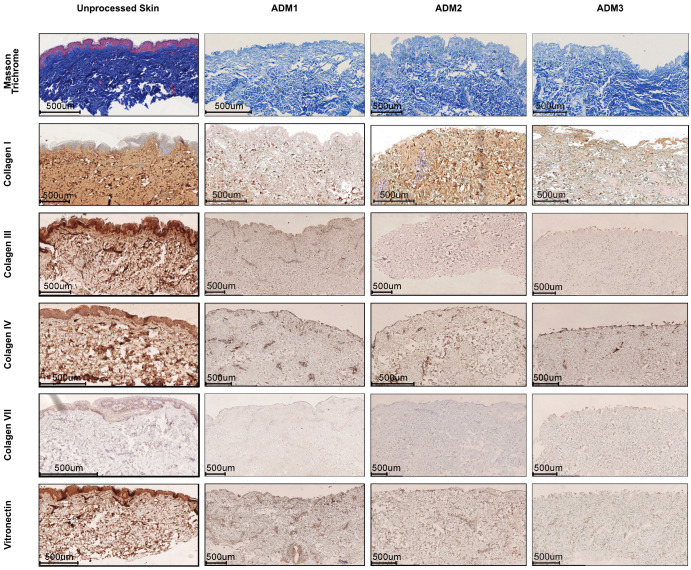
Figure 2.
Different decellularization protocols preserve matrix structure but change the presence of collagen fibers. Representative histochemical (Masson Trichrome) and immunohistochemical (Collagen I, II, III, IV, VII, and Vitronectin) staining of ADMs derived from abdominoplasty skin decellularized with ADM1, ADM2, or ADM3 protocol. Representative pictures of five independent analyses. Size bar—500 μm.

