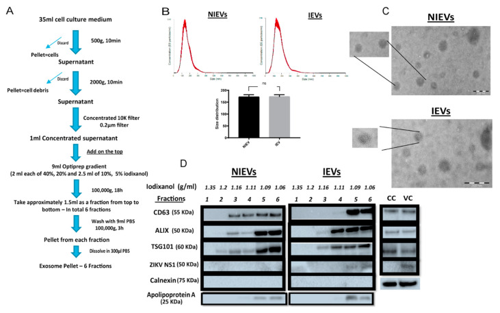
Figure 1.
Isolation and characterization of non-infected and ZIKV-infected cell-derived EVs. (A) Experimental workflow for extracellular vesicle (EV) isolation from non-infected/ZIKV-infected human brain microvascular endothelial cells (hcMEC/D3). (B) Nanoparticle tracking analysis reveals no significant differences in size and number of particles between non-infected (NIEVs) and ZIKV-infected EVs (IEVs). (C) Electron microscopy indicates the presence of membrane-enclosed vesicles smaller than 200 nm in both non-infected (upper) and infected (bottom) EV preparations. Scale bar graph: 200 nm. (D) Western blot analysis demonstrates the presence of distinct profiles of several EV- and non-EV markers among fractions of both NIEVs and IEVs. Thirty microliters of each fraction (density is indicated in g/mL) were loaded into the SDS polyacrylamide gel. Lysates from non-infected (CC) and ZIKV-infected cells (VC) were also included.

