Abstract
Neurotrophin receptors such as the tropomyosin receptor kinase A receptor (TrkA) and the low-affinity binding p75 neurotrophin receptor p75NTR play a critical role in neuronal survival and their functions are altered in Alzheimer’s disease (AD). Changes in the dynamics of receptors on the plasma membrane are essential to receptor function. However, whether receptor dynamics are affected in different pathophysiological conditions is unexplored. Using live-cell single-molecule imaging, we examined the surface trafficking of TrkA and p75NTR molecules on live neurons that were derived from human-induced pluripotent stem cells (hiPSCs) of presenilin 1 (PSEN1) mutant familial AD (fAD) patients and non-demented control subjects. Our results show that the surface movement of TrkA and p75NTR and the activation of TrkA- and p75NTR-related phosphoinositide-3-kinase (PI3K)/serine/threonine-protein kinase (AKT) signaling pathways are altered in neurons that are derived from patients suffering from fAD compared to controls. These results provide evidence for altered surface movement of receptors in AD and highlight the importance of investigating receptor dynamics in disease conditions. Uncovering these mechanisms might enable novel therapies for AD.
Keywords: Alzheimer’s disease, TrkA, p75NTR, receptor dynamics, live-cell single-molecule imaging, neuronal, human-induced pluripotent stem cell
1. Introduction
Changes in lateral diffusion of receptors on the plasma membrane are crucial in determining their functional status. The altered surface movements can result in the formation of membrane protein complexes that activate signaling molecules [1]. For instance, single-molecule tracking experiments showed that the neurotrophin receptors, such as the tropomyosin receptor kinase A receptor (TrkA), also known as high-affinity nerve growth factor receptor, have two distinct motility states on the membrane surface, defined as mobile and immobile phases [1]. The membrane recruitment of downstream intracellular signaling proteins occurs when TrkA is in the immobile phase [1]. It has also been shown that the diffusion coefficient (D) that is calculated from the cell surface movement of receptors provides a good estimate for the effect of a ligand on G protein-coupled receptors [2]. Consequently, the functions of receptors can be predicted by measuring the surface movement parameters of receptor molecules. A large number of diseases are initiated by dysregulation of the receptor-related signaling pathways such as Parkinson’s disease, schizophrenia, and cancer [3,4,5]. Although the expression level and pattern of various receptors have been extensively studied in disease models [6,7] the surface movement of receptors in different diseases has been scarcely investigated. It has recently been published that the surface diffusion of AMPA receptors is disturbed in rodent models of Huntington’s disease and the restoration of the AMPA receptor diffusion rescues memory dysfunction [8]. These data indicate that revealing receptor dynamics in different diseases may contribute to developing new treatments for neurodegenerative diseases such as Alzheimer’s disease (AD).
Alzheimer’s disease is the most common cause of dementia which is characterized by extracellular deposition of beta-amyloid (Aβ) and intracellular neurofibrillary tangles [9]. The familial form of AD (fAD) is caused by genetic mutations, such as mutations in genes for encoding presenilin (PSEN1 or PSEN2) proteins [10]. In AD the unbalanced signaling through the low-affinity binding p75 neurotrophin receptor (p75NTR) versus TrkA related signaling [6] plays a pivotal role in changes of altered cellular functions. TrkA and p75NTR activate signaling pathways that are essential to decide the fate of neurons [6,11].
TrkA and p75NTR are frequently co-expressed and interact with each other and they cooperate in mediating signals to survival [12,13]. TrkA alone stimulates pro-survival signaling pathways such as phospholipase C gamma (PLCγ), extracellular signal-regulated kinase 1/2 (ERK1/2), and phosphoinositide-3-kinase (PI3K)/serine/threonine-protein kinases (AKT) pathways [12,14]. On the other hand, in nerve growth factor (NGF) deficiency, TrkA switches from a pro-survival to a pro-apoptotic receptor [15]. p75NTR alone promotes apoptosis via the activation of sphingomyelinase/ceramide or c-jun N-terminal kinase (JNK) pathway but can signal survival when triggering NF-ƙB and PI3/AKT pathways [16,17,18].
Our understanding of AD pathogenesis is currently limited by difficulties in obtaining live neurons from patients, especially in the earlier stages of the disease. Furthermore, the culture of primary human neuronal cells is particularly challenging because of their limited life span [19]. Also, biopsies are often taken from patients at the end-stage of the disease and suitable control tissues that are taken from healthy individuals might be inaccessible due to ethical concerns and potential health risks [20]. Human-induced pluripotent stem cell (hiPSCs) based technology overcomes many of these limitations [21], providing a suitable cellular model for AD. Human iPSCs can be cultured for an unlimited time and differentiated into any type of cell of the human body, such as neurons of the central nervous system (CNS) [22]. We have successfully established and applied AD patient-derived hiPSC lines for modeling AD neuropathology [23,24].
Here, we applied these hiPSCs and performed single-molecule tracking approaches to examine the surface movement of p75NTR and TrkA in living human neurons from PSEN1 mutant patients and non-demented control subjects. We also examined how downstream signaling pathways, such as ERK1/2, AKT, and JNK1/2 that play essential roles in fAD, could be linked to TrkA and p75NTR activation change in PSEN1 mutant neurons.
2. Results
2.1. Characterization of Neuronal Properties of hiPSC-Derived Cultures
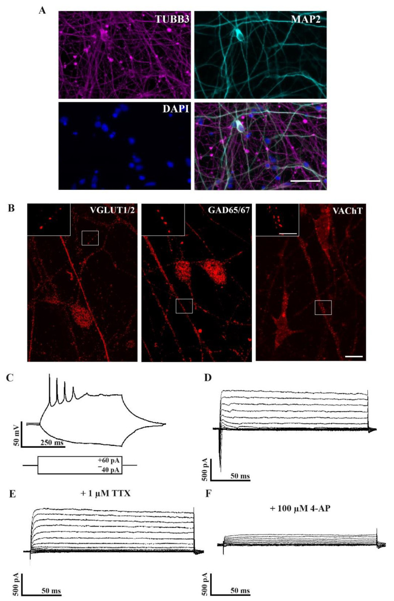
hiPSC-derived seven week old neurons were tested to see if they showed neuronal characteristics. All of the cell lines possessed neuron-like morphology and were positively stained for the neuronal markers, β–III tubulin (TUBB3) and microtubule-associated protein 2 (MAP2) (Figure 1A), representing the neuronal differentiation of iPSC derived cell cultures.
Figure 1.
Neuronal phenotypes of differentiated hiPSCs. Immunofluorescence staining of microtubule-associated protein 2 (MAP2, red) and β–III tubulin (TUBB3, green) indicating that the hiPSC-derived neuronal cultures show neuronal phenotypes by week seven (A). Nuclei were labeled with DAPI (blue). Scale bar = 50 µm. Images depict VGLUT1/2, GAD65/67, and VAChT immunoreactivity in the cytoplasm and neurites of seven week old human iPSC-derived neurons (B). Inserts demonstrate VGLUT1/2, GAD65/67, and VAChT immunoreactive dots on neurites of hiPSCs. IPSC lines: Ctrl-2, fAD-1. Scale bar = 10 µm, insert = 5 µm. Whole-cell patch-clamp recordings show that maturing neurons (seven weeks old) generate action potentials (C) and display Na+ and K+ currents (D) that can be blocked by TTX (E) or 4-AP (F), respectively; n = 12.
The identification of the neuronal phenotype of the hiPSC-derived neurons was conducted by immunolabeling them for glutamatergic (VGLUT1/2), GABAergic (GAD65/67), and cholinergic (VAChT) markers. Our results showed that hiPSC-derived neurons represent a phenotypically mixed population (Figure 1B) in agreement with our earlier published results [24].
In the next step, we examined whether our neuronal differentiation protocol resulted in cells with electrophysiological properties similar to mature neurons. Using the whole-cell patch-clamp technique, the passive electrophysiological parameters of 12 neurons were measured in the cultures. The resting membrane potential and the input resistance of the patched neurons were −64.75 ± 3.9 mV and 742.18 ± 64.59 MΩ, respectively (values are represented as mean ± SEM). Action potentials and voltage-gated Na+ and K+ currents were also recorded from these cells. As evidence for maturation, the cells exhibited either single or repetitive action potentials in response to positive current injection (Figure 1C). Furthermore, a series of 10 mV depolarization voltage steps from −90 mV resulted in the opening of Na+ and K+ channels, indicating fast inward and slow outward components, respectively (Figure 1D). Tetrodotoxin (TTX) blocked the inward, while 4-AP blocked the outward currents representing the activation of voltage-gated Na+ and K+ channels (Figure 1E,F). Overall, the seven week old hiPSC-derived cultures resembled mature neuronal cultures with mixed phenotypes.
2.2. Expression and Single-Molecule Detection of TrkA and p75NTR in Control and fAD hiPSC-Derived Neurons
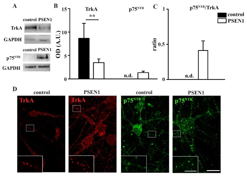
As the balance of TrkA and p75NTR signaling is disturbed in fAD [6], the total TrkA and p75NTR levels in hiPSC-derived neuronal cultures that were obtained from non-demented controls and fAD patients with the PSEN1 mutation were examined by Western blot. Our results demonstrated that the expression of TrkA significantly decreased in PSEN1 fAD hiPSC neurons (Figure 2A,B). In contrast, the p75NTR signal was below the detection limit in the cultures of control lines but was abundant in PSEN1 fAD hiPSC neurons (Figure 2A,B). Consequently, the ratio of p75NTR/TrkA was elevated in PSEN1 mutant neurons (Figure 2C). Next, immunocytochemical stainings for TrkA and p75NTR on fixed hiPSC-derived neuronal cultures that were obtained from non-demented control and PSEN1 mutant cell lines were performed. The results showed that both neurites and somata expressed TrkA and p75NTR (Figure 2D). In contrast to fixed immunocytochemistry, TrkA and p75NTR molecules were only observed on processes of live cells (Supplementary Movies S1 and S2). To reveal that the moving TrkA and p75NTR molecules are only detected on the neurites, we carried out correlated live-cell single-molecule (TrkA or p75NTR) imaging and fixed cell immunocytochemistry (TUBB3) experiments (Supplementary Movie S3).
Figure 2.
Characterization of TrkA and p75NTR expression in control and PSEN1 mutant neurons. Representative blots represent TrkA and p75NTR expression in PSEN1 mutant cultures compared to controls (A). IPSC lines: Ctrl-2, fAD-1; n = 6. Histograms demonstrate TrkA (A), p75NTR (B) expression in PSEN1 mutant neuronal cultures compared to controls and their ratio (C) (** p < 0.01). Images show TrkA (red) and p75NTR (green) expression in the cytoplasm and neurites of seven week old controls (Ctrl-2) and PSEN1 mutant neurons (fAD-1) (D). The data are expressed in optical densities (pixel/area) ± SEM. Scale bar = 20 µm, scale bar insert = 5 µm.
2.3. Surface Movement Characteristics of TrkA and p75NTR in Control hiPSC Neurons
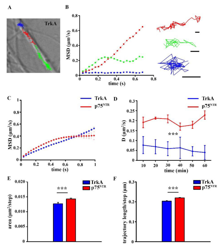
Live-cell imaging experiments were performed to determine the surface movement parameters of TrkA and p75NTR molecules in the plasma membrane of neurons using TIRF microscopy. After fluorescent labeling of TrkA and p75NTR receptors, 10 s long videos were recorded, single molecule movements were tracked, and then analyzed. Before comparing the diffusion parameters of TrkA and p75NTR in the control and fAD hiPSC neurons, we analyzed the surface dynamics of TrkA and p75NTR in hiPSC neurons that were derived from healthy subjects. Based on the MSD functions, TrkA and p75NTR molecules exhibited two main types of movements on hiPSC neurons that were obtained from non-demented control cell lines: Brownian diffusion, when receptors are moving freely between plasma membrane barriers, and confined motion when the surface movement of receptors are restricted to a small area (Figure 3A,B). On average, p75NTR molecules showed confined movements, while TrkA molecules exhibit Brownian diffusion (Figure 3C). The evaluated average diffusion coefficients of TrkA and p75NTR did not change significantly during the measurement either in the control or PSEN1 mutant samples (Figure 3D). However, the average diffusion coefficient of TrkA and p75NTR on healthy individual-derived neurites indicated that p75NTRs were moving significantly faster on neurites than TrkAs (Figure 3D; Table S1).
Figure 3.
Single-molecule imaging of TrkA and p75NTR in control neurons. Representative trajectories of TrkA molecules on neurites (A). Scale bar: 5 µm. The mean square displacement (MSD) functions represent TrkA molecules with different diffusion modes (B). Scale bars: 0.5 µm. MSD-Δt plots show the diffusion mode of TrkA and p75NTR in the control neurites (C). The diffusion coefficient of TrkA and p75NTR are shown on healthy individual-derived neurites at different time points (D). Histograms display the diffusion area (E) and the trajectory length (F) of TrkA and p75NTR molecules in control neurites. The data are expressed as mean ± SEM (*** p < 0.001). The number of trajectories of TrkA in the control and PSEN1 mutant neurons = 1154; 1246, the number of trajectories of p75NTR in control and PSEN1 mutant neurons = 1830; 2148. IPSC lines: Ctrl-1, Ctrl-2, Ctrl-3; fAD-1; fAD-2, fAD-3.
The diffusion area of p75NTR molecules was more extended on neurites that were derived from healthy individuals than that of TrkA (Figure 3E; Table S1). Accordingly, the length of p75NTR trajectories was significantly longer (Figure 3F; Table S1).
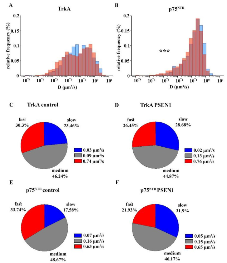
To identify the diffusion states of TrkA and p75NTR, we used Variational Bayesian-Hidden Markov model (VB-HMM) clustering analysis [25] on the trajectories for each neurite. The analysis showed that the diffusion of both TrkA and p75NTR molecules can be classified into three phases: slow, medium, and fast. Our results suggested that the percentage of the fast fraction increased, while the proportion of slow and medium phases did not change significantly when comparing p75NTR to TrkA molecules (Figure 5C,E; Table S2). Nevertheless, the average diffusion coefficient of the slow and medium fractions of p75NTR significantly increased compared to TrkA (Figure 5C,E; Table S2).
2.4. Comparison of the Surface Movement of TrkA and p75NTR in Control and PSEN1 fAD hiPSC-Derived Neurons
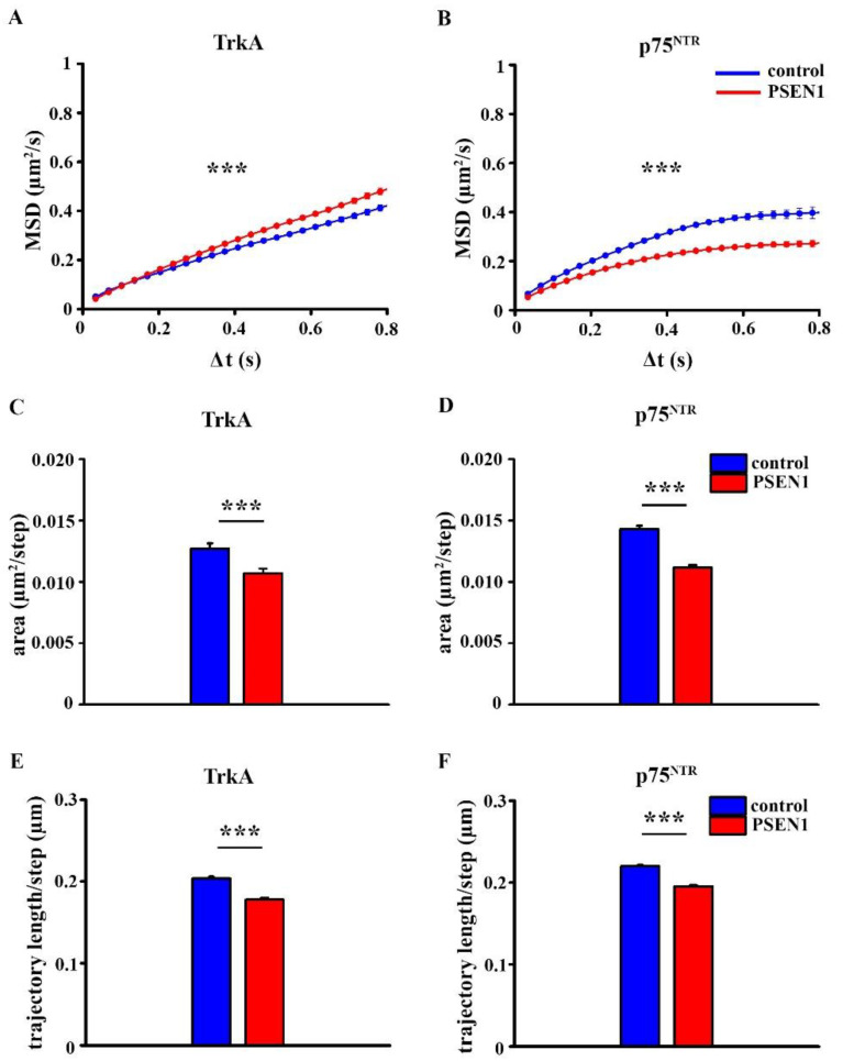
As surface movement of receptors determine their activation state [26,27], we compared the diffusion parameters of TrkA and p75NTR in healthy individual-derived and PSEN1 fAD hiPSC neurons. MSD-Δt plots of the trajectories showed that the surface movement of TrkA molecules were less confined in the PSEN1 fAD hiPSC neurons when compared with the non-demented control samples (Figure 4A). On the other hand, p75NTR molecules exhibited a more confined diffusion mode in the PSEN1 fAD hiPSC neurons (Figure 4B).
Figure 4.
MSD-Δt plots, diffusion area, and trajectory length of TrkA and p75NTR in the control and PSEN1 mutant neurons. MSD curves show the diffusion modes of TrkA (A) and p75NTR (B) in non-demented and PSEN1 mutant neurites. Graphs display the diffusion area of TrkA (C) and p75NTR (D) in the control and PSEN1 mutant neurites. Histograms exhibit the trajectory length of TrkA (E) and p75NTR (F) in control and PSEN1 mutant neurites. The data are expressed as mean ± SEM (*** p < 0.001). The number of trajectories of TrkA in control and PSEN1 mutant neurons = 1154; 1246, the number of trajectories of p75NTR in control and PSEN1 mutant neurons = 1830; 2148. IPSC lines: Ctrl-1, Ctrl-2, Ctrl-3; fAD-1; fAD-2, fAD-3.
The diffusion area was smaller in the PSEN1 fAD hiPSC neurons for both TrkA and p75NTR molecules (Figure 4C,D; Table S1). In addition, the trajectory length that was normalized to steps was also smaller in the case of both TrkA and p75NTR molecules in fAD neurons (Figure 4E,F; Table S1).
The average diffusion coefficient of TrkA molecules did not change in PSEN1 fAD hiPSC neurites compared to the non-demented control neurites (Figure 5A; Table S1). In contrast, the surface movement of p75NTR receptor molecules significantly decreased in the PSEN1 fAD hiPSC neurites (Figure 5B; Table S1).
Figure 5.
Distribution of the diffusion coefficients and the percentage of slow, medium, and fast fractions of TrkA and p75NTR in the control and PSEN1 mutant neurons. Logarithmic distribution histograms show the relative frequency of different diffusion intervals in the range of 10−5–10 µm2/s for TrkA (A) and p75NTR (B) in control (blue bars) and PSEN1 mutant neurites (red bars). The number of trajectories of TrkA in control and PSEN1 mutant neurons = 1154; 1246, p75NTR: the number of trajectories of p75NTR in control and PSEN1 mutant neurons = 1830; 2148. Pie charts demonstrate the percentage of slow, medium, and fast fractions of the trajectories of TrkA (C,D) and p75NTR (E,F) per neurite and their average diffusion coefficients in the control (C,E) and PSEN1 mutant neurons (D,F). The data are expressed as mean ± SEM (*** p < 0.001). The number of neurites in the control and PSEN1 mutant cultures for TrkA = 142; 197, the number of neurites in the control and PSEN1 mutant cultures for p75NTR = 154; 219. IPSC lines: Ctrl-1, Ctrl -2, Ctrl -3; fAD-1; fAD-2, fAD-3.
The percentage of slow, medium, and fast fractions of TrkA movement remained unchanged when comparing fAD and control neurites (Figure 5C,D; Table S2. The average diffusion coefficient of the slow phase decreased, the medium fraction increased, while the fast phase did not change in fAD neurites (Figure 5C,D; Table S2).
In contrast to TrkA, the proportion of slow trajectories of p75NTR increased while the percentage of fast trajectories decreased in the PSEN1 fAD hiPSC neurites (Figure 5E,F; Table S2). The average diffusion coefficient of the slow phase of p75NTR decreased in fAD, but the average diffusion coefficients of the other two fractions remained unchanged in the neurites of PSEN1 mutant hiPSC neurons (Figure 5E,F; Table S2).
2.5. Expression of the ERK1/2, AKT and JNK1/2 Signaling Pathways in Non-Demented Control and PSEN1 fAD hiPSC-Derived Neurons
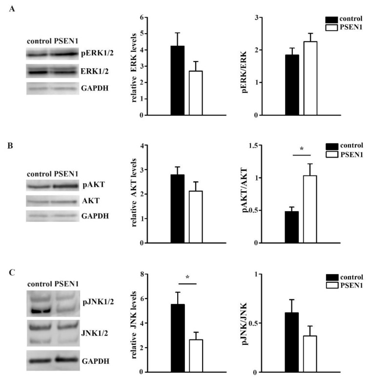
Since changes in surface movements of receptors lead to functional changes via the modification of signaling transduction [1], the activation of signaling pathways were examined. To investigate whether TrkA and p75NTR-related downstream signaling were altered in PSEN1 fAD hiPSC neurons, we examined the ERK1/2, AKT, and JNK1/2 signaling pathways. Our Western blot analysis revealed that the expression levels of ERK1/2 and AKT did not change, while the JNK1/2 levels significantly decreased in the cultures of PSEN1 fAD samples (Figure 6A–C). The activation of ERK1/2 and JNK1/2 pathway was not altered but the AKT signaling pathway was shown to be more active in the PSEN1 fAD cultures (Figure 6A–C).
Figure 6.
Signaling pathways in the control and PSEN1 mutant neurons. Representative blots illustrate the expression levels of pERK1/2, ERK1/2, pAKT, AKT, pJNK1/2, and JNK1/2 signaling molecules and GAPDH in PSEN1 mutant and control cultures. Histograms show the expression of ERK (A), AKT (B), JNK (C) normalized to GAPDH and their ratio of pERK/ERK, pAKT/AKT, and pJNK/JNK in the PSEN1 mutant cultures compared to controls. The data are expressed in optical densities (pixel/area) ± SEM (* p < 0.05), IPSC lines: Ctrl-2, fAD-1; n = 6.
3. Discussion
Our results showed that the surface trafficking of TrkA and p75NTR are altered in hiPSC-derived neurons that are differentiated from PSEN1 mutant fAD patients. The surface movement of TrkA molecules was less confined in PSEN1 mutant neurites. Contrarily, the trafficking of p75NTR molecules was more confined in the fAD neurites. The movement of TrkA and p75NTR both covered smaller areas. The average diffusion coefficient of TrkA did not change, while the average diffusion coefficient of p75NTR decreased in fAD neurites. We revealed that the diffusion state of both TrkA and p75NTR molecules can be classified into three diffusion states: slow, medium, and fast phases. While the percentage of these diffusion states of TrkA was unaffected in the neurons that were derived from PSEN1 mutant patients, the percentage of the slow component of p75NTR increased and its fast component decreased. In addition, we found that the AKT signaling pathway was more active in PSEN1 fAD cultures.
We have characterized the neuronal phenotype of the hiPSC-derived cultures. Our results demonstrated that the seven week old cultures expressed neuronal markers such as TUBB3 and MAP2 and showed electrophysiological characteristics of mature neurons. In addition, we tested their neuronal phenotype, revealing that hiPSC-derived neuronal cultures form a mixed population of glutamatergic, GABAergic, and cholinergic neurons when dual-SMAD inhibition-based neuronal differentiation is used [22,24]. We have also validated the fAD patient’s iPSC-based cellular pathology model that was used in this study. Our results showed that PSEN1 mutant hiPSC neurons produced more Aβ1-40 and Aβ1-42 and that Aβ1-42/Aβ1-40 ratio increased compared to the control cells. Previously, we have confirmed that hyperphosphorylation of TAU protein and an increased level of active glycogen synthase kinase 3 beta (GSK3B), a physiological kinase of TAU, is detected in PSEN1 mutant patient groups [24]. In our previous findings we also demonstrated that γ-secretase inhibitor DAPT treatment on control and PSEN1 mutant iPSC-derived neurons resulted in reduced endogenous amyloid levels and intracellular accumulation of an AβPP-C-terminal fragment [23].
Next, we have examined the expression pattern of TrkA and p75NTR on the control and PSEN1 mutant iPSC-derived neurons as TrkA and p75NTR play a critical role in the progress of AD. Although the expression levels of TrkA and p75NTR are differently affected depending on the examined brain area and the progression stage of AD [28], their ratio is essential in determining the functional outcome [6,29]. Our data show that the TrkA levels are decreased, while p75NTR levels are elevated in the PSEN1 mutant neurites. Consequently, the ratio of p75NTR/TrkA is higher in PSEN1 mutant neurons compared to neurons that were derived from non-demented control cell lines suggesting that the balance is shifted toward the p75NTR activated intracellular signaling in PSEN1 mutant neurons.
Although the expression pattern of TrkA and p75NTR, their interaction, and stimulated signaling pathways leading to AD progression are being extensively studied [6,30], their surface movements in AD have remained unexplored so far. Therefore, in the current study, we investigated the surface trafficking of TrkA and p75NTR on control and PSEN1 mutant iPSC-derived neurons.
Our data demonstrate that TrkA and p75NTR exhibited different diffusion modes in the control hiPSC-derived cultures. MSD-Δt plots of receptor trajectories indicated that TrkA molecules show Brownian diffusion, while p75NTR molecules display confined motion, and these diffusion modes are altered in PSEN1 mutant fAD neurons. The lateral diffusion of TrkA molecules was less confined in fAD neurites compared to the non-demented controls, while the diffusion of p75NTR was significantly more confined in fAD neurites. Our finding also shows that the diffusion area of both neurotrophin receptors is smaller in the PSEN1 mutant neurites. It is now evident that various mechanisms, including the corralling and tethering effect of membrane skeleton [31], lipid complexity [32], lipid-protein interaction [33], and crowding effect of membrane proteins [32,34,35] regulate the dynamics of membrane receptors and ultimately the cell functions. The actin cytoskeleton forms barriers making the membrane compartmentalized. The membrane domains that are generated by the actin fence deviate the dynamics of membrane proteins away from Brownian diffusion [36,37]. Axons and dendrites have a unique membrane cytoskeleton, a periodic cortical actin-spectrin network [38,39,40,41,42] that plausibly also creates special membrane domains causing anomalous diffusion [43]. AD is associated with a disruption of membrane properties such as alterations in membrane lipid composition [44] or increased spectrin proteolysis [45] that may contribute to changes that are seen in the altered diffusion mode and area of TrkA and p75NTR in PSEN1 mutant neurites.
The surface movements of receptors also indicate their activation state [26,27]. Signal transduction starts with receptor activation, which, in turn, can be precisely described by the changes of their surface trafficking in the plasma membrane [26,27]. Here we show that p75NTR moves faster along the neurites than TrkA in control hiPSCs. In PSEN1 mutant neurites the average diffusion coefficient of TrkA does not change while p75NTR moves slower. As described earlier, TrkA is immobilized upon ligand binding, which is related to the start of signal transduction [1]; p75NTR has also been shown to slow down and start signalization upon ligand binding [46]. Based on these results our data indicate that TrkA might be less active, while p75NTR might be more active in PSEN1 mutant neurons as it is also suggested by the increased expression of p75NTR/TrkA. The clustering analysis that was based on VB-HMM showed that the percentage of slow, medium, and fast fractions of TrkA did not change, whereas the proportion of slow components of p75NTR molecules increased, their fast fraction decreased, and there was no change in the percentage of the medium fraction. As the average diffusion coefficients of the three fractions did not differ from the values that were calculated for p75NTR in healthy individual-derived neurites, we assumed that the decrease in the average diffusion coefficient of p75NTR in fAD was due to a shift of fast-moving molecules to the slow state.
TrkA and p75NTR are associated with downstream signaling molecules that are important in determining the fate of neurons [6,11,47]. TrkA is generally responsible for signaling survival in response to ligand by inducing ERK1/2 and AKT phosphorylation [12]. On the other hand, p75NTR activation can lead to survival via the AKT signaling pathway [18] but can stimulate apoptotic pathways as well via the JNK pathway [18]. As receptor dynamics determine signaling pathways activation, we examined the changes in the activation of the above-mentioned most relevant TrkA- and p75NTR-related signaling pathways in fAD. We found that the AKT pathway is more active in PSEN1 mutant neurons. This finding is in accordance with a study that performed global transcriptomic analysis of human iPSC lines carrying PSEN1 mutations and reported that the genes of (PI3K)-AKT signaling pathway are among the most enriched [48].
Even though the activation of the ERK1/2 and JNK pathway did not show any differences between the control and PSEN1 mutant neurons, we uncovered a significant decrease in the expression levels of JNK and pJNK in fAD. The actual changes of the intracellular downstream actions and finding the connection between the receptor dynamics of TrkA/p75NTR and the intracellular signaling pathways, however, require further investigation and are beyond the scope of this study.
In summary, our data provide evidence that the surface trafficking of TrkA and p75NTR is altered in PSEN1 mutant hiPSC-derived neurons compared to those of non-demented control hiPSC-derived neurons. These results draw attention to the significance of the investigation of receptor dynamics in disease conditions. Understanding these mechanisms may provide novel therapeutic strategies for the prevention and/or treatment of AD.
4. Materials and Methods
4.1. Generation of hiPSC-Derived Neurons
A total of three Alzheimer’s disease (AD) patient-derived iPSC lines were used in our experiments. The first one, BIOT-7183-PSEN1 (referred as BIOTi001-A in hPSCreg; https://hpscreg.eu/, accessed on 7 December 2021), bearing a mutation in PSEN1 gene (PSEN1 c.265G > C, p.V89L; was already thoroughly characterized (fAD-1; female, age: 55) [49]. The other two iPSC lines were generated from two male siblings bearing the same PSEN1 c.449T > C, p.L150P mutation (fAD-3 and fAD-4; males, age: 58) as published previously [50]. The patients were clinically diagnosed and characterized by the Institute of Genomic Medicine and Rare Disorders, Semmelweis University, Budapest, Hungary or at the Danish Dementia Research Centre, Rigshospitalet, University of Copenhagen, Denmark, as described previously [24,49,50]. Non-demented volunteers (assessed by clinical evaluation) were used as controls (3 individuals: Ctrl-1, female, age:33; Ctrl-2, female, age:36; Ctrl-5, female, age:56) and all iPSC lines were established, characterized, and maintained under the same conditions, as we published earlier [24]. Neural progenitor cells (NPCs) were generated from each of the hiPSCs by dual inhibition of the SMAD signaling pathway [22] (See details in Supplementary Materials). NPCs then were terminally differentiated into cortical neurons (see details in Supplementary Materials). The extracellular Aβ1-40 and Aβ1-42 levels were measured in the cultures (Supplementary Figure S1) and the neuronal differentiation was validated with immunocytochemistry and electrophysiology (see Supplementary Materials, Supplementary Figure S2 and Table S3 for details). Expression of TrkA and p75NTR on hiPSC-derived neurons was identified with immunocytochemistry (Figure 2D, Supplementary Movies S4 and S5).
4.2. Single-Molecule Imaging and Analysis of TrkA and p75NTR Molecule Surface Movements in Live Neurons Using Total Internal Reflection Fluorescence (TIRF) Microscopy
Live-cell immunofluorescent labeling was performed to detect TrkA and p75NTR molecules in the plasma membrane of the neurons. Compared to conventional epifluorescence imaging where high background and out-of-focus light is observable, total internal reflection fluorescence (TIRF) microscopy provides a better signal, lower light intensity, and a higher resolution which makes the precise superficial membrane detections and live-cell imaging possible. After the measurement, the diffusion parameters such as the diffusion coefficients; the percentage of slow, medium, and fast fractions; the diffusion area; and the trajectory length of TrkA and p75NTR molecules were calculated. Maximum likelihood estimation [51] was applied to obtain the corresponding diffusion coefficient for each trajectory. Clustering analysis, that was based on a Variational Bayesian for Hidden Markov model, was used to determine the percentage of the slow, medium, and fast fractions of trajectories per neurites [25]. The diffusion area and the length of a molecule trajectory were calculated by a MATLAB script (see Supplementary Materials and Supplementary Figure S3 for details).
4.3. Investigation of the Activation of Signaling Pathways in hiPSC-Derived Neuronal Cultures
The activation of TrkA and p75NTR-related downstream signaling pathways (ERK1/2, AKT, JNK) were examined in representative samples of a PSEN1 mutant and a non-demented individual (control) using Western blot (see Supplementary Materials and Table S4 for details).
4.4. Statistical Analysis
Values are expressed as mean ± S.E.M. Statistical differences were considered significant at * p < 0.05; ** p < 0.01; *** p < 0.001. All results were analyzed using Statistica 13.3 for Windows (TIBCO) (see details in the Supplementary Materials).
Acknowledgments
The research was performed in collaboration with Edina Szabo-Meleg, and the Nano-Bio-Imaging Core Facility at the Szentágothai Research Centre of the University of Pécs. The authors wish to thank Catherine M. Fuller (The University of Alabama at Birmingham) for English language editing.
Supplementary Materials
The following are available online at https://www.mdpi.com/article/10.3390/ijms222413260/s1.
Author Contributions
Conceptualization, A.D. and I.M.Á.; Formal analysis, S.G., D.E., and T.Z.J.; Funding acquisition, A.D. and I.M.Á.; Investigation, K.B. and J.K.; Methodology, K.B., J.K., T.K., M.K., C.V., T.Z.J., T.F., A.K., and A.T.; Resources, A.D. and I.M.Á.; Supervision, I.M.Á.; Visualization, K.B. and J.K.; Writing—original draft, K.B. and J.K.; Writing—review & editing, A.D. All authors have read and agreed to the published version of the manuscript.
Funding
This work was supported by the Hungarian Brain Research Program (KTIA_NAP_13-2014-0001, 20017-1.2.1-NKP-2017-00002), the Hungarian Scientific Research Fund (OTKA; 112807), and the European Union and was co-financed by the European Social Fund under the following grants: EFOP-3.6.1.-16-2016-00004 (Comprehensive Development for Implementing Smart Specialization Strategies at the University of Pécs), EFOP 3.6.2-16-2017-00008 (The Role of Neuro-inflammation in Neurodegeneration: From Molecules to Clinics), and the Higher Education Institutional Excellence Program of the Ministry for Innovation and Technology in Hungary, within the framework of the 5. thematic program of the University of Pécs and ÚNKP-18-3-III (New National Excellence Program of the Ministry of Human Capacities) and FP7-PEOPLE-2012-IAPP Grant No. 324451 (STEMMAD).
Institutional Review Board Statement
Written informed consent was obtained from the subjects who provided samples for iPSC derivation. Ethical approval was obtained from the competent authority to establish and maintain hiPSC lines (in Hungary: Medical Research Council (in Hungarian: Egészségügyi Tudományos Tanács—Tudományos és Kutatásetikai Bizottság; ETT-TUKEB) ETT-TUKEB 834/PI/09, 8-333/2009-1018EKU; In Denmark: De Videnskabsetiske Komiteer for Region Hovedstaden; StemMad and BrainStem: Approval: H-4-2011-157).
Informed Consent Statement
Informed consent was obtained from all subjects involved in the study.
Data Availability Statement
Additional files are made available online along with the manuscript.
Conflicts of Interest
J.K. and A.T. were the employee of BioTalentum Ltd. at the time when the published experiments were performed. A.D. is the director and owner of BioTalentum Ltd.
Footnotes
Publisher’s Note: MDPI stays neutral with regard to jurisdictional claims in published maps and institutional affiliations.
References
- 1.Shibata S.C., Hibino K., Mashimo T., Yanagida T., Sako Y. Formation of signal transduction complexes during immobile phase of NGFR movements. Biochem. Biophys. Res. Commun. 2006;342:316–322. doi: 10.1016/j.bbrc.2006.01.126. [DOI] [PubMed] [Google Scholar]
- 2.Yanagawa M., Hiroshima M., Togashi Y., Abe M., Yamashita T., Shichida Y., Murata M., Ueda M., Sako Y. Single-molecule diffusion-based estimation of ligand effects on G protein–coupled receptors. Sci. Signal. 2018;11:eaao1917. doi: 10.1126/scisignal.aao1917. [DOI] [PubMed] [Google Scholar]
- 3.Bohush A., Niewiadomska G., Filipek A. Role of Mitogen Activated Protein Kinase Signaling in Parkinson’s Disease. Int. J. Mol. Sci. 2018;19:2973. doi: 10.3390/ijms19102973. [DOI] [PMC free article] [PubMed] [Google Scholar]
- 4.Albert-Gascó H., Ros-Bernal F., Castillo-Gómez E., Olucha-Bordonau F.E. MAP/ERK Signaling in Developing Cognitive and Emotional Function and Its Effect on Pathological and Neurodegenerative Processes. Int. J. Mol. Sci. 2020;21:4471. doi: 10.3390/ijms21124471. [DOI] [PMC free article] [PubMed] [Google Scholar]
- 5.Hers I., Vincent E.E., Tavaré J.M. Akt signalling in health and disease. Cell. Signal. 2011;23:1515–1527. doi: 10.1016/j.cellsig.2011.05.004. [DOI] [PubMed] [Google Scholar]
- 6.Fahnestock M., Shekari A. ProNGF and Neurodegeneration in Alzheimer’s Disease. Front. Neurosci. 2019;13:129. doi: 10.3389/fnins.2019.00129. [DOI] [PMC free article] [PubMed] [Google Scholar]
- 7.Rangel-Barajas C., Coronel I., Florán B. Dopamine receptors and neurodegeneration. Aging Dis. 2015;6:349–368. doi: 10.14336/AD.2015.0330. [DOI] [PMC free article] [PubMed] [Google Scholar]
- 8.Zhang H., Zhang C., Vincent J., Zala D., Benstaali C., Sainlos M., Grillo-Bosch D., Daburon S., Coussen F., Cho Y., et al. Modulation of AMPA receptor surface diffusion restores hippocampal plasticity and memory in Huntington’s disease models. Nat. Commun. 2018;9:4272. doi: 10.1038/s41467-018-06675-3. [DOI] [PMC free article] [PubMed] [Google Scholar]
- 9.O’Brien R.J., Wong P.C. Amyloid precursor protein processing and Alzheimer’s disease. Annu. Rev. Neurosci. 2011;34:185–204. doi: 10.1146/annurev-neuro-061010-113613. [DOI] [PMC free article] [PubMed] [Google Scholar]
- 10.Piaceri I., Nacmias B., Sorbi S. Genetics of familial and sporadic Alzheimer’s disease. Front. Biosci. 2013;5:167–177. doi: 10.2741/E605. [DOI] [PubMed] [Google Scholar]
- 11.Capsoni S., Tiveron C., Vignone D., Amato G., Cattaneo A. Dissecting the involvement of tropomyosin-related kinase A and p75 neurotrophin receptor signaling in NGF deficit-induced neurodegeneration. Proc. Natl. Acad. Sci. USA. 2010;107:12299–12304. doi: 10.1073/pnas.1007181107. [DOI] [PMC free article] [PubMed] [Google Scholar]
- 12.Huang E.J., Reichardt L.F. Trk Receptors: Roles in Neuronal Signal Transduction. Annu. Rev. Biochem. 2003;72:609–642. doi: 10.1146/annurev.biochem.72.121801.161629. [DOI] [PubMed] [Google Scholar]
- 13.Mamidipudi V., Wooten M.W. Review Dual Role for p75 NTR Signaling in Survival and Cell Death: Can Intracellular Mediators Provide an Explanation? J. Neurosci. Res. 2002;68:373–384. doi: 10.1002/jnr.10244. [DOI] [PubMed] [Google Scholar]
- 14.Segal R.A. SELECTIVITY IN NEUROTROPHIN SIGNALING: Theme and Variations. Annu. Rev. Neurosci. 2003;26:299–330. doi: 10.1146/annurev.neuro.26.041002.131421. [DOI] [PubMed] [Google Scholar]
- 15.Mok S.-A., Lund K., Campenot R.B. Neurotrophin-regulated retrograde apoptotic signal 546 A retrograde apoptotic signal originating in NGF-deprived distal axons of rat sympathetic neurons in compartmented cultures. Cell Res. 2009;19:546–560. doi: 10.1038/cr.2009.11. [DOI] [PubMed] [Google Scholar]
- 16.Bhakar A.L., Howell J.L., Paul C.E., Salehi A.H., Becker E.B.E., Said F., Bonni A., Barker P.A. Apoptosis induced by p75NTR overexpression requires Jun kinase-dependent phosphorylation of Bad. J. Neurosci. 2003;23:11373–11381. doi: 10.1523/JNEUROSCI.23-36-11373.2003. [DOI] [PMC free article] [PubMed] [Google Scholar]
- 17.Foehr E.D., Lin X., O’Mahony A., Geleziunas R., Bradshaw R.A., Greene W.C. NF-kappa B signaling promotes both cell survival and neurite process formation in nerve growth factor-stimulated PC12 cells. J. Neurosci. 2000;20:7556–7563. doi: 10.1523/JNEUROSCI.20-20-07556.2000. [DOI] [PMC free article] [PubMed] [Google Scholar]
- 18.Roux P.P., Bhakar A.L., Kennedy T.E., Barker P.A. The p75 Neurotrophin Receptor Activates Akt (Protein Kinase B) through a Phosphatidylinositol 3-Kinase-dependent Pathway. J. Biol. Chem. 2001;276:23097–23104. doi: 10.1074/jbc.M011520200. [DOI] [PubMed] [Google Scholar]
- 19.Yuan T., Liao W., Feng N.-H., Lou Y.-L., Niu X., Zhang A.-J., Wang Y., Deng Z.-F. Human induced pluripotent stem cell-derived neural stem cells survive, migrate, differentiate, and improve neurological function in a rat model of middle cerebral artery occlusion. Stem Cell Res. Ther. 2013;4:73. doi: 10.1186/scrt224. [DOI] [PMC free article] [PubMed] [Google Scholar]
- 20.Hossini A.M., Megges M., Prigione A., Lichtner B., Toliat M.R., Wruck W., Schröter F., Nuernberg P., Kroll H., Makrantonaki E., et al. Induced pluripotent stem cell-derived neuronal cells from a sporadic Alzheimer’s disease donor as a model for investigating AD-associated gene regulatory networks. BMC Genom. 2015;16:84. doi: 10.1186/s12864-015-1262-5. [DOI] [PMC free article] [PubMed] [Google Scholar]
- 21.Takahashi K., Yamanaka S. Induction of pluripotent stem cells from mouse embryonic and adult fibroblast cultures by defined factors. Cell. 2006;126:663–676. doi: 10.1016/j.cell.2006.07.024. [DOI] [PubMed] [Google Scholar]
- 22.Chambers S.M., Fasano C.A., Papapetrou E.P., Tomishima M., Sadelain M., Studer L. Highly efficient neural conversion of human ES and iPS cells by dual inhibition of SMAD signaling. Nat. Biotechnol. 2009;27:275–280. doi: 10.1038/nbt.1529. [DOI] [PMC free article] [PubMed] [Google Scholar]
- 23.Lo Giudice M., Mihalik B., Turi Z., Dinnyés A., Kobolák J. Calcilytic NPS 2143 Reduces Amyloid Secretion and Increases sAβPPα Release from PSEN1 Mutant iPSC-Derived Neurons. J. Alzheimer’s Dis. 2019;72:885–899. doi: 10.3233/JAD-190602. [DOI] [PMC free article] [PubMed] [Google Scholar]
- 24.Ochalek A., Mihalik B., Avci H.X., Chandrasekaran A., Téglási A., Bock I., Giudice M.L., Táncos Z., Molnar K., Laszlo L., et al. Neurons derived from sporadic Alzheimer’s disease iPSCs reveal elevated TAU hyperphosphorylation, increased amyloid levels, and GSK3B activation. Alzheimer’s Res. Ther. 2017;9:90. doi: 10.1186/s13195-017-0317-z. [DOI] [PMC free article] [PubMed] [Google Scholar]
- 25.Hiroshima M., Pack C.-G., Kaizu K., Takahashi K., Ueda M., Sako Y. Transient Acceleration of Epidermal Growth Factor Receptor Dynamics Produces Higher-Order Signaling Clusters. J. Mol. Biol. 2018;430:1386–1401. doi: 10.1016/j.jmb.2018.02.018. [DOI] [PubMed] [Google Scholar]
- 26.Murakoshi H., Iino R., Kobayashi T., Fujiwara T., Ohshima C., Yoshimura A., Kusumi A. Single-molecule imaging analysis of Ras activation in living cells. Proc. Natl. Acad. Sci. USA. 2004;101:7317–7322. doi: 10.1073/pnas.0401354101. [DOI] [PMC free article] [PubMed] [Google Scholar]
- 27.Makino H., Malinow R. AMPA receptor incorporation into synapses during LTP: The role of lateral movement and exocytosis. Neuron. 2009;64:381–390. doi: 10.1016/j.neuron.2009.08.035. [DOI] [PMC free article] [PubMed] [Google Scholar]
- 28.Counts S.E., Nadeem M., Wuu J., Ginsberg S.D., Saragovi H.U., Mufson E.J. Reduction of cortical TrkA but not p75NTR protein in early-stage Alzheimer’s disease. Ann. Neurol. 2004;56:520–531. doi: 10.1002/ana.20233. [DOI] [PubMed] [Google Scholar]
- 29.Costantini C., Weindruch R., Della Valle G., Puglielli L. A TrkA-to-p75NTR molecular switch activates amyloid beta-peptide generation during aging. Biochem. J. 2005;391:59–67. doi: 10.1042/BJ20050700. [DOI] [PMC free article] [PubMed] [Google Scholar]
- 30.Sycheva M., Sustarich J., Zhang Y., Selvaraju V., Geetha T., Gearing M., Babu J.R. Pro-Nerve Growth Factor Induces Activation of RhoA Kinase and Neuronal Cell Death. Brain Sci. 2019;9:204. doi: 10.3390/brainsci9080204. [DOI] [PMC free article] [PubMed] [Google Scholar]
- 31.Sako Y., Nagafuchi A., Tsukita S., Takeichi M., Kusumi A. Cytoplasmic regulation of the movement of E-cadherin on the free cell surface as studied by optical tweezers and single particle tracking: Corralling and tethering by the membrane skeleton. J. Cell Biol. 1998;140:1227–1240. doi: 10.1083/jcb.140.5.1227. [DOI] [PMC free article] [PubMed] [Google Scholar]
- 32.Duncan A., Reddy T., Koldsø H., Hélie J., Fowler P.W., Chavent M., Sansom M.S.P. Protein crowding and lipid complexity influence the nanoscale dynamic organization of ion channels in cell membranes. Sci. Rep. 2017;7:16647. doi: 10.1038/s41598-017-16865-6. [DOI] [PMC free article] [PubMed] [Google Scholar]
- 33.Corradi V., Sejdiu B.I., Mesa-Galloso H., Abdizadeh H., Noskov S.Y., Marrink S.J., Tieleman D.P. Emerging Diversity in Lipid-Protein Interactions. Chem. Rev. 2019;119:5775–5848. doi: 10.1021/acs.chemrev.8b00451. [DOI] [PMC free article] [PubMed] [Google Scholar]
- 34.Löwe M., Kalacheva M., Boersma A.J., Kedrov A. The more the merrier: Effects of macromolecular crowding on the structure and dynamics of biological membranes. FEBS J. 2020;287:5039–5067. doi: 10.1111/febs.15429. [DOI] [PubMed] [Google Scholar]
- 35.Guigas G., Weiss M. Effects of protein crowding on membrane systems. Biochim Biophys. Acta-Biomembr. 2016;1858:2441–2450. doi: 10.1016/j.bbamem.2015.12.021. [DOI] [PubMed] [Google Scholar]
- 36.Kusumi A., Sako Y. Cell surface organization by the membrane skeleton. Curr. Opin. Cell Biol. 1996;8:566–574. doi: 10.1016/S0955-0674(96)80036-6. [DOI] [PubMed] [Google Scholar]
- 37.Kusumi A., Nakada C., Ritchie K., Murase K., Suzuki K., Murakoshi H., Kasai R.S., Kondo J., Fujiwara T. Paradigm Shift of the Plasma Membrane Concept from the Two-Dimensional Continuum Fluid to the Partitioned Fluid: High-Speed Single-Molecule Tracking of Membrane Molecules. Annu. Rev. Biophys. Biomol. Struct. 2005;34:351–378. doi: 10.1146/annurev.biophys.34.040204.144637. [DOI] [PubMed] [Google Scholar]
- 38.Xu K., Zhong G., Zhuang X. Actin, spectrin, and associated proteins form a periodic cytoskeletal structure in axons. Science. 2013;339:452–456. doi: 10.1126/science.1232251. [DOI] [PMC free article] [PubMed] [Google Scholar]
- 39.Zhong G., He J., Zhou R., Lorenzo D., Babcock H.P., Bennett V., Zhuang X. Developmental mechanism of the periodic membrane skeleton in axons. eLife. 2014;3:e04581. doi: 10.7554/eLife.04581. [DOI] [PMC free article] [PubMed] [Google Scholar]
- 40.He J., Zhou R., Wu Z., Carrasco M.A., Kurshan P.T., Farley J.E., Simon D.J., Wang G., Han B., Hao J., et al. Prevalent presence of periodic actin-spectrin-based membrane skeleton in a broad range of neuronal cell types and animal species. Proc. Natl. Acad. Sci. USA. 2016;113:6029–6034. doi: 10.1073/pnas.1605707113. [DOI] [PMC free article] [PubMed] [Google Scholar]
- 41.Leterrier C., Potier J., Caillol G., Debarnot C., Rueda Boroni F., Dargent B. Nanoscale Architecture of the Axon Initial Segment Reveals an Organized and Robust Scaffold. Cell Rep. 2015;13:2781–2793. doi: 10.1016/j.celrep.2015.11.051. [DOI] [PubMed] [Google Scholar]
- 42.D’Este E., Kamin D., Göttfert F., El-Hady A., Hell S.W. STED Nanoscopy Reveals the Ubiquity of Subcortical Cytoskeleton Periodicity in Living Neurons. Cell Rep. 2015;10:1246–1251. doi: 10.1016/j.celrep.2015.02.007. [DOI] [PubMed] [Google Scholar]
- 43.Unsain N., Stefani F.D., Cáceres A. The Actin/Spectrin Membrane-Associated Periodic Skeleton in Neurons. Front. Synaptic Neurosci. 2018;10:10. doi: 10.3389/fnsyn.2018.00010. [DOI] [PMC free article] [PubMed] [Google Scholar]
- 44.Walter J., van Echten-Deckert G. Cross-talk of membrane lipids and Alzheimer-related proteins. Mol. Neurodegener. 2013;8:56. doi: 10.1186/1750-1326-8-34. [DOI] [PMC free article] [PubMed] [Google Scholar]
- 45.Yan X.-X., Jeromin A., Jeromin A. Spectrin Breakdown Products (SBDPs) as Potential Biomarkers for Neurodegenerative Diseases. Curr. Transl. Geriatr. Exp. Gerontol. Rep. 2012;1:85–93. doi: 10.1007/s13670-012-0009-2. [DOI] [PMC free article] [PubMed] [Google Scholar]
- 46.Marchetti L., Bonsignore F., Gobbo F., Amodeo R., Calvello M., Jacob A., Signore G., Spagnolo C.S., Porciani D., Mainardi M., et al. Fast-diffusing p75(NTR) monomers support apoptosis and growth cone collapse by neurotrophin ligands. Proc. Natl. Acad. Sci. USA. 2019;116:21563–21572. doi: 10.1073/pnas.1902790116. [DOI] [PMC free article] [PubMed] [Google Scholar]
- 47.Costantini C., Scrable H., Puglielli L. An aging pathway controls the TrkA to p75NTR receptor switch and amyloid β-peptide generation. EMBO J. 2006;25:1997–2006. doi: 10.1038/sj.emboj.7601062. [DOI] [PMC free article] [PubMed] [Google Scholar]
- 48.Kwart D., Gregg A., Scheckel C., Murphy E., Paquet D., Duffield M., Fak J., Olsen O., Darnell R.B., Tessier-Lavigne M. A Large Panel of Isogenic APP and PSEN1 Mutant Human iPSC Neurons Reveals Shared Endosomal Abnormalities Mediated by APP β-CTFs, Not Aβ. Neuron. 2019;104:256–270.e5. doi: 10.1016/j.neuron.2019.07.010. [DOI] [PubMed] [Google Scholar]
- 49.Nemes C., Varga E., Táncos Z., Bock I., Francz B., Kobolák J., Dinnyés A. Establishment of PSEN1 mutant induced pluripotent stem cell (iPSC) line from an Alzheimer’s disease (AD) female patient. Stem Cell Res. 2016;17:69–71. doi: 10.1016/j.scr.2016.05.019. [DOI] [PubMed] [Google Scholar]
- 50.Tubsuwan A., Pires C., Rasmussen M.A., Schmid B., Nielsen J.E., Hjermind L.E., Hall V.J., Nielsen T.T., Waldemar G., Hyttel P., et al. Generation of induced pluripotent stem cells (iPSCs) from an Alzheimer’s disease patient carrying a L150P mutation in PSEN-1. Stem Cell Res. 2016;16:110–112. doi: 10.1016/j.scr.2015.12.015. [DOI] [PubMed] [Google Scholar]
- 51.Zwiernik P., Uhler C., Richards D. Maximum likelihood estimation for linear Gaussian covariance models. J. R. Stat. Soc. Ser. B Stat. Methodol. 2017;79:1269–1292. doi: 10.1111/rssb.12217. [DOI] [Google Scholar]
Associated Data
This section collects any data citations, data availability statements, or supplementary materials included in this article.
Supplementary Materials
Data Availability Statement
Additional files are made available online along with the manuscript.


