Zhang, Y., Li, C, Qin, Y., Cepparulo, P., Millman, M., Chopp, M., Kemper, A., Szalad, A, Lu, X., Wang, L., & Zhang, Z. G. (2021). Small extracellular vesicles ameliorate peripheral neuropathy and enhance chemotherapy of oxaliplatin on ovarian cancer, Journal of Extracellular Vesicle, 10(5), e12073. https://doi.org/10.1002/jev2.12073
In the initially published version of this article some of the labels in the figures were incorrect. The erratum is listed below
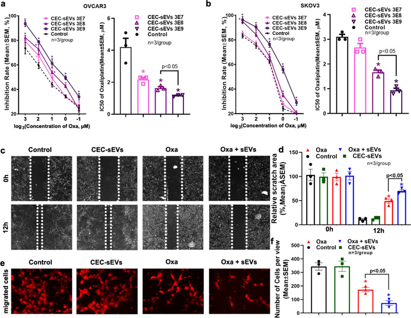
The x‐axis labels and legends in Figure 2a and 2b were incorrect. The labels on the x‐axis were “‐3, ‐2, ‐1, 0, 1”. This should have been “3, 2, 1, 0, ‐1”. The legends for the x‐axis were “log 3(Concentration of Oxa)”. This should have been "log3(Concentration of Oxa, μM).
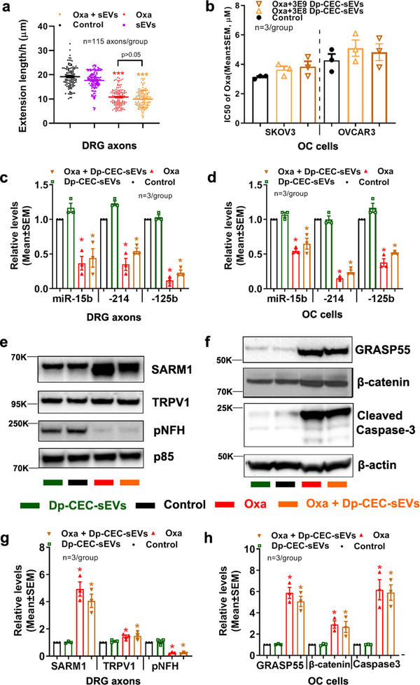
The x‐axis labels in Figure 10g and 10h were incorrect. In Figure 10g, the x‐axis labels were “miR‐15b, ‐214, ‐125b”. This should have been “SARM1, TRPV1, pNFH”. In Figure 10h, the x‐axis labels were “miR‐15b, ‐214, ‐125b”. This should have been “GRASP55, β‐catenin, Caspase3”.
FIGURE 2.

CEC‐sEVs enhanced anti‐cancer effects of oxaliplatin in OC cells. Quantitative data of MTT cell viability assays on OVCAR3 (a) and SKOV3 (b) cells show the inhibition rates and corresponding IC50, respectively, of oxaliplatin in combination with different concentrations of CEC‐sEVs. Representative images (c) and quantitative data (d) show the results of a 12h‐period wound healing assay of OVCAR3 cells treated with PBS (control), CEC‐sEVs, oxaliplatin (oxa) and CEC‐sEVs in combination with oxaliplatin (oxa + sEVs). Representative images (e) and quantitative data (f) show the results of the Transwell migration assay of OVCAR3 cells treated with different conditions for 24h. N indicates the replications. One‐way ANOVA with Tukey's multiple comparisons test was used. * p<0.05 vs control; Error bars indicate the standard error of the mean (SEM).
FIGURE 10.

Dp‐CEC‐sEVs do not promote axonal growth and sensitize OC cells to oxaliplatin, and do not alter oxaliplatin‐induced the changes of miRNAs and proteins in axons and OC cells. Quantitative data in a show growth cone extension of DRG axons within 60 minutes and treated with PBS (con), Oxaliplatin (oxa), Dp‐CEC‐sEVs and oxaliplatin in combination with Dp‐CEC‐sEVs (oxa+ Dp‐CEC‐sEVs). Quantitative data in b show IC50 of oxaliplatin in combination with different concentration of Dp‐CEC‐sEVs on SKOV3 and OVCAR3 cells, respectively. qRT‐PCR results show the levels of miR‐15b, 214 and 125b in axons of DRG neurons (c) and in SKOV3 cells (d), respectively, which were received different treatments. Representative Western blots results and their quantitative data show the levels of proteins in axons of DRGs (e, g) and in SKOV3 cells (f, h), respectively, which received different treatments. N in b‐d, g and h indicate the number of replications. K indicates the molecular weight KDa. One‐way ANOVA with Tukey's multiple comparisons test was used. * p<0.05, *** p<0.001 vs control; #, p<0.05 vs oxa. Error bars indicate the standard error of the mean (SEM).
Below are corrected figures:


