Abstract
Cat scratch disease (CSD) is a frequent clinical outcome of Bartonella henselae infection in humans. Recently, two case reports indicated Bartonella clarridgeiae as an additional causative agent of CSD. Both pathogens have been isolated from domestic cats, which are considered to be their natural reservoir. B. clarridgeiae and B. henselae can be distinguished phenotypically by the presence or absence of flagella, respectively. Separation of the protein content of purified flagella of B. clarridgeiae by sodium dodecyl sulfate-polyacrylamide gel electrophoresis and immunoblot analysis indicated that the flagellar filament is mainly composed of a polypeptide with a mass of 41 kDa. N-terminal sequencing of 20 amino acids of this protein revealed a perfect match to the N-terminal sequence of flagellin (FlaA) as deduced from the sequence of the flaA gene cloned from B. clarridgeiae. The flagellin of B. clarridgeiae is closely related to flagellins of Bartonella bacilliformis and several Bartonella-related bacteria. Since flagellar proteins are often immunodominant antigens, we investigated whether antibodies specific for the FlaA protein of B. clarridgeiae are found in patients with CSD or lymphadenopathy. Immunoblotting with 724 sera of patients suffering from lymphadenopathy and 100 healthy controls indicated specific FlaA antibodies in 3.9% of the patients' sera but in none of the controls. B. clarridgeiae FlaA is thus antigenic and expressed in vivo, providing a valuable tool for serological testing. Our results further indicate that B. clarridgeiae might be a possible etiologic agent of CSD or lymphadenopathy. However, it remains to be clarified whether antibodies to the FlaA protein of B. clarridgeiae are a useful indicator of acute infection.
Cat scratch disease (CSD) caused by Bartonella henselae is the most common Bartonella infection worldwide. In its typical form, CSD is a self-limiting regional lymphadenopathy, although atypical clinical courses with severe disease manifestations can also occur (1, 3). Recently, Bartonella clarridgeiae was suggested as an additional causative agent of CSD. This pathogen was initially isolated from the cat of a human immunodeficiency virus (HIV)-positive patient with CSD. The blood culture from the patient himself, however, grew only Bartonella henselae (7). B. clarridgeiae is closely related to the other Bartonella species, with similarities in the 16S rRNA ranging from 97.4 to 98.5% (17). B. clarridgeiae carries flagella (17) as previously described for Bartonella bacilliformis (33), but not for any other Bartonella species described up to now (9). Evidence for the involvement of B. clarridgeiae in causing CSD comes from two recent case reports. Kordick et al. (15) described a case of lymphadenopathy after a cat bite. B. clarridgeiae was isolated from a blood culture of the patient's cat, and antibodies against this agent were found in the patient's sera during the acute and convalescent phases by indirect fluorescent-antibody (IFA) test. The second case of CSD suspected to be caused by B. clarridgeiae was described in 1998 by Margileth and Baehren (19). The symptoms of the 35-year-old patient (fever, chills, night sweat, headaches, and somnolence) were interpreted as possible CSD. From an abscess of the chest wall, however, pneumococci had been isolated. Retrospective examination of the patient's serum showed a titer of 1:128 only against B. clarridgeiae, and this species was isolated from a blood culture of his cat.
Both species, B. henselae and B. clarridgeiae, have been isolated from domestic cats, which are considered to be the natural reservoir of these bacteria (5, 6, 12–14, 20, 22, 30). Attempts to isolate Bartonella spp. from immunocompetent patients suffering from CSD usually lack a positive culture. Currently, CSD is diagnosed serologically in most cases. Serological investigations for antibodies against Bartonella sp. showed a cross-reactivity between B. henselae and Bartonella quintana of 95 to 100% in many studies (10, 11, 23, 31). Similar cross-reactions have also been observed in our laboratory with B. clarridgeiae as antigen (IFA based on infected Vero cells; unpublished data). Therefore, a specific marker was needed for confirmation of suspected B. clarridgeiae infections. It was shown that B. henselae may have pili (4), whereas B. clarridgeiae possesses multiple unipolar flagella (17). Besides B. bacilliformis, B. clarridgeiae is the only human pathogenic Bartonella species which is flagellated. It was the aim of this study to characterize the B. clarridgeiae flagellin subunit (FlaA) and to examine its usefulness for serological detection of B. clarridgeiae infections.
MATERIALS AND METHODS
Bacterial strains and growth conditions.
The B. clarridgeiae strain used in this study was isolated from the blood of a free-ranging cat from the city of Nancy, France (13). Cultures were grown on Columbia agar (Difco Laboratories, Augsburg, Germany) supplemented with 5% defibrinated sheep blood. The plates were incubated at 37°C in a humid atmosphere containing 5% carbon dioxide. Growth was usually observed after 3 days of incubation, and the cultures were harvested at 6 days postinoculation. B. bacilliformis ATCC 35685T was grown on glucose-yeast extract-cysteine-containing blood agar (Sifin GmbH, Berlin, Germany), and plates were incubated at 26°C in a humid atmosphere.
Isolation and purification of flagella.
Early passages of B. clarridgeiae from 15 to 20 agar plates were harvested into 5 ml of phosphate-buffered saline (PBS). This suspension was blended in a commercial blender (KS 10; Bühler, Tübingen, Germany) for 30 min at room temperature to shear off the flagella. Bacteria were sedimented by centrifugation at 4,000 × g for 40 min, and the resulting supernatant was incubated for 12 h with saturated ammonium sulfate (vol/vol). This preparation was centrifuged at 4,000 × g for 40 min, and the resulting pellet was suspended in 1 ml of phosphate-buffered saline (PBS) and dialyzed four times for 30 min at 4°C against 2,000 ml of PBS to remove the ammonium sulfate. The same process was used to obtain a crude flagella preparation from B. bacilliformis.
SDS-PAGE.
Sodium dodecyl sulfate-polyacrylamide gel electrophoresis (SDS-PAGE) was performed by the method of Laemmli (16). The stacking and separating gels contained 4.5 and 10% acrylamide, respectively. Gel lanes were loaded with 15 to 20 μg each of protein and run in a Mini-Protean II cell (Bio-Rad) at 100 V for 2 h. Protein bands were stained with Coomassie brilliant blue R-250 (Serva). The molecular masses of the proteins were calculated from a calibration curve prepared with a low-molecular-mass calibration kit (Amersham Pharmacia Biotech, Freiburg, Germany). Protein concentrations were determined by the method described by Peterson (27) with bovine serum albumin as a standard.
N-terminal amino acid sequencing of B. clarridgeiae flagellin.
A preparative SDS-polyacrylamide gel was performed with the isolated flagella fraction. The proteins were electrophoretically transferred overnight in a borate buffer (50 mM boric acid, 10% [vol/vol] methanol, 0.02% mercaptoethanol [pH 8.6]) to a 0.45-μm-pore-size polyvinylidene difluoride membrane (Immobilon-P; Millipore Corporation, Bedford, Mass.) in a Transblot-Cell (Bio-Rad), washed in distilled water, and visualized by staining with Ponceau-S. The suspected 41-kDa flagellin band was excised from the gel, and the first 20 N-terminal amino acids were sequenced by Edman degradation with a gas-phase protein sequencer 477A (Applied Biosystems) equipped with online analysis of the amino acid derivatives.
Cloning and sequencing of the flaA gene of B. clarridgeiae and B. bacilliformis.
The flaA gene of B. clarridgeiae and B. bacilliformis was PCR amplified with primer pairs prCHD127 and prCHD128 and prCHD126 and prCHD128, respectively, which were delineated from the published sequence of the B. bacilliformis flaA gene (accession no. L20677) (Table 1). Hotstart PCR amplification was performed with 0.5 μg of CsCl-purified bacterial DNA, 100 pmol of each primer, 1× reaction buffer (Stratagene), 25 μM deoxynucleoside triphosphates (dNTPs) (Pharmacia), 2.5 U of Taq polymerase (BRL Gibco), and 2.5 U of Taq extender (Stratagene) in a total volume of 100 μl. The reaction mixture was heated to 95°C for 30 s prior to addition of Taq polymerase. Twenty-five cycles of amplification for 10 s at 95°C, 10 s at 50°C, and 7 min at 72°C were performed, followed by a 10-min incubation at 72°C. The generated PCR products were isolated by electrophoresis in a 1% TAE-agarose gel and purified from an excised gel fragment by a Geneclean kit I (Bio101, Dianova). The purified PCR fragments were cloned into the vector pTOPO-TA by using the TOPO TA cloning kit (Invitrogen), resulting in plasmid pCD387 for B. bacilliformis flaA and plasmid pCD388 for B. clarridgeiae flaA. DNA sequencing of the insert of pCD387 and pCD388 was performed with an ABI 377 DNA sequencer (Applied Biosystems). Sequencing reactions were performed by the AmpliTaq BigDye Terminator Cycle Sequencing Ready Reaction Kit (Perkin-Elmer) with primers prCHD126, prCHD128, prCHD151, M13-forward, and M13-reverse for pCD387 and primers prCHD127, prCHD128, M13-forward, and M13-reverse for pCD388 (Table 1). The sequence obtained for pCD387 confirmed the published sequence of the flaA gene of B. bacilliformis (accession no. L20677), except for a deletion of two cytosines at positions 1264 and 1265. The sequence obtained for pCD388 contained most of the flaA open reading frame (ORF) of B. clarridgeiae, including 29 bp of 5′-untranslated sequences, while the very 3′ end of this ORF was not contained in the amplicon and was sequenced by using chromosomal DNA as a template. Chromosomal sequencing was performed with a LiCor automatic sequencing system (MWG Biotech) with the infrared dye-labeled (IR 800) primer prCHD153 (Table 1). The sequencing reactions were carried out by using 10 μg of sheared chromosomal DNA and the Thermo Sequenase fluorescence-labeled primer cycle sequencing kit (Amersham Life Science) according to the manufacturer's instructions. From the resulting chromosomal sequence, primer prCHD165 was deduced and used for the generation of the PCR products prCHD151/165 and pcr127/165, which encompass the 3′ end of flaA (Table 1). Geneclean-purified PCR fragments were used as a template for AmpliTaq sequencing reactions with primers prCHD151, prCHD165, prCHD168, and prCHD169 (Table 1) to complete double-stranded sequencing of the B. clarridgeiae flaA gene.
TABLE 1.
Primer sequences for amplification and sequencing of the flaA gene of B. clarridgeiae and B. bacilliformis
| Primer name | Primer sequence | Specificity |
|---|---|---|
| prCHD126 | 5′-GATTTCTAGATTTAAGAAGGAGATATACATATGGGT TCTAGTATATTAACTAATAGA-3′ | Forward, positions 180–206 of B. bacilliformis flaA (accession no. L20677); additional sequences including XbaI and ribosome binding site at 5′ (highlighted in boldface) |
| prCHD127 | 5′-CGGGATCCCTGTAAATGCAGCATGTCCCTT-3′ | Forward, positions 130–151 of B. bacilliformis flaA (accession no. L20677); additional sequences including EcoRI site at 5′ (highlighted in boldface) |
| prCHD128 | 5′-CCCAAGCTTACGGAACAAACCTAAAATATTCTG-3′ | Reverse, positions 1329–1306 of B. bacilliformis flaA (accession no. L20677); additional sequences including HindIII site at 5′ (highlighted in boldface) |
| prCHD151 | 5′-AATATCCTTTCCAATGGTGGT-3′ | Forward, positions 548–568 of B. bacilliformis flaA (accession no. L20677); |
| prCHD153 | 5′-CAGCAACAGCTTGGTATTCAGGCTC-3′ | Forward, positions 1152–1176 of B. clarridgeiae flaA (this work) |
| prCHD165 | 5′-CAAATAAGGGAGAACAACATTGTA-3′ | Reverse, 136–113 bp downstream of B. clarridgeiae flaA (this work) |
| prCHD168 | 5′-CTTGGCGAGTTACATTCAGAC-3′ | Forward, positions 915–935 of B. clarridgeiae flaA (this work) |
| prCHD169 | 5′-AGTCTGACCTCCATTGGAAAG-3′ | Reverse, positions 455–435 of B. clarridgeiae flaA (this work) |
| M13-forward | 5′-TGTAAAACGACGGCCAGT-3′ | |
| M13-reverse | 5′-CAGGAAACAGCTATGACC-3′ |
Human sera.
Sera from 724 patients with lymphadenopathy, for which CSD was considered in the differential diagnosis, and 100 sera from healthy controls have been investigated for detection of antibodies to the B. clarridgeiae FlaA protein. Evidence of infection with B. henselae was determined by IFA as described previously (31). Ten sera from patients with Salmonella enterica serovar Typhi infections, 9 sera from patients with Yersinia enterocolitica infections, 19 sera from patients with Borrelia burgdorferi infections, 15 sera from patients with H. pylori infections, 2 sera from patients suffering from brucellosis, and 1 serum from an HIV-positive patient suffering from an Agrobacterium radiobacter infection were tested for cross-reactivity to B. clarridgeiae FlaA protein. All patients' sera having FlaA antibodies against B. clarridgeiae were additionally tested against the FlaA protein of B. bacilliformis.
An antiserum produced in rabbit against B. clarridgeiae (Y. Piémont, Strasbourg, France, unpublished data) served as a positive control, and an antiserum produced in rabbit against Rhizobium meliloti flagellin (kindly provided by R. Schmitt, Regensburg, Germany) (28) was tested for cross-reactivity with the FlaA protein of B. clarridgeiae.
Western blot analysis.
For Western blot analysis, the polyacrylamide gel was prepared as described above and the proteins were electrophoretically transferred to a polyvinylidene difluoride membrane (Immobilon-P; Millipore Corporation, Bedford, Mass.) as described by Towbin et al. (35). The membrane was blocked with 3% nonfat dry milk in PBS containing 0.05% Tween 20 (PBST), subsequently washed, incubated with a 1:100 dilution of the test sera in PBST–3% milk solution for 1 h at room temperature, washed three times in PBST, and incubated with a 1:1,000 dilution of alkaline phosphatase-conjugated antihuman immunoglobulin G (IgG) (heavy and light chains) (Jackson ImmunoResearch Laboratories, Inc., West Grove, Pa.) for 1 h at room temperature. After three additional washes, antibodies were detected with BCIP/NBT (5-bromo-4-chloro-3-indolyl phosphate–nitroblue tetrazolium; Sigma Chemical Company, St. Louis, Mo.).
Nucleotide sequence accession number.
The nucleotide sequence of the flaA locus of B. clarridgeiae (bases 1 to 1260) has been assigned database accession no. AJ251711.
RESULTS
Isolation and identification of B. clarridgeaie flagella.
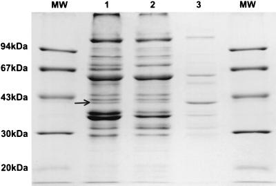
Flagella were sheared off from the whole B. clarridgeiae cells by intensive shaking. This crude flagellum preparation showed on a Coomassie brilliant blue-stained SDS-PAGE gel two prominent bands at 41 and 60 kDa (Fig. 1). Some additional minor bands at approximately 98, 35, 32, and 29 kDa were seen in the flagellum preparation, which were also present as major bands in the untreated cells and in the cellular debris. Their identity, however, remains unknown. The efficiency of the method was tested by isolation of flagella from various other flagellated bacteria, including B. bacilliformis, Proteus mirabilis, Salmonella enterica serovar Enteritidis, and Salmonella enterica serovar Paratyphi A (results not shown). No comparable proteins were found in supernatants from the unflagellated species B. henselae after similar treatment (results not shown).
FIG. 1.
Analysis of B. clarridgeiae flagellin preparation by SDS-PAGE and Coomassie brilliant blue staining. Lanes: MW, molecular mass marker; 1, whole-cell lysates of B. clarridgeiae; 2, cellular debris after shearing off the flagella; 3, crude flagellar protein. The arrow indicates the position of the 41-kDa band identified as the FlaA protein.
N-terminal amino acid sequencing of B. clarridgeiae flagellin.
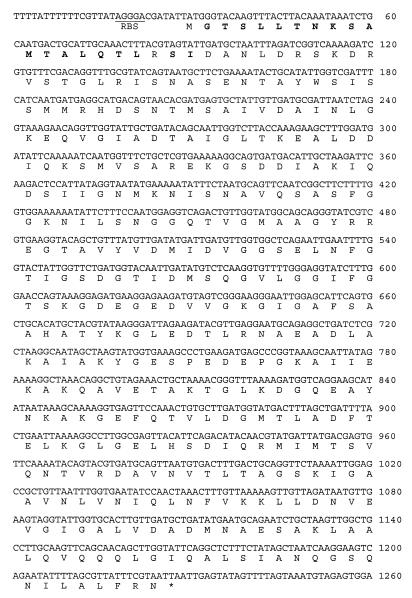
The first 20 N-terminal amino acids of the flagellin subunit were found to be GTSLLTNKSAMTALQTLXSI and were compared with known flagellin sequences (BLAST Search). Sixteen residues were exact matches with those of B. bacilliformis flagellin, and 4 residues were conservatively replaced. The following sequencing of the flaA gene confirmed the amino acid sequence of the N terminus (Fig. 2).
FIG. 2.
DNA sequence and protein translation of the B. clarridgeiae flagellin locus (flaA). The DNA sequence and translation of the flaA ORF are shown. The putative ribosome binding site (RBS) is underlined, and the N-terminal amino acids determined by protein sequencing are in boldface.
Cloning and sequencing of the B. clarridgeiae flagellin gene.
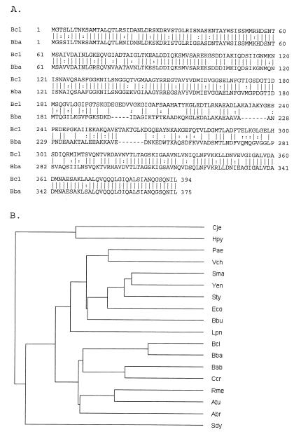
Heterologous primers deduced from the published sequence of B. bacilliformis flagellin (flaA) locus were used to amplify a major part of the flaA gene of B. clarridgeiae. The amplicon was cloned and sequenced, and the missing part at the very 3′ end of this gene was determined by chromosomal sequencing and confirmed by sequencing of longer-range PCR products generated with primers deduced from the chromosomal sequence. A total of 1,260 bp of double-stranded sequence was determined, encompassing 29 bp of 5′-untranslated sequence, the ORF, and 37 bp of 3′-untranslated sequence (Fig. 2). The ORF of 1,197 bp encodes a protein of 399 amino acids. The 20 N-terminal amino acids of the major flagellin subunit determined by protein sequencing are perfectly matched, suggesting that the cloned flaA gene of B. clarridgeiae encodes the major flagellar subunit characterized before (Fig. 2). The deduced amino acid sequence of B. clarridgeiae is most closely related to flagellin encoded by B. bacilliformis (accession no. L20677) (Fig. 3A) with 68% identity and 80% similarity. Resequencing of the B. bacilliformis flaA gene revealed an error in the published sequence, resulting in a reading frame shift close to the stop codon (see Materials and Methods). An average distance tree generated after alignment of flagellin protein sequences of various bacterial species is illustrated in Fig. 3B. Related bacteria of the α2-subgroup of proteobacteria, such as B. clarridgeiae and B. bacilliformis, Brucella abortus, Caulobacter crescentus, Rhizobium meliloti, Agrobacterium tumefaciens, and Azospirillum brasiliense are clustered in one clade, while the majority of flagellated bacterial pathogens, such as Legionella pneumophila, Pseudomonas aeruginosa, Vibrio cholerae, Serratia marcescens, Yersinia enterocolitica, S. enterica serovar Typhimurium, Escherichia coli, and Borrelia burgdorferi, fall into a separate clade, and Campylobacter jejuni and Helicobacter pylori are even less related (Fig. 3B).
FIG. 3.
Comparison of the protein sequence of B. clarridgeiae FlaA with those of other known flagellins. (A) Protein sequence comparison of FlaA from B. clarridgeiae (this work) and B. bacilliformis (accession no. L20677 [sequence corrected by resequencing]) by BLAST search. Identities and similarities are indicated by vertical bars and colons, respectively. (B) Average distance tree of a protein sequence alignment (CLUSTAL W) of flagellins from various bacteria. Abr, Azospirillum brasiliense Laf1 (accession no. U26679); Atu, Agrobacterium tumefaciens FlaD (accession no. U95165); Bba, Bartonella bacilliformis flagellin (accession no. L20677); Bcl, Bartonella clarridgeiae (this work); Bbu, Borrelia burgdorferi flagellar filament 41-kDa core protein (flagellin) (accession no. P11089), Bab, Brucella abortus FliC (accession no. AF019251); Ccr, Caulobacter crescentus flagellin Fljm (accession no. 052529); Cje, Campylobacter jejuni flagellin A (accession no. AAC25643); Eco, Escherichia coli flagellin (accession no. AB028473); Hpy, Helicobacter pylori flagellin B (accession no. AE001449); Lpn, Legionella pneumophila flagellin (accession no. X83232); Pae, Pseudomonas aeruginosa flagellin (accession no. AF034764); Rme, Rhizobium meliloti flagellin flaA (accession no. A39436); Sma, Serratia marcescens flagellin (accession no. D32256); Sty, Salmonella enterica serovar Typhimurium flagellin (accession no. AAB33952); Vch, Vibrio cholerae flagellin (accession no. AF007121); Yen, Yersinia enterocolitica thermoregulated motility protein (accession no. L33468).
Altogether, the cloned flaA locus of B. clarridgeiae encodes a major flagellar subunit, which is well conserved among the α2 proteobacteria, but clearly different from flagellins of other bacteria.
Western blot analysis.
Immunoblot analysis with rabbit anti-B. clarridgeiae antiserum demonstrated the immunogenicity of the 41-kDa protein. The FlaA protein was recognized by the antiserum against B. clarridgeiae in both the whole-cell lysate of B. clarridgeiae and the flagellar preparation. This flagellin subunit of B. clarridgeiae was further recognized by positive immunoblot reactions with an antiserum against both Agrobacterium radiobacter and Rhizobium meliloti, two bacteria closely related to Bartonella spp. (21). These results indicated that FlaA might be an immunogenic protein during B. clarridgeiae infections. However, it must be considered that sera containing antibodies to B. bacilliformis, A. radiobacter, or R. meliloti may cross-react with this protein.
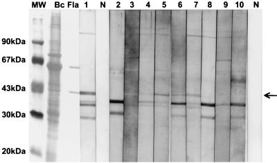
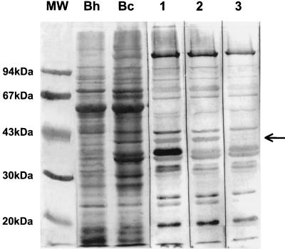
The potential applicability of the FlaA protein as an antigen for detection of B. clarridgeiae infections in humans was examined by Western blot analysis of 724 sera of patients with lymphadenopathy and suspected CSD. Based on an IFA titer against B. henselae of 1:≥512, CSD was diagnosed serologically in 156 patients. Nearly one-third (229) of all patients had low antibody titers of 1:64 to 1:256, indicating the onset or the end of CSD or simply contact with Bartonella species in the past. B. clarridgeiae antiflagellin antibodies were present in 3.9% (28 of 724) of the serum samples (Table 2). Typical reactions of some of these sera with the B. clarridgeiae FlaA protein are shown in Fig. 4. The 60-kDa protein seems not to be of immunologic importance in all these sera. All serological results, including the detection of antiflagellin antibodies to B. clarridgeiae by Western blot analysis and the antibody titers against B. henselae measured in the IFA assay are given in Table 2. There was no correlation between the sera containing antiflagellin antibodies and the IFA titer against B. henselae. Seven patients with antibodies against the FlaA protein had clinically and serologically (B. henselae titer of ≥1:512 in the IFA) confirmed CSD, 12 had low antibody titers, and 9 had no antibodies to B. henselae in the IFA. No antibodies against the 41-kDa protein were found in the 100 sera of the healthy control group. Sera of patients with antibodies against flagellated bacteria like Salmonella enterica serovar Typhi (10 sera), Brucella spp. (2 sera), Yersinia enterocolitica (9 sera), Helicobacter pylori (15 sera), and Borrelia burgdorferi (19 sera) were tested for cross-reactivity to the FlaA protein of B. clarridgeiae. None of these 55 sera reacted with the 41-kDa protein. In contrast, the FlaA protein band was recognized by the human serum with an A. radiobacter infection and by the rabbit R. meliloti antiserum.
TABLE 2.
IFA titers (IgG) of anti-B. henselae antibodies and antibody recognition of the 41-kDa (FlaA) band in sera of 724 patients with lymphadenopathy and 100 healthy controls
| IFA titer | No. of sera investigated | No. (%) of sera B. clarridgeiae immunoblot positivea |
|---|---|---|
| Patients | ||
| <64 | 339 | 9 (2.6) |
| 64–256 | 229 | 12 (5.2) |
| ≥512 | 156 | 7 (4.5) |
| Total | 724 | 28 (3.9) |
| Healthy controls | ||
| <64 | 100 | 0 |
Positive for the 41-kDa band.
FIG. 4.
Examination of human sera for antibodies to B. clarridgeiae FlaA protein by immunoblot analysis. Lanes: MW, molecular mass marker; Bc and Fla, amido black-stained B. clarridgeiae whole-cell lysate and flagellin preparation, respectively; 1, positive control (rabbit antiserum); N, negative control without serum; 2, 3, 4, 8, 9, and 10, negative sera; 5, 6, and 7, positive reactive sera containing antiflagellin antibodies. The arrow indicates the position of the 41-kDa FlaA protein.
The positive reaction of the 28 sera against the 41-kDa protein of B. clarridgeiae was not changed by absorption of these sera with B. henselae cells. However, it was considerably removed in all cases by absorbing with B. clarridgeiae whole-cell antigen, indicating the specificity of the antiflagellin antibodies (Fig. 5). Absorption of the sera with B. henselae whole cells resulted in a strongly diminished reaction of some other protein bands (with exception of the 41-kDa band) in the immunoblot, which confirms the cross-reactivity between B. henselae and B. clarridgeiae, as already seen in the IFA. Additionally, 7 of the 28 anti-B. clarridgeiae FlaA-positive sera did recognize the FlaA protein of B. bacilliformis, indicating a cross-reactivity between the two Bartonella species. The FlaA protein band of B. bacilliformis migrated faster than that of B. clarridgeiae in each of our flagellin preparations as well as in the protein profile of the whole bacteria (data not shown). We determined an apparent molecular mass of 40 kDa for the B. bacilliformis flagellin as opposed to 42 kDa reported previously (33).
FIG. 5.
Immunoblot analysis of a representative anti-FlaA antibody-positive serum showing the specificity of the anti-B. clarridgeiae-flagellin antibodies. Lanes: MW, molecular mass marker; Bh and Bc, amido black-stained B. henselae and B. clarridgeiae whole-cell antigen, respectively; 1, unabsorbed serum; 2, serum absorbed with whole cells of B. henselae; 3, serum absorbed with whole cells of B. clarridgeiae.
DISCUSSION
Some Bartonella spp. cause identical clinical symptoms (e.g., B. quintana or B. henselae in bacillary angiomatosis), and in these cases, differential diagnosis by serology proves difficult because of considerable antigenic cross-reactions (10, 11, 23, 31). Similar problems certainly arise with B. clarridgeiae as an additional causative agent of CSD. This problem will be even more complicated by the fact that both B. henselae and B. clarridgeiae have their reservoirs in cats, and humans injured by cats can be infected either by only one of the two agents (7) or by both. In such a situation, the presence of a specific antigen in one of the related species could provide a valuable tool for a specific test.
Although the flagellins of B. bacilliformis and B. clarridgeiae are, according to our results, closely related, cross-reactions are less likely to occur because of the geographical restriction of B. bacilliformis to the Andean regions of South America. However, in 25% (7 of 28) of the B. clarridgeiae anti-FlaA-positive sera, a cross-reaction was seen with the FlaA protein of B. bacilliformis. This must be considered when sera from patients living in areas in which B. bacilliformis is endemic are investigated. Other closely Bartonella-related bacteria, like Azospirillum, Caulobacter crescentus, or Rhizobium meliloti, are not pathogenic for humans, except for some Brucella species and also Agrobacterium radiobacter (tumefaciens), which has been shown to be an opportunistic pathogen in immunocompromised individuals (29, 34). We could demonstrate that an antiserum against Agrobacterium radiobacter and against Rhizobium meliloti strongly cross-reacted with the 41-kDa FlaA protein of B. clarridgeiae. However, such a cross-reactivity was not observed with human sera from patients suffering from brucellosis.
Dendrogram analysis of several flagellin sequences has shown that the Bartonella flagella together with Azospirillum, R. meliloti, A. tumefaciens, and C. crescentus flagellins form a cluster that is separated from all other flagellins, including that of E. coli (Fig. 3B) (24). Flagellar proteins of other pathogenic bacteria are clearly different, and with sera of patients suffering from salmonellosis or Lyme borreliosis, for example, we could not detect cross-reactions to the 41-kDa protein. In a comparison of the protein profiles of the human pathogenic Bartonella species like B. henselae, B. quintana, B. elizabethae, B. bacilliformis, and B. clarridgeiae, a 41-kDa protein band is found only for the flagellated species B. clarridgeiae (32; data not shown). In our investigations, the FlaA protein band of B. bacilliformis was found to migrate with an apparent molecular mass of 40 kDa as opposed to 42 kDa reported previously (33).
The simultaneous occurrence of antibodies against both B. henselae and B. clarridgeiae provides a diagnostic problem, which only can be solved by careful diagnostic observation of patients with acute CSD. This could be done by serologic follow-up in the acute and convalescent phases of disease or identification of the infecting agents by other methods, like culture or PCR. Unfortunately only one lymph node was available from our 28 patients with antibodies against the 41-kDa protein, and the PCR in this case indicated an infection with B. henselae rather than with B. clarridgeiae (results not shown). Similar observations were made by Lawson and Collins (17) when they first isolated B. clarridgeiae from a patient's cat, although the patient himself, however, suffered from a B. henselae septicemia. It cannot be excluded that either the infection with B. clarridgeiae is less symptomatic than the typical B. henselae infection or a simultaneous infection with both agents from the same source may occur. Alternatively, since B. clarridgeiae is also found in cats (12, 13, 14, 20), patients injured by cat bites or scratches in particular may suffer from double infections with these Bartonella species.
A large number of studies have been carried out with various flagellated human pathogens, demonstrating the immunogenic effect of the flagellins (2, 8, 18, 25, 26, 33, 36). For some infections, FlaA is a major antigen and anti-FlaA antibodies can be detected in the serum of infected hosts (18, 26). In contrast, for Borrelia burgdorferi, FlaA was found not to be an immunodominant antigen in mammalian hosts (mouse, rabbit, and rhesus monkey), but it was expressed in some patients with Lyme disease (36). Anti-FlaB is usually the first detectable antibody found in the acute stage of Lyme disease and is consistently present during the entire infection (8, 36). A study with sera of patients with culture-proven Legionella pneumophila serogroup 1 pneumonia demonstrated that antibodies against flagella appeared later in the course of infection (2), a situation contrary to infections with Borrelia burgdorferi (8).
Up to now, no culture-proven or at least PCR-proven B. clarridgeiae infection in humans has been reported. Since the culturing of Bartonella species from human specimens is very difficult, serologic testing for antibodies will remain the main diagnostic procedure for Bartonella infections. Our results suggest that B. clarridgeiae indeed might be a possible, but rare, agent of CSD. However, whether antibodies to the FlaA protein of B. clarridgeiae are a useful indicator of acute infection remains to be clarified. Because of the lack of a “gold standard” (e.g., culture), serologic results should be interpreted critically until the role of B. clarridgeiae and the detection of anti-FlaA antibodies in CSD have been sufficiently confirmed.
ACKNOWLEDGMENTS
We greatly thank Stefan Bereswill for helpful suggestions, Tanja Schülin and Daniela Huzly for providing the Agrobacterium radiobacter antiserum, and Rüdiger Schmitt for kindly providing the Rhizobium meliloti antiserum. We also thank Karin Oberle, Ina Wagner, and Vera Augsburger for excellent technical assistance.
REFERENCES
- 1.Anderson B E, Neuman M A. Bartonella spp. as emerging human pathogens. Clin Microbiol Rev. 1997;10:203–219. doi: 10.1128/cmr.10.2.203. [DOI] [PMC free article] [PubMed] [Google Scholar]
- 2.Bangsborg J M, Shand G, Høiby N. Antibody response to major cross-reactive Legionella antigens during infection. In: Barbaree J M, Breiman R F, Dufour H P, editors. Legionella—current status and emergent prospectives. Washington, D.C.: American Society for Microbiology; 1993. pp. 26–29. [Google Scholar]
- 3.Bass J W, Vincent J M, Person D A. The expanding spectrum of Bartonella infections. II. Cat-scratch disease. Pediatr Infect Dis J. 1997;16:163–179. doi: 10.1097/00006454-199702000-00002. [DOI] [PubMed] [Google Scholar]
- 4.Batterman H J, Peek J A, Loutit J S, Falkow S, Tompkins L S. Bartonella henselae and Bartonella quintana adherence to and entry into cultured human epithelial cells. Infect Immun. 1995;63:4553–4556. doi: 10.1128/iai.63.11.4553-4556.1995. [DOI] [PMC free article] [PubMed] [Google Scholar]
- 5.Bergmans A M C, de Jong C M A, van Amerongen G, Schot C S, Schouls L M. Prevalence of Bartonella species in domestic cats in The Netherlands. J Clin Microbiol. 1997;35:2256–2261. doi: 10.1128/jcm.35.9.2256-2261.1997. [DOI] [PMC free article] [PubMed] [Google Scholar]
- 6.Chomel B B, Carlos E T, Kasten R W, Yamamoto K, Chang C-C, Carlos R S, Abenes M V, Pajares C M. Bartonella henselae and Bartonella clarridgeiae infection in domestic cats from the Philippines. Am J Trop Med Hyg. 1999;60:593–597. doi: 10.4269/ajtmh.1999.60.593. [DOI] [PubMed] [Google Scholar]
- 7.Clarridge J E, III, Raich T J, Pirwani D, Simon B, Tsai L, Rodriguez-Barradas M C, Regnery R, Zollo A, Jones D C, Rambo C. Strategy to detect and identify Bartonella species in routine clinical laboratory yields Bartonella henselae from human immunodeficiency virus-positive patient and unique Bartonella strain from his cat. J Clin Microbiol. 1995;33:2107–2113. doi: 10.1128/jcm.33.8.2107-2113.1995. [DOI] [PMC free article] [PubMed] [Google Scholar]
- 8.Craft J E, Fischer D K, Shimamoto G T. Antigens of Borrelia burgdorferi recognized during Lyme disease. J Clin Investig. 1986;78:934–939. doi: 10.1172/JCI112683. [DOI] [PMC free article] [PubMed] [Google Scholar]
- 9.Dehio C, Sander A. Bartonella as emerging pathogens. Trends Microbiol. 1999;7:226–228. doi: 10.1016/s0966-842x(99)01523-1. [DOI] [PubMed] [Google Scholar]
- 10.Dupon M, Delarclause A M S, Brouqui P, Drancourt M, Raoult D, Demascarel A, Lacut J Y. Evaluation of serological response to Bartonella henselae, Bartonella quintana and Afipia felis antigens in 64 patients with suspected cat scratch disease. Scand J Infect Dis. 1996;28:361–366. doi: 10.3109/00365549609037920. [DOI] [PubMed] [Google Scholar]
- 11.Engbaek K, Koch C. Immunoelectrophoretic characterization and cross-reactivity of Rochalimaea henselae, Rochalimaea quintana and Afipia felis. APMIS. 1994;102:931–942. [PubMed] [Google Scholar]
- 12.Gurfield A N, Boulouis H-J, Chomel B B, Heller R, Kasten R W, Yamamoto K, Piemont Y. Coinfection with Bartonella clarridgeiae and Bartonella henselae and different Bartonella henselae strains in domestic cats. J Clin Microbiol. 1997;35:2120–2123. doi: 10.1128/jcm.35.8.2120-2123.1997. [DOI] [PMC free article] [PubMed] [Google Scholar]
- 13.Heller R, Artois M, Xemar V, De Briel D, Gehin H, Jaulhac B, Monteil H, Piemont Y. Prevalence of Bartonella henselae and Bartonella clarridgeiae in stray cats. J Clin Microbiol. 1997;35:1327–1331. doi: 10.1128/jcm.35.6.1327-1331.1997. [DOI] [PMC free article] [PubMed] [Google Scholar]
- 14.Kordick D L, Breitschwerdt E B. Persistent infection of pets within a household with three Bartonella species. Emerg Infect Dis. 1998;4:325–328. doi: 10.3201/eid0402.980225. [DOI] [PMC free article] [PubMed] [Google Scholar]
- 15.Kordick D L, Hilyard E J, Hadfield T L, Wilson K H, Steigerwalt A G, Brenner D J, Breitschwerdt E B. Bartonella clarridgeiae, a newly recognized zoonotic pathogen causing inoculation papules, fever, and lymphadenopathy (cat scratch disease) J Clin Microbiol. 1997;35:1813–1818. doi: 10.1128/jcm.35.7.1813-1818.1997. [DOI] [PMC free article] [PubMed] [Google Scholar]
- 16.Laemmli U K. Cleavage of structural proteins during the assembly of the head of bacteriophage T4. Nature (London) 1970;227:680–685. doi: 10.1038/227680a0. [DOI] [PubMed] [Google Scholar]
- 17.Lawson P A, Collins M D. Description of Bartonella clarridgeiae sp. nov. isolated from the cat of a patient with Bartonella henselae septicemia. Med Microbiol Lett. 1996;5:64–73. [Google Scholar]
- 18.Li Z, Dumas F, Dubreuil D, Jacques M. A species-specific periplasmatic flagellar protein of Serpulina (Treponema) hyodysenteriae. J Bacteriol. 1993;175:8000–8007. doi: 10.1128/jb.175.24.8000-8007.1993. [DOI] [PMC free article] [PubMed] [Google Scholar]
- 19.Margileth A M, Baehren D F. Chest-wall abscess due to cat-scratch disease (CSD) in an adult with antibodies to Bartonella clarridgeiae: case report and review of the thoracopulmonary manifestations of CSD. Clin Infect Dis. 1998;27:353–357. doi: 10.1086/514671. [DOI] [PubMed] [Google Scholar]
- 20.Marston E L, Finkel B, Regnery R L, Winoto I L, Ross Graham R, Wignal S, Simanjuntak G, Olson J G. Prevalence of Bartonella henselae and Bartonella clarridgeiae in an urban Indonesian cat population. Clin Diagn Lab Immunol. 1999;6:41–44. doi: 10.1128/cdli.6.1.41-44.1999. [DOI] [PMC free article] [PubMed] [Google Scholar]
- 21.Marston E L, Sumner J W, Regnery R L. Evaluation of intraspecies genetic variation within the 60 kDa heat-shock protein gene (groEL) of Bartonella species. Int J Syst Bacteriol. 1999;49:1015–1023. doi: 10.1099/00207713-49-3-1015. [DOI] [PubMed] [Google Scholar]
- 22.Maruyama S, Tanaka S, Sakai T, Katsube Y. Prevalence of Bartonella species among pet cats in Japan. 1st International Conference on Bartonella as Emerging Pathogens. Tubingen, Germany, March 5–7. J Microbiol Methods. 1999;37:284–285. . (Abstract.) [PubMed] [Google Scholar]
- 23.McGill S L, Regnery R L, Karem K L. Characterization of human immunoglobulin (Ig) isotype and IgG subclass response to Bartonella henselae infection. Infect Immun. 1998;66:5915–5920. doi: 10.1128/iai.66.12.5915-5920.1998. [DOI] [PMC free article] [PubMed] [Google Scholar]
- 24.Moens S, Michiels K, Keijers V, Van Leuven F, Vanderleyden J. Cloning, sequencing, and phenotypic analysis of laf1, encoding the flagellin of the lateral flagella of Azospirillum brasilense Sp7. J Bacteriol. 1995;177:5419–5426. doi: 10.1128/jb.177.19.5419-5426.1995. [DOI] [PMC free article] [PubMed] [Google Scholar]
- 25.Neumeister B, Susa M, Nowak B, Straube E, Ruckdeschel G, Hacker J, Marre R. Enzyme immunoassay for detection of antibodies against isolated flagella of Legionella pneumophila. Eur J Clin Microbiol Infect Dis. 1995;14:764–767. doi: 10.1007/BF01690990. [DOI] [PubMed] [Google Scholar]
- 26.Norris S J Treponema pallidum Polypeptide Research Group. Polypeptides of Treponema pallidum: progress toward understanding their structural, functional, and immunologic roles. Microbiol Rev. 1993;57:750–779. doi: 10.1128/mr.57.3.750-779.1993. [DOI] [PMC free article] [PubMed] [Google Scholar]
- 27.Peterson G L. A simplification of the protein assay method of Lowry et al. which is more generally applicable. Anal Biochem. 1977;83:346–356. doi: 10.1016/0003-2697(77)90043-4. [DOI] [PubMed] [Google Scholar]
- 28.Pleier E, Schmitt R. Expression of two Rhizobium meliloti flagellin genes and their contribution to the complex filament structure. J Bacteriol. 1991;173:2077–2085. doi: 10.1128/jb.173.6.2077-2085.1991. [DOI] [PMC free article] [PubMed] [Google Scholar]
- 29.Salavert M, Breton J R, Perez-Tamarit D, Perez-Belles C, Gobernado M. Persistent bacteremia and infection of an intravascular device by Agrobacterium radiobacter (tumefaciens) in a boy with AIDS. Enferm Infect Microbiol Clin. 1997;15:496–497. [PubMed] [Google Scholar]
- 30.Sander A, Bühler C, Pelz K, von Cramm E, Bredt W. Detection and identification of two Bartonella henselae variants in domestic cats in Germany. J Clin Microbiol. 1997;35:584–587. doi: 10.1128/jcm.35.3.584-587.1997. [DOI] [PMC free article] [PubMed] [Google Scholar]
- 31.Sander A, Posselt M, Oberle K, Bredt W. Seroprevalence to Bartonella henselae in patients with cat scratch disease and in healthy controls: evaluation and comparison of two commercial serological tests. Clin Diagn Lab Immunol. 1998;5:486–490. doi: 10.1128/cdli.5.4.486-490.1998. [DOI] [PMC free article] [PubMed] [Google Scholar]
- 32.Sander A. Microbiological diagnosis of Bartonella species and Afipia felis. In: Schmidt A, editor. Bartonella and Afipia species emphasizing Bartonella henselae. Vol. 1. Basel, Switzerland: Karger; 1998. pp. 98–112. [Google Scholar]
- 33.Scherer D C, DeBuron-Connors I, Minnick M F. Characterization of Bartonella bacilliformis flagella and effect of antiflagellin antibodies on invasion of human erythrocytes. Infect Immun. 1993;61:4962–4971. doi: 10.1128/iai.61.12.4962-4971.1993. [DOI] [PMC free article] [PubMed] [Google Scholar]
- 34.Southern P M., Jr Bacteremia due to Agrobacterium tumefaciens (radiobacter). Report of infection in a pregnant woman and her stillborn fetus. Diagn Microbiol Infect Dis. 1996;24:43–45. doi: 10.1016/0732-8893(95)00200-6. [DOI] [PubMed] [Google Scholar]
- 35.Towbin H, Staehelin T, Gordon J. Electrophoretic transfer of proteins from polyacrylamide gels to nitrocellulose sheets: procedure and some applications. Proc Nat Acad Sci USA. 1979;76:4350–4354. doi: 10.1073/pnas.76.9.4350. [DOI] [PMC free article] [PubMed] [Google Scholar]
- 36.Yigong G E, Charon N W. FlaA, a putative flagellar outer sheath protein, is not an immunodominant antigen associated with Lyme disease. Infect Immun. 1997;65:2992–2995. doi: 10.1128/iai.65.7.2992-2995.1997. [DOI] [PMC free article] [PubMed] [Google Scholar]


