

Abstract
Sebaceous differentiation can be normally seen in salivary glands. An entity first described by Hamperi. Sebaceous components are present in several salivary gland tumors like Warthin’s tumor and mucoepidermoid tumor. But, if the sebaceous component predominates, it is known as a sebaceous adenoma. If lymphatic stroma in the background is prominent, it qualifies as sebaceous lymphadenoma. The term was coined and properly described by McGavran et al., differentiating it from similar appearing tumors like Warthin’s tumor, sebaceous adenoma, and mucoepidermoid tumor. Clinicians, as well as pathologists, need to be aware of this entity as it is known to undergo malignant degeneration. Since it is a benign entity, the most common symptom is painless parotid swelling. The patient in this case also presented with painless parotid swelling and underwent preoperative FNAC and MRI. But, the diagnosis was made after surgical excision. The patient recovered well after surgery and is being followed. Since this disease is known for malignant degeneration and recurrence careful diagnosis is required. Histopathological picture may be confused with other common entities like Warthin also known as papillary cystadenoma lymphomatosum since both have sebaceous and lymphoid components. McGavran differentiated between both entities. Sebaceous lymphadenoma can convert to sebaceous adenocarcinoma and is thus important to know the correct diagnosis, even if it is postoperative since recurrence may be associated with malignant changes. Sometimes, the picture may be confused with lymphoma and in that case, immunotyping can come as a rescue for diagnosis. The authors of this report intend to report this rare entity and emphasize the need for clinicians and pathologists being aware of it and keeping it as a differential while dealing with a similar parotid tumor.
Keywords: Parotid tumor, Sebaceous lymphadenoma, Sebaceous adenoma, Salivary gland tumor
Introduction
Sebaceous lymphadenoma of parotid is a rare entity, accounting for less than 1% of all parotid tumors [1]. The presence of sebaceous cells is common in the normal parotid gland and thus can be found in several parotid tumors like Warthin’s tumor. But, predominant presences of the sebaceous component entitle it to be considered under salivary tumor with sebaceous differentiation [2]. Clinically, it present like any other parotid tumor with swelling. Owing to its benign nature, facial nerve palsy is seldom seen. Diagnosis on FNAC is difficult and confirmation can be done only after a biopsy. Surgery alone is often curative and despite being benign, recurrences have been reported. Clinicians and pathologists must be aware of this condition and its malignant counterpart. This case report intends to report the incidence of one such case.
Case Report
A 62-year-old male presented with swelling in the left parotid region for 4–5 months, gradually increasing in size. There were no signs or symptoms suggestive of facial nerve involvement. No overlying skin involvement (Fig. 1). MRI of the head and neck showed an exophytic lesion of 3.4 × 3.0 × 5.4 cm arising from the tail of the left parotid. The lesion was hyperintense compared to surrounding muscles on T1 images. No significant enhancement noted with gadolinium, although there were significant areas of scattered enhancement in the center of the lesion. No extension of the lesion in the deeper lobe (Fig. 2 a and b). Likely radiological diagnosis of pleomorphic adenoma was considered with keeping in mind differentials like Warthin/mucoepidermoid tumor.
Fig. 1.
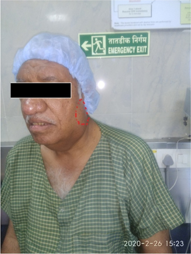
Clinical picture of patient preoperatively
Fig. 2.

a Axial T1-weighted image of the tumor showing hyperintensity compared to surrounding muscles. b Coronal T1-weighted image of the tumor.
FNAC from the parotid lesion was performed. It showed many lymphoid cells with fluid background and few monolayered epithelial cells were seen. No atypical cells were found. Thus, a cytological diagnosis of benign lymphoepithelial lesion was given.
The patient underwent a superficial parotidectomy. Postoperative recovery was normal. There was mild facial nerve weakness which gradually improved over the next few days. Histopathological analysis was done. Grossly, it was a well-circumscribed lobulated tumor with a yellowish-brown cut surface (Fig. 3). Microscopically, the tumor showed nests and glandular structures of benign epithelial cells with sebaceous differentiation associated with abundant lymphoid tissue with germinal centers. Features were suggestive of sebaceous lympadenoma (Fig. 4 a and b).
Fig. 3.

Gross specimen of superficial parotidectomy showing yellowish-brown tumor
Fig. 4.

a Low power magnification: islands of sebaceous cells in the reactive lymphoid stroma. b High magnification view: sebaceous cells with no atypia and lymphoid stroma
Since it is a benign lesion, no further treatment was offered and the patient is being followed routinely.
Discussion
Sebaceous glands can be normally present in salivary glands. In order of frequency, it is found in parotid, submandibular, and sublingual. Hamperi first described sebaceous differentiation in salivary glands. He found it in four submandibular and one parotid gland. In literature, sebaceous differentiation has been reported in 11% and 6% of the normal parotid and submandibular glands respectively [3]. The mere presence of sebaceous differentiation can be seen in several salivary gland tumors like Warthin, mixed tumor, mucoepidermoid tumor, and adenoid cystic carcinoma. But, when the sebaceous component is predominant, it qualifies as sebaceous adenoma. These sebaceous cells are similar to cutaneous sebaceous glands evident on electron microscope examination and thin-layer chromatography of lipid material. If predominant sebaceous material is accompanied by prominent lymphoid stroma, it is designated sebaceous lymphadenoma [2]. Rarely, their malignant counterparts can also be encountered, i.e., sebaceous adenocarcinoma. WHO classification of salivary gland tumors 2017 has removed the entity sebaceous lymphadenocarcinoma and just kept the term sebaceous adenocarcinoma [4].
In 1950, Rawson et al. reported this tumor [5]. He described it along with other lymphoepithelial tumors of the salivary gland. The term sebaceous lymphadenoma was coined by McGavran et al. in 1959 [6]. They differentiated it from papillary cystadenoma lymphomatosum (Warthin’s tumor). McGavran explained that although sebaceous components can be present in Warthin’s tumor, the tumor originally described by Rawson had a predominant sebaceous element; thus, it cannot be classified as papillary cystadenoma lymphomatosum. The sebaceous component predominant salivary tumor was also described by Foote and Frazell at Memorial Centre for Cancer and Allied Disease, New York in 1953 [7]. But, this tumor lacked lymphoid background, thus classified it as sebaceous adenoma, and due to low occurrence, the author actually doubted its occurrence. McGavran differentiated it from sebaceous lymphadenoma.
It is a disease of the 5th or 6th decade and more common in males, probably because of more preponderance of sebaceous glands in males [8]. Although the disease occurs in old age, it is argued that sebaceous differentiation pace up post-puberty under influence of probably androgen similar to what happens to cutaneous sebaceous glands [9]. The presence of lymphoid stroma in the tumor is quite a mystery. Gnepp et al. reported a series of 21 cases of sebaceous lymphadenoma and demonstrated the presence of germinal center and subcapsular marginal sinuses in the tumor. Thus, his conclusion was that the tumor arises from the ectopic salivary gland in lymphoid tissues [3].
Preoperative diagnosis of the disease is difficult with imaging and FNAC [10]. Sebaceous lymphadenoma has no characteristic radiological finding and is thus confused with other benign lesions. Similarly, FNAC can be confused with other benign lesions, although FNAC is helpful to determine if it is a benign lesion or not since in parotid tumors treatment is dictated based on the benign and malignant nature of the tumor. Also, this tumor can rarely undergo malignant degeneration and thus, FNAC is must preoperatively. Sometimes, FNA may be confused with lymphoma owing to the predominant lymphoid population [11]. Characteristic prevalence of the sebaceous component can easily differentiate sebaceous lymphadenoma from various lymphoma, but if doubt exists, immunophenotyping can be employed.
Clinically, it is an indolent tumor with a benign course. Complete excision is the only and sufficient treatment. Recurrences are common if excision is suboptimal, and thus, care must be taken to excise with the utmost care and having adequate margin. Chances of malignant transformation are rare but present.
Conclusion
Sebaceous lymphadenoma is a rare entity and its etiopathogenesis and histogenesis are not clear. The course is benign and following adequate surgical resection, chances of recurrence are low. Failure to identify a lesion preoperatively does not alter the prognosis as long as the disease is benign. Since malignant degeneration is known excision is a must. Thus, on a cytology report showing epithelial and lymphoid components along with a common diagnosis of Warthin’s and mucoepidermoid, a differential of sebaceous lymphadenoma must be kept in mind.
Abbreviations
- MRI
Magnetic resonance imaging
- FNAC
Fine needle aspiration cytology
Footnotes
Publisher's Note
Springer Nature remains neutral with regard to jurisdictional claims in published maps and institutional affiliations.
Contributor Information
Gajanan Kanitkar, Email: gakanitkar@gmail.com.
Prasant Chandra, Email: dr.prashantchandra@gmail.com.
Anirudha Puntambekar, Email: anirudhabhu05@gmail.com.
References
- 1.Al- EM. Sebaceous lymphadenoma of parotid gland: a case report of a unique presentation in an immunocompromised patient. J Family Med Prim Care. 2020;9:1202–1205. doi: 10.4103/jfmpc.jfmpc_1115_19. [DOI] [PMC free article] [PubMed] [Google Scholar]
- 2.Rosai J (2011) Major and minor salivary glands (Chapter 12) in Rosai and Ackerman’s Surgical Pathology, 10th edn, vol 1. MOSBY Elsevier, p 829
- 3.Gnepp DR, Brannon R (1984) Sebaceous neoplasms of salivary gland origin. Report of 21 cases. Cancer 53: 2155–2170. 10.1002/1097-0142(19840515)53:10%3C2155::aid-cncr2820531026%3E3.0.co;2-f [DOI] [PubMed]
- 4.Hellquist H, Paiva-Correia A, Vander Poorten V, et al. Analysis of the clinical relevance of histological classification of benign epithelial salivary gland tumours. Adv Ther. 2019;36(8):1950–1974. doi: 10.1007/s12325-019-01007-3. [DOI] [PMC free article] [PubMed] [Google Scholar]
- 5.Rawson AJ, Horn RC. Sebaceous glands containing of the salivary gland. Surgery. 1950;1954:93–101. [PubMed] [Google Scholar]
- 6.McGavran MH, Bauer WC, Ackerman LV (1960) Sebaceous lymphadenoma of the parotid salivary gland. Cancer 13:1185–7. https://doi.org/10.1002/1097-0142(196011/12)13:6%3C1185::aid-cncr2820130605%3E3.0.co;2-p [DOI] [PubMed]
- 7.Foote FW Jr, Frazell EL (1953) Tumors of the major salivary glands. Cancer 6(6):1065–1133. https://doi.org/10.1002/1097-0142(195311)6:6<1065::aid-cncr2820060602>3.0.co;2-0. [DOI] [PubMed]
- 8.Shekarkhar G, Soleimanpour H, Jafari SH et al (2018) Case report sebaceous lymphadenoma of parotid: imaging, cytological and histological findings in detail”, Hindawi. Case Rep Pathol. 10.1155/2018/2915907 [DOI] [PMC free article] [PubMed]
- 9.Linhartova A. Sebaceous glands in salivary gland tissue. Arch Pathol. 1974;98:320. doi: 10.1155/2018/2915907. [DOI] [PubMed] [Google Scholar]
- 10.Hayashi D, Tysome JR, Boyei E, Gluckman P, Barbaccia C. Sebaceous lymphadenoma of the parotid gland: report of two cases and review of the literature. Acta Otorhinolaryngol Ital. 2007;27(3):144–146. [PMC free article] [PubMed] [Google Scholar]
- 11.Sun L, ZhurC,tong J, , et al. BMJ Case Rep. 2018;11:e224975. doi: 10.1136/bcr-2018-224975. [DOI] [PMC free article] [PubMed] [Google Scholar]