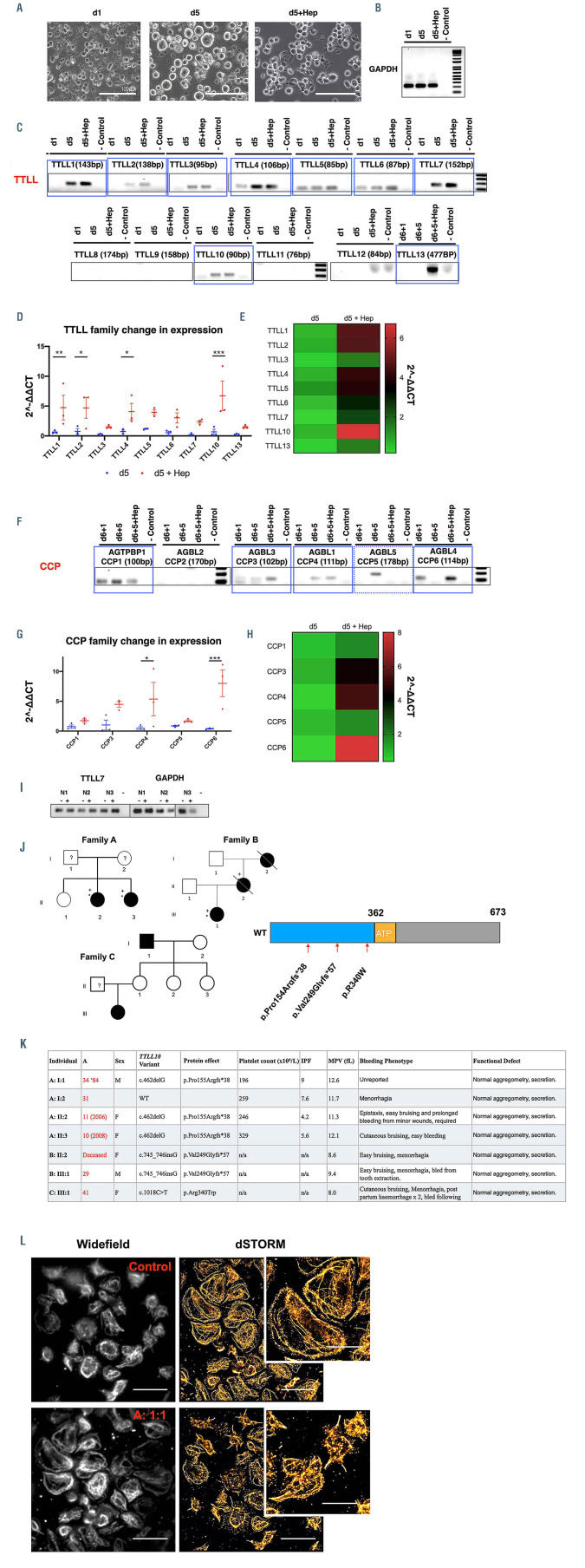
Figure 6.
Induced pluripotent stem cell megacaryocytes and platelets differentially express TTLL and CCP enzymes to regulate polymodifications during megakaryocyte maturation and platelet production. The loss of tubulin tyrosine ligase like (TTL) TTLL10 is possibly linked to a bleeding phenotype in unrelated human patients. (A) RNA was generated from induced pluripotent stem cell megacaryocytes (iPSC-MK) at different stages of terminal differentiation. Day 1 (d1) day 5 (d5) day 5 cells treated with heparin to induce proplatelet formation (d5+Hep) were used to determine whether an upregulation of these enzymes is evident on platelet production. (B) Samples were amplified with housekeeping GAPDH primers (C) A number of TTLL family enzymes were observed, including TTLL1, 2, 3, 4, 5, 6, 7, 10, and 13. Of these, a number appeared to be upregulated in mature and proplatelet forming cells. (D and E) These samples were taken forward and expression was quantified over multiple differentiations using the ΔΔCT method TTLL1, 2, 4, and 10 expression was found to be significantly upregulated on treatment with heparin (**P= 0.0081, *P= 0.0105, *P= 0.0260, ***P=0.0004 respectively). (F to H) A similar panel was performed on cytosolic carboxypeptidase (CCP) enzymes which reverse polymodifications, with expression of CCP1, 3, 4, 5, and 6 was observed. Statistically significant upregulation of CCP4, and CCP6 were observed on proplatelet production (*P=0.0130, ***P=0.0009). (I) In resting (-) and Creactive protein (CRP) activated (+) platelets from three healthy donors TTLL7 was the only modifying enzyme found to be consistently expressed. (J) Three unrelated families were identified within the Genotyping and Phenotyping of Platelets Study (GAPP) cohort, two with frameshift truncations and one with a missense substitution (p.Pro15Argfs*38, p.Val249Glyfs*57, and p.Arg340Trp respectively, and as shown on the protein schematic of TTLL10). (K) All three families present with normal platelet counts and function, but demonstrate an elevated mean platelet volume (MPV) and consistent histories of bleeding including cutaneous bruising and menorrhagia. (L) Patient A:1:1 was recalled and demonstrated abnormally large platelets on spreading on fibrinogen when imaged by widefield and single molecule localisation microscopy (tubulin staining, 10 μm scale bar, 5 μm in cropped image). (n=3, standard deviation. Two-Way ANOVA with multiple comparisons performed. Complete unedited gels found in the Online Supplementary Figures S9 and S10).

