Abstract
Objective
The Chinese medicine Huangqi Guizhi Wuwu Decoction (HGWD) has been reported to improve the clinical symptoms and restore nerve function after ischemic stroke; however, its active ingredients are not well-determined. Therefore, this study aimed to investigate the bioactive compounds of HGWD and explore the possible mechanism of action.
Methods
The methods, including live HT22 cells, solid-phase extraction, and HPLC-MS/MS were utilized. The potential ingredients were identified through comparisons with literature and monomer compounds. Then, oxygen-glucose deprivation reperfusion (OGD/R)-treated HT22 cells were utilized to investigate the effect of HGWD components with specific binding affinities. Reactive oxygen species (ROS), superoxide dismutase (SOD), lactate dehydrogenase (LDH), and Tunel staining were used as testing indexes to analyze the protective effects of potential active ingredients on OGD/R-induced damage.
Results
Eleven compounds with specific binding affinities were identified as calycosin-7-O-glucoside, calycosin, formononetin, cinnamic alcohol, cinnamic acid, betaine, dl-2-phenylpropionic acid, 4-hydroxycinnamic acid, 6-methylcoumarin, wogonin, and paeoniflorin. Among them, six compounds had a protective effect on OGD/R-treated HT22 cells. Furthermore, calycosin-7-O-glucoside, calycosin, paeoniflorin, 4-hydroxycinnamic acid, wogonin, and formononetin could regulate oxidative stress and apoptosis to attenuate the cell damage caused by OGD/R.
Conclusion
The mechanism of action of HGWD to promote neurological recovery after ischemic stroke was related to the regulation of oxidative stress and apoptosis. This study suggested that cell membrane affinity chromatography combined with HPLC-MS/MS could be applied to screen potential active components in traditional Chinese medicines (TCM).
Keywords: Huangqi Guizhi Wuwu Decoction, HGWD, solid-phase extraction, HT22 cells, traditional Chinese medicines, stroke
Introduction
Ischemic stroke, which accounts for the majority of all strokes, is characterized by the blockage or narrowing of an artery that supplies blood to the brain; therefore, it is associated with high morbidity, high mortality, high recurrence rate, and several complications.1 Also known as Cerebral Artery Thrombosis, ischemic stroke occurs because of plaque formation and build-up in the arteries (atherosclerosis [AS]), thereby triggering brain energy metabolism disorders, excitatory amino acid toxicity, oxidative/nitrification stress damage, inflammation, cell apoptosis, autophagy, and a series of pathological reactions.2 The brain is susceptible to oxidative damage due to its high lipid content and oxygen consumption.3 Most studies have shown that oxidative stress and lipid peroxidation in the early stage of cerebral ischemia induce neuroinflammation, leading to brain damage.4,5 Under normal physiological conditions, oxygen-free radicals produced by mitochondria can be eliminated by antioxidant systems, such as superoxide dismutase (SOD) and glutathione peroxidase (GSH-Px) in the body. However, in cerebral ischemia, energy depletion leads to the accumulation of lactic acid in the cells, thereby resulting in acidosis and death of neurons.6 Therefore, reducing neuronal injuries caused by oxidative stress can be adopted as a promising strategy for the treatment of stroke.
Huangqi Guizhi Wuwu Decoction (HGWD) is a classical Chinese prescription described in Jingui yaolue, written by Zhang Zhongjing during the Eastern Han Dynasty.7 It is composed of Radix Astragali (Huangqi), Ramulus Cinnamomi (Guizhi), Paeoniae Radix Alba (Baishao), Zingiberis Rhizoma Recens (Shengjiang), and Jujubae Fructus (Dazhao). Clinical studies have shown that HGWD can promote the recovery of patients’ nerve function, improve cerebral blood flow, and exert multiple therapeutic effects.8 However, the scientific basis and potential pharmacological mechanisms of HGWD in the treatment of ischemic stroke remains unclear.
Modern pharmacological studies have demonstrated that the interaction between a drug and the target protein (drug–receptor interaction) on the cell membrane is a critical step in determining the pharmacological activity of a molecule.9 Cell membrane chromatography is a well-developed biological chromatographic technique in which cell membranes containing receptors are used as the stationary phase and has been utilized for screening active components from complex medicines, such as traditional Chinese medicines (TCM). This method is based on the incubation of TCM extracts with live cells followed by solid-phase affinity chromatography.10 Our subject group has successfully employed this method to screen six potential angiogenesis-promoting compounds from Buyang Huanwu Decoction in a previous research.11,12
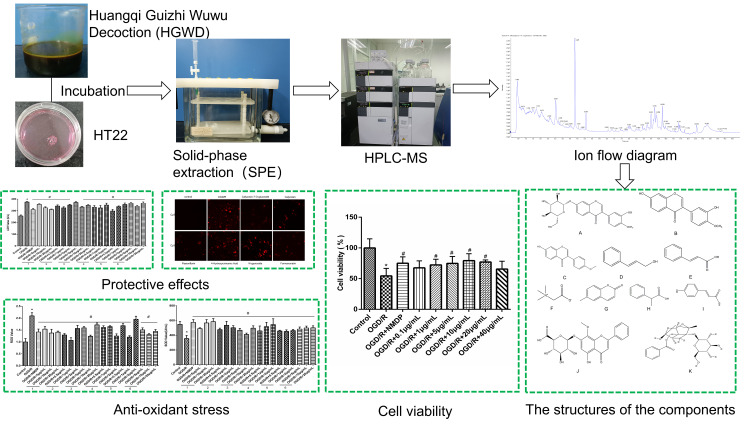
This study aims to identify the potential therapeutic components of HGWD based on the method of cell membrane solid-phase chromatography. First, an optimized solid-phase extraction (SPE) method was used to concentrate the dissociation solution of HT22 cells treated with the HGWD extract, followed by the identification of HT22-binding compounds by high-performance liquid chromatography-mass spectroscopy/mass spectroscopy (HPLC-MS/MS). Then, cell viability assay was applied to evaluate the effect of the identified compounds on protecting HT22 cells from oxygen-glucose deprivation reperfusion (OGD/R) damage. Finally, the potential mechanism of the active ingredients of HGWD for the treatment of stroke was elucidated by detecting the expression levels of reactive oxygen species (ROS), SOD, and lactate dehydrogenase (LDH). The detailed workflow is depicted in Figure 1.
Figure 1.
The workflow of the experimental procedures; *P<0.05 vs control group, #P < 0.05 vs OGD/R group.
Abbreviations: HGWD, Huangqi Guizhi Wuwu Decoction; SPE, solid-phase extraction.
Materials and Methods
Materials
HGWD, composed of five Chinese herbal medicines (Table 1), was purchased from Kangmei Pharmaceutical Co., Ltd (Guangzhou, China) and identified by Professor Danyan Zhang from Guangzhou University of Chinese Medicine (Guangzhou, China). Calycosin-7-O-glucoside, calycosin, formononetin, cinnamic alcohol, cinnamic acid, betaine, dl-2-phenylpropionic acid, 4-hydroxycinnamic acid, 6-methylcoumarin, wogonoside, and paeoniflorin were purchased from Chengdu Keloma Biotechnology Co., Ltd. (Chengdu, China). Nimodipine was purchased from Shanghai Yuanye Bio-Technology Co., Ltd. The purity of all the reference standards was above 98%. Bovine serum albumin (BSA), DMEM medium (Dulbecco’s Modified Eagle Medium), trypsin (1:250), and phosphate-buffered saline (PBS) buffer were obtained from GIBCO (Invitrogen Corporation, Carlsbad, CA, USA). Solid-phase extraction (SPE) columns were purchased from Phenomenex Inc. (Torrance, CA, USA). Cell Counting Kit-8 (CCK-8) was obtained from Dojindo Laboratories (Kumamoto, Japan). LDH, SOD, and ROS assay kits were obtained from Nanjing Jiancheng Bioengineering Institute (Nanjing, China). HT22 cells were obtained from Shanghai Kanglang Biotechnology Co., Ltd.
Table 1.
Components of Huangqi Guizhi Wuwu Decoction (HGWD)
| Latin Name | English Name | Chinese Name | Grams, g | Used Part |
|---|---|---|---|---|
| Astragalus membranaceus (Fisch.) Bge. var. mongholicus (Bge.) Hsiao or Astragalus membranaceus (Fisch.) Bge. | Radix Astragali | Huangqi | 9 | Root |
| Cinnamomum cassia (L.) J.Presl | Ramulus Cinnamomi | Guizhi | 9 | Dry shoots |
| Paeonia lactiflora PalL or Paeonia veitchii Lynch | Paeoniae Radix Alba | Baishao | 9 | Root |
| Zingiber officinale Roscoe | Zingiberis Rhizoma Recens | Shengjiang | 18 | Rhizome |
| Ziziphus jujuba Mill | Jujubae Fructus | Dazhao | 4 pieces | Ripe fruit |
Preparation of HGWD
The HGWD extract was prepared according to our previous study.13 For this, Astragali Radix (9 g), Ramulus Cinnamomi (9 g), Paeoniae Radix Alba (9 g), Zingiberis Rhizoma Recens (18 g), and Jujubae Fructus (4 pieces) were mixed and immersed in distilled water (1:10 w/v) for 30 min and extracted by refluxing for 1 h. The obtained filtrate was combined and concentrated to 50 mL, after which, the concentrate was adsorbed by D101 macroporous resin. Water-soluble components were removed with water. Then, 20%, 40%, and 70% ethanol eluates were collected and concentrated under reduced pressure to obtain a 1.08 g/mL sample solution. The sample was filtered through a 0.22 µm microporous membrane for further analysis.
Quality Control of HGWD
The quality control of HGWD was carried out with HPLC, and the content of HGWD was determined with reference to paeoniflorin, calycosin-7-O-glucoside, and calycosin.
The following chromatographic conditions were used: Waters C18 column (4.6 mm × 250 mm, 5 µm); mobile phase, acetonitrile (A) and 0.025% formic acid in water (B); column temperature, 30 °C; flow rate, 1.0 mL/min; and injection volume, 10 µL. The elution conditions are compiled in Table S1. The method was validated in terms of specificity, precision, accuracy, extraction recovery, and stability.
Biospecific Live-Cell-Based Isolation of HGWD Components
HT22 cells (Shanghai Kanglang Biotechnology Co., Ltd.) were cultured in a 10 cm culture dish. When the growth density reached 80%, HGWD extract was added to the HGWD group, and the same amount of medium was added to the control group. To facilitate the binding of active components to their corresponding receptors, both groups were incubated at 37°C for 4 h. Afterward, the cells were washed five times with PBS to remove unbound components; the bound components were dissociated from HT22 by sodium citrate buffer (pH 4.0). The collected dissociation sample was enriched and purified with an SPE column. Then, the eluents were dried using nitrogen, dissolved in 1 mL of 50% methanol, and filtered through a 0.22 µm membrane for HPLC-MS/MS analysis. The experiment was repeated three times.
HPLC-MS/MS
HPLC was performed on an instrument equipped with an SIL-82A autosampler, LC-20A pump, and SPD-20A detector (Shimadzu, Japan). The following chromatographic conditions were used: Kinetex C18 column (50 mm × 2.1 mm, 2.6 µm); mobile phase A, acetonitrile, and mobile phase B, 0.025% formic acid in water; column temperature, 35 °C; flow rate, 0.3 mL/min; and injection volume, 3 µL. The elution condition was as follows: 10–30% A at 0–5.5 min, 30–60% A at 5.5–9 min, and 60–90% A at 9–15 min.
MS analysis was performed by AB SCIEX Triple TOF 5600+ system (Framingham, MA, USA). Following MS conditions were used: ESI ion source, the scan range of 50–1200 Da, and scanning in positive and negative ion mode. The optimal MS parameter settings were as follows: ion spray voltage floating (ISVF), 4500 V; ion source gas 1(GS1), 55 psi; ion source gas 2(GS1), 55 psi; curtain gas (CUR), 35 psi; collision energy (CE), 45 V; temperature, 600 °C. The results were analyzed using Peak View software to extract information, such as fragment ions. The sample information was also compared with standard materials and literature for further identification.
Establishment of OGD/R Model and Drug Treatment
HT22 cells were cultured in a DMEM medium containing 10% FBS in an incubator (37 °C, 5% CO2). When the cell density reached 90%, the medium was removed, a glucose-free medium was added, and the plate was incubated in an incubator (95% N2 and 5% CO2) at 37 °C for 8 h to induce OGD/R injury. Then, the medium was replaced with DMEM medium, and the cells were further incubated at 37 °C for 6 h. To determine the effect of the identified components on HT22 cells with OGD/R injury, experiment groups were formed with or without identified components. Test groups consisted of cells treated with calycosin-7-O-glucoside, calycosin, formononetin, cinnamic alcohol, cinnamic acid, betaine, dl-2-phenylpropionic acid, 4-hydroxycinnamic acid, 6-methylcoumarin, wogonoside, and paeoniflorin dissolved in DMSO (0.1%) and diluted with DMEM medium to appropriate concentrations. The control group was treated with DMEM medium (0.1% DMSO), while nimodipine was used as the positive control (5 µg/mL).
Cell Viability
To determine the viability of cells after treating with different drugs, CCK-8 kit was employed. After treatment with different drugs, a complete medium containing 10% CCK-8 was added to HT22 cells (1 × 10⁴cells per well) seeded into 96-well plates and incubated for 2 h. The OD value at 450 nm of each group was measured using a microplate reader (Thermo Scientific, Rockford, IL, United States). Cell viability was calculated based on the following formula: Cell viability = OD450 of the test group/OD450 of the control group × 100%.
Release Assay
The release of LDH was measured by an LDH assay kit (Nanjing Jiancheng Institute of Biological Engineering, Nanjing, China). The cells were first treated with OGD/R and then incubated with different drugs; the cell supernatant obtained after this step was incubated with substrate solution at 37 °C for 15 min. To this, 2,4-dinitrophenylhydrazine was added and incubated for another 15 min. The absorbance signal was detected on a microplate reader at 450 nm. Each experiment was carried out in sextuplicate.
Detection of ROS
The levels of ROS of each group were evaluated by a kit utilizing the fluorescent probe 2ʹ,7ʹ-dichlorofluorescein-diacetate (DCFH-DA). The cells were cultured in 96-well plates for 24 h and treated with OGD/R, except for the normal group. OGD/R-treated HT22 cells were incubated with calycosin-7-O-glucoside (5,10, and 20 µg/mL), calycosin (0.5,1, and 2 µg/mL), paeoniflorin (5,10, and 20 µg/mL), wogonoside (0.5,1, and 2 µg/mL), and formononetin (5,10, and 20 µg/mL) for 24 h. Then, the DCFH-DA fluorescent probe working solution was added, and the plates were incubated at 37 °C in the dark for 30 min. The intensity of ROS was measured at an OD of 525 nm. Each experiment was performed in sextuplicate.
Determination of SOD Content
SODs are a group of detoxification enzymes as they are powerful antioxidants. This enzyme can scavenge free radicals, such as superoxide anion, thereby protecting cells from damage caused by free radicals. The SOD activity was measured utilizing WST-1 that produces a water-soluble formazan dye upon reduction with superoxide anion. The OGD/R-treated cells were incubated with different drugs; the cell supernatant obtained after this step was incubated with enzyme working solution and substrate application solution at 37°C for 20 min. The fluorescence intensity was then detected using a microplate reader at 450 nm. Each experiment was carried out in sextuplicate.
TUNEL Assay
The experiment was carried out according to the manufacturer’s instructions using the One Step TUNEL Apoptosis Assay Kit (Beyotime, Shanghai, China). After drug treatment, HT22 cells were fixed with 4% paraformaldehyde and permeabilized with 0.3% Triton X-100 for 20 min. Next, the cells were incubated with a fluorescein TUNEL-TdT reaction mixture incubated at room temperature for 1 h in the dark. After washing, the apoptotic cells were detected by fluorescence microscope (Olympus Corporation) and analyzed via ImageJ analysis software. Each experiment was conducted in triplicate.
Statistical Analysis
All experiments were repeated at least three times. The generated data were presented as mean ±standard deviation (SD). Between-group comparisons of the results were made by Student’s t-test (P < 0.05 was considered significant). Statistical analysis was performed using SPSS 22.0 software, and graphs were plotted by GraphPad Prism 7.0 software.
Results and Discussion
Quality Control of HGWD
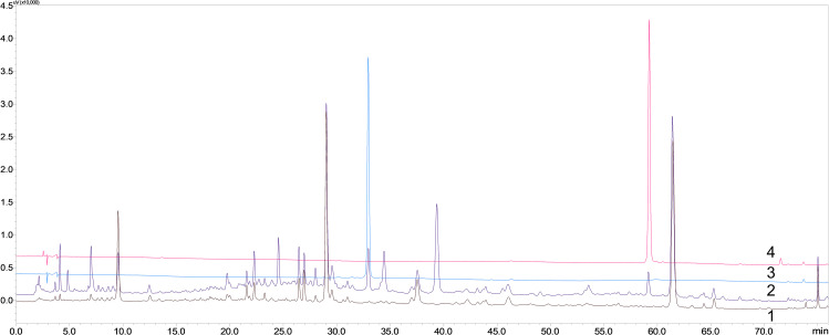
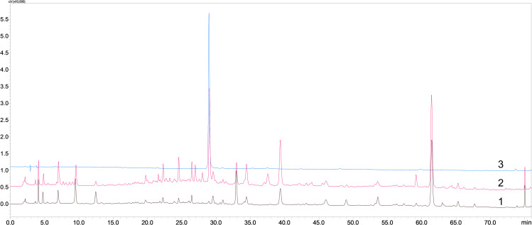
The quality control tests, including validation of specificity, linearity, and lower limit of quantitation (LLOQ), precision and accuracy, stability, and extraction recovery, were executed. The typical chromatograms of calycosin-7-O-glucoside, calycosin, and paeoniflorin were used as references (Figures 2 and 3). There was no interference in the corresponding absorption peaks, indicating that the method has good specificity. The calibration curves exhibited linearity over the concentration range of 2.50–25.00 µg/mL for calycosin-7-O-glucoside, 0.25–2.51 µg/mL for calycosin, and 95.78–2394.50 µg/mL for paeoniflorin, with the correlation coefficients of calycosin-7-O-glucoside, calycosin, and paeoniflorin being y = 19113x-0905 (R2 = 0.9980), y = 26469x-2066.2 (R2 = 0.9998), and y = 813.34x-13,570 (R2 = 0.9990), respectively. The precision and accuracy results of the three analytes demonstrated that the precision of the instrument was good (Table S2). The results of stability are shown in Table S3. The sample recovery test results showed that the average recoveries were 100.04%, 103.99%, and 106.47% for calycosin-7-O-glucoside, calycosin, and paeoniflorin, respectively, and RSD values were 1.97%, 3.05%, and 2.58%, respectively (Table S4). Based on the linear relationship, the average contents of calycosin-7-O-glucoside, calycosin, and paeoniflorin in HGWD were calculated to be 1884.33 µg, 250.41 µg, and 114,040.05 µg, respectively (Table S5).
Figure 2.
The specific HPLC chromatograms of calycosin-7-O-glucoside and calycosin (1. HGWD without Radix Astragali (negative sample); 2. HGWD; 3. Calycosin-7-O-glucoside; 4. Calycosin).
Figure 3.
The specific HPLC chromatograms of paeoniflorin (1. HGWD without Paeoniae Radix Alba (negative sample); 2. HGWD; 3. paeoniflorin).
Biospecific Live-Cell-Based Isolation and HPLC-MS/MS Identification of HGWD Components
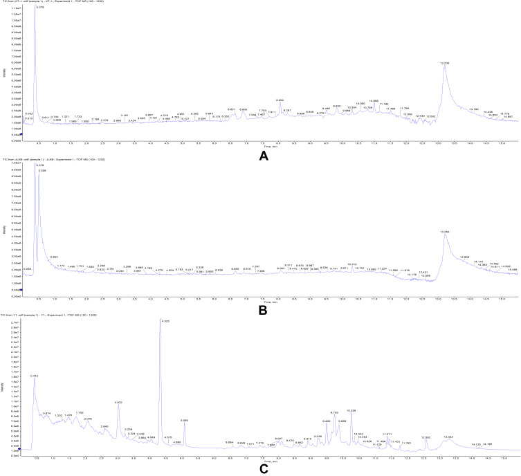
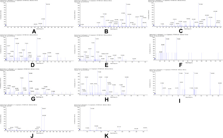
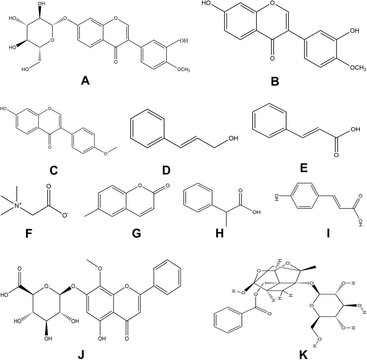
Biospecific live-cell-based isolation of active compounds, which is the combination of life sciences and chromatographic separation, from medicinal extracts is an emerging technology with tremendous prospects. Here, living cells are used as the stationary phase, which can specifically bind to the active ingredients of TCM. The bound components are eluted and determined with modern analytical techniques, such as HPLC or LC/MS.13 Compared with Figure 4C, few peaks were detected by HPLC-MS in the fifth wash solution and blank dissociation solution (Figure 4A and B). With the help of Peak View software, a total of eleven compounds were identified.14 Among them, eight compounds were identified under the positive ion mode, namely calycosin-7-O-glucoside (C22H22O10), calycosin (C16H12O5), formononetin (C16H12O4), cinnamic alcohol (C9H10O), cinnamic acid (C9H8O2), betaine (C5H11NO2), dl-2-phenylpropionic acid (C9H10O2), and 6-methylcoumarin (C10H8O2); three components were identified in negative ion mode, including 4-hydroxycinnamic acid (C9H8O3), wogonoside (C22H20O11), and paeoniflorin (C23H28O11). The secondary mass spectrum was shown in Figure 5 and Table 2. To further determine the chemical structures of the compounds, the data was compared with the standard product and literature. The MS data of calycosin-7-O-glucoside,14 calycosin,15 formononetin,16 cinnamic alcohol,17 cinnamic acid,18 betaine,19 wogonoside,20 and paeoniflorin21 were consistent with previous reports. The structures of dl-2-phenylpropionic acid, 6-methylcoumarin, and 4-hydroxycinnamic acid were determined in comparison with the standard product. In short, the eleven compounds that had a binding affinity to HT22 were identified as calycosin-7-O-glucoside, calycosin, formononetin, cinnamic alcohol, cinnamic acid, betaine, dl-2-phenylpropionic acid, 4-hydroxycinnamic acid, 6-methylcoumarin, wogonin, and paeoniflorin (Figure 6).
Figure 4.
Chromatograms of the fifth washing solution, blank washing dissociation solution, and Huangqi Guizhi Wuwu Decoction (HGWD) dissociation solution in negative ion mode. (A) The fifth washing solution; (B) Blank group dissolution solution; (C) Huangqi Guizhi Wuwu Decoction (HGWD) dissociation solution.
Figure 5.
MS spectra of eleven affinity components (A) calycosin-7-O-glucoside; (B) calycosin; (C) formononetin; (D) cinnamic alcohol; (E) cinnamic acid; (F) betaine; (G) 6-methylcoumarin; (H) dl-2-phenylpropionic acid; (I) 4-hydroxycinnamic acid; (J) wogonoside; (K) paeoniflorin.
Table 2.
Fragment Ion Information of Affinity Component in the Dissociation Solution
| Name | Formula | Selected Ion | Extraction Mass (Da) | Found At mass (Da) | Error (ppm) | MS/MS |
|---|---|---|---|---|---|---|
| Calycosin-7-O-glucoside | C22H22O10 | [M+H] + | 447.1286 | 447.1272 | −3.13 | 285.0729, 270.0502, 253.0481, 225.0531 |
| Calycosin | C16H12O5 | [M+H] + | 285.0751 | 285.0758 | 2.46 | 285.0763, 270.0503, 253.0477, 225.0526 |
| Formononetin | C16H12O4 | [M+H] + | 269.0808 | 269.0803 | −1.86 | 269.0814, 254.0554, 225.0548, 181.0643 |
| Cinnamic alcohol | C9H10O | [M+H] + | 135.0804 | 135.0800 | −2.96 | 135.1000, 79.0604 |
| Cinnamic acid | C9H8O2 | [M+H] + | 149.0597 | 149.0592 | −3.35 | 133.0706, 103.0567, 79.0580 |
| Betaine | C5H11NO2 | [M+H] + | 118.0863 | 118.0861 | −1.69 | 73.0492, 61.0335, 58.0664, 55.9391 |
| 6-Methylcoumarin | C10H8O2 | [M+H] + | 161.0597 | 161.0592 | −3.10 | 131.0529, 117.0410, 115.0547, 103.0558 |
| DL-2-phenylpropionic acid | C9H10O2 | [M+H] + | 151.0754 | 151.0751 | −1.99 | 122.9656, 113.9601, 103.9584, 79.0535 |
| 4-Hydroxycinnamic acid | C9H8O3 | [M-H]− | 163.0401 | 163.0400 | −0.61 | 162.8900, 117.0529, 93.0361, 91.0361 |
| Wogonoside | C22H20O11 | [M-H]− | 459.09328 | 459.0928 | −1.05 | 283.0580, 268.0357, 113.0243, 71.0174 |
| Paeoniflorin | C23H28O11 | [M+COOH]− | 525.1602 | 525.1606 | 0.76 | 525.3073, 449.1495, 327.1114, 121.0305 |
Figure 6.
The structures of the identified affinity components (A) calycosin-7-O-glucoside; (B) calycosin; (C) formononetin; (D) cinnamic alcohol; (E) cinnamic acid; (F) betaine; (G) 6-methylcoumarin; (H) dl-2-phenylpropionic acid; (I) 4-hydroxycinnamic acid; (J) wogonoside; (K) paeoniflorin.
Cell Viability Assay
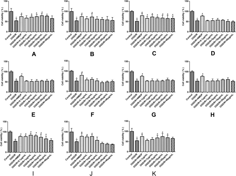
The results indicate a significant reduction in the survival rate of the OGD/R group compared with the control group (P < 0.05), indicating that OGD/R caused damage to HT22 cells. After the intervention with different concentrations of calycosin-7-O-glucoside (1, 5, 10, and 20 µg/mL), the survival rates were significantly different from that of the OGD/R group (P < 0.05), indicating that calycosin-7-O-glucoside could significantly alleviate the OGD/R-induced cell damage (Figure 7). In addition, calycosin (1 µg/mL), paeoniflorin (5–10 µg/mL), 4-hydroxycinnamic acid (0.1–20 µg/mL), formononetin (0.1–40 µg/mL), and wogonoside (0.1–20 µg/mL) could significantly enhance the cell viability of OGD/R-treated cells (P < 0.05). However, as compared to the OGD/R-treated group (P>0.05), no significant changes in survival rates were observed in the groups treated with cinnamic alcohol, cinnamic acid, betaine, dl-2-phenylpropionic acid, and 6-methylcoumarin. Studies have shown that calycosin-7-O-glucoside can reduce the injury induced by OGD/R in HT22 cells through the SIRT1/FOXO1/PGC-1α pathway.22 Calycosin can improve the ischemic stroke injury in rats by transforming microglia from a resting state to an active “ameba-like state” through the BDNF/Trkb pathway.23 It can also prevent autophagy, apoptosis, cell death, and inflammation caused by oxidative stress, which is essential for neuroprotection.24 Studies have demonstrated that formononetin may enhance neuronal differentiation and synaptic plasticity through PI3K/AKT/ERK pathway.25 Paeoniflorin is the active component of the red peony root, which can reduce cerebral ischemic damage by regulating the Ca2+/CaMKII/CREB signaling pathway.26 4-Hydroxycinnamic acid is a neuroprotective agent on account of its strong antioxidant and anti-apoptotic features.27 The results of this study indicated that calycosin-7-O-glucoside, calycosin, paeoniflorin, 4-hydroxycinnamic acid, formononetin, and wogonoside, have a protective effect on OGD/R-treated cells at the determined concentration range, while cinnamic alcohol, cinnamic acid, betaine, dl-2-phenylpropionic acid, and 6-methylcoumarin have no such effect. Therefore, calycosin-7-O-glucoside, calycosin, paeoniflorin, 4-hydroxycinnamic acid, formononetin, and wogonoside may be the active ingredients of HGWD for promoting nerve function recovery after stroke.
Figure 7.
Effects of the identified compounds on the survival rate of OGD/R-treated HT22 cells. (A) calycosin-7-O-glucoside; (B) calycosin; (C) formononetin; (D) cinnamic alcohol; (E) cinnamic acid; (F) betaine; (G) dl-2-phenylpropionic acid; (H) 6-methylcoumarin; (I) 4-hydroxycinnamic acid; (J) wogonoside; (K) paeoniflorin (*P < 0.05 vs control group, #P < 0.05 vs OGD/R group; n = 6).
The Protective Effects of Calycosin-7-O-Glucoside, Calycosin, Paeoniflorin, 4-Hydroxycinnamic Acid, Wogonoside, and Formononetin in HT22 Cells Against OGD/R-Induced Injury
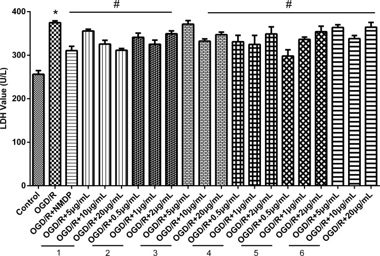
Under the pathological state of ischemia and hypoxia, excessive accumulation of lactic acid inhibits the activity of LDH in cells, leading to the death of neurons.28 The results of LDH content determination (Figure 8) indicate that compared with the control group, the LDH activity of the OGD/R-treated cells increased significantly. Compared with the OGD/R group (P<0.05), a significant reduction in LDH activity was observed after the treatment of OGD/R-treated cells with calycosin-7-O-glucoside, calycosin, paeoniflorin, 4-hydroxycinnamic acid, wogonoside, and formononetin.
Figure 8.
Effects of the identified compounds on LDH activity in cell supernatant. (1) calycosin-7-O-glucoside; (2) calycosin; (3) paeoniflorin; (4) 4-hydroxycinnamic acid; (5) wogonoside; (6) formononetin. (*P < 0.05 vs control group, #P < 0.05 vs OGD/R group; n = 6).
Calycosin-7-O-Glucoside, Calycosin, Paeoniflorin, 4-Hydroxycinnamic Acid Wogonoside, and Formononetin Alleviates OGD/R-Induced Oxidative Stress
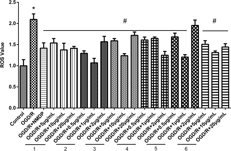
Oxidative stress is the imbalance of oxygen-containing free radicals and antioxidants in the body, which means that the removal rate of ROS is much lower than the production rate, leading to lipid peroxidation and damage to tissues and organs. Oxidative stress caused by reperfusion after cerebral ischemia is one of the pathological mechanisms of ischemic brain injury.3 The results of the ROS content determination (Figure 9) indicate that compared with the control group, the fluorescence intensity of the OGD/R group was significantly higher (P < 0.05). However, after drug intervention, the intracellular fluorescence intensity of each group was significantly reduced compared with that of the OGD/R group (P < 0.05).
Figure 9.
Effects of the identified compounds on ROS levels in cell supernatant. (1) calycosin-7-O-glucoside; (2) calycosin; (3) paeoniflorin; (4) 4-hydroxycinnamic acid; (5) wogonoside; (6) formononetin. (*P < 0.05 vs control group, #P < 0.05 vs OGD/R group; n = 6).
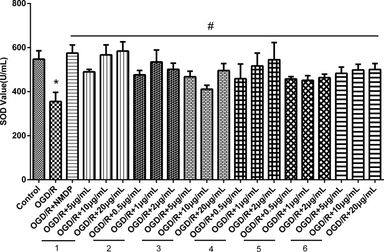
The results of SOD content determination indicated that compared with the control group, the fluorescence intensity of the OGD/R group was significantly higher (P<0.05), indicating that OGD/R caused oxidative stress damage. Also, the protective effect of calycosin-7-O-glucoside and 4-hydroxycinnamic acid, wogonoside, and formononetin increased with the increase of the drug concentration, reflecting dose-dependency. Calycosin and paeoniflorin exerted the strongest protective effects at the concentration of 1 µg/mL and 20 µg/mL, respectively (Figure 10). These results showed that the above ingredients could alleviate damage induced by oxidative stress, thereby exerting a neuroprotective effect against OGD/R injury.
Figure 10.
Effects of the identified compounds on SOD levels in cell supernatant. (1) calycosin-7-O-glucoside; (2) calycosin; (3) paeoniflorin; (4) 4-hydroxycinnamic acid; (5) wogonoside; (6) formononetin. (*P < 0.05 vs control group, #P < 0.05 vs OGD/R group; n = 6).
Calycosin-7-O-Glucoside, Calycosin, Paeoniflorin, 4-Hydroxycinnamic Acid Wogonoside, and Formononetin Induces Apoptosis
Tunel staining were used to evaluate the effect of drugs on apoptosis in HT22 cells. Tunel staining showed that the apoptosis cell was significantly increased in OGD/R group compared with the control group (P < 0.01), and after drug treatment, apoptosis cell was significantly reduced compared with the OGD/R group (P < 0.01) (Figure 11). These results suggested that calycosin-7-O-glucoside, calycosin, paeoniflorin, 4-hydroxycinnamic acid wogonoside, and formononetin were able to attenuate OGD/R-mediated apoptosis. Therefore, we hypothesized that the mechanism of action of HGWD to promote neurological recovery may be related to the regulation of apoptosis.
Figure 11.
TUNEL (red) staining showing the apoptotic HT22 cells after different treatments (A and B). (Scale bar: 100 μm. Data are shown as means ± SD, #P < 0.01 vs control group; *P < 0.01 vs OGD/R group; n = 3).
Conclusions
In this study, we established a cell membrane solid-phase chromatography method to screen eleven active components from HGWD. The survival rate of HT22 cells after OGD/R injury was investigated to explore the activity of the identified compounds. Among them, six components had different degrees of protective effect on OGD/R-treated HT22 cells. The protective effect was measured by the levels of ROS, SOD and LDH and the degree of apoptosis. The results showed that calycosin-7-O-glucoside, calycosin, paeoniflorin, 4-hydroxycinnamic acid, wogonoside, and formononetin could alleviate the oxidative stress injury and apoptosis induced by OGD/R. Therefore, HGWD can promote neurological recovery after ischemic stroke by regulating oxidative stress and apoptosis. Current knowledge on the treatment of ischemic stroke using HGWD is majorly derived from clinical research and not from experimental research. This experimental study, based on cell membrane solid-phase chromatography, is the first to explore the possible mechanisms of the therapeutic effects of HGWD. However, due to the complexity of HGWD composition, some compounds with higher response values remain unidentified according to the total ion chromatogram of positive and negative samples. Therefore, further research is required for the identification of these compounds.
Funding Statement
This work was supported by the National Natural Science Foundation of China (grant number 81373973), Natural Science Foundation of China (grant number 81573872). All authors have declared no conflict of interest.
Abbreviations
HGWD, Huangqi Guizhi Wuwu Decoction; OGD/R, oxygen-glucose deprivation reperfusion; ROS, reactive oxygen species; SOD, superoxide dismutase, LDH, lactate dehydrogenase; TCM, traditional Chinese medicines; BSA, bovine serum albumin; DMEM, Dulbecco’s Modified Eagle; PBS, phosphate-buffered saline; SPE, solid-phase extraction; CCK-8, Cell Counting Kit-8.
Author Contributions
All authors made a significant contribution to the work reported, whether that is in the conception, study design, execution, acquisition of data, analysis, and interpretation, or in all these areas; took part in drafting, revising or critically reviewing the article; gave final approval of the version to be published; have agreed on the journal to which the article has been submitted; and agree to be accountable for all aspects of the work.
Disclosure
The authors declare no conflicts of interest.
References
- 1.Morsi N, Ghorab D, Badie H. Brain targeted solid lipid nanoparticles for brain ischemia: preparation and in vitro characterization. Pharm Dev Technol. 2013;18(3):736–744. doi: 10.3109/10837450.2012.734513 [DOI] [PubMed] [Google Scholar]
- 2.Zhen Y, Ding C, Sun J, et al. Activation of the calcium-sensing receptor promotes apoptosis by modulating the JNK/p38 MAPK pathway in focal cerebral ischemia-reperfusion in mice. Am J Transl Res. 2016;8(2):911–921. [PMC free article] [PubMed] [Google Scholar]
- 3.Adibhatla R, Hatcher J. Lipid oxidation and peroxidation in CNS health and disease: from molecular mechanisms to therapeutic opportunities. Antioxid Redox Signal. 2010;12(1):125–169. doi: 10.1089/ars.2009.2668 [DOI] [PubMed] [Google Scholar]
- 4.Chamorro Á, Dirnagl U, Urra X, et al. Neuroprotection in acute stroke: targeting excitotoxicity, oxidative and nitrosative stress, and inflammation. Lancet Neurol. 2016;15(8):869–881. doi: 10.1016/s1474-4422(16)00114-9 [DOI] [PubMed] [Google Scholar]
- 5.Cobley J, Fiorello M, Bailey D. 13 reasons why the brain is susceptible to oxidative stress. Redox Biol. 2018;15:490–503. doi: 10.1016/j.redox.2018.01.008 [DOI] [PMC free article] [PubMed] [Google Scholar]
- 6.Saeed S, Shad K, Saleem T, et al. Some new prospects in the understanding of the molecular basis of the pathogenesis of stroke. Exp Brain Res. 2007;182(1):1–10. doi: 10.1007/s00221-007-1050-9 [DOI] [PubMed] [Google Scholar]
- 7.Hua W, Bu P. Treatment of 68 cases of diabetic peripheral neuropathy with integrated traditional Chinese and western medicine. Jiangsu Tradit Chin Med. 2008;11:65–66. [Google Scholar]
- 8.Xu L, Wang Z, Xia Y. Effect of Huangqi Guizhi Wuwu decoction on patients with cerebral infarction in recovery stage. Pract Clin Med Integr Tradit Chin Western Med. 2019;19(11):55–57. [Google Scholar]
- 9.Hu Q, Bu Y, Cao R, et al. Stability designs of cell membrane cloaked magnetic carbon nanotubes for improved life span in screening drug leads. Anal Chem. 2019;91(20):13062–13070. doi: 10.1021/acs.analchem.9b03268 [DOI] [PubMed] [Google Scholar]
- 10.Wu S, Wang N, He Q, et al. The establishment of the method of cell biochromatograpy and analysis of the active ingredients from TongQiaoHuoXue decoction acting on the neurocytes. Chem Pharm Bull. 2018;66(10):983–991. doi: 10.1248/cpb.c18-00455 [DOI] [PubMed] [Google Scholar]
- 11.Liao F, Meng Y, Zheng H, et al. Biospecific isolation and characterization of angiogenesis-promoting ingredients in Buyang Huanwu decoction using affinity chromatography on rat brain microvascular endothelial cells combined with solid-phase extraction, and HPLC-MS/MS. Talanta. 2018;179:490–500. doi: 10.1016/j.talanta.2017.11.018 [DOI] [PubMed] [Google Scholar]
- 12.Yan X, Wang S, Yu A, et al. Cell chromatography-based screening of the active components in Buyang Huanwu Decoction promoting axonal regeneration. Biomed Res Int. 2019;2019:6970198. doi: 10.1155/2019/6970198 [DOI] [PMC free article] [PubMed] [Google Scholar]
- 13.Zheng H, Shen X, He Y, et al. Pharmacokinetic analysis of Huangqi Guizhi Wuwu Decoction on blood and brain tissue in rats with normal and cerebral ischemia-reperfusion injury by microdialysis with HPLC-MS/MS. Drug Des Devel Ther. 2020;14:2877–2888. doi: 10.2147/dddt.s257020 [DOI] [PMC free article] [PubMed] [Google Scholar]
- 14.Liao F, Yu A, Yu J, et al. Identification of active ingredients mediating anti-platelet aggregation effects of BuyangHuanwu decoction using a platelet binding assay, solid phase extraction, and HPLC-MS/MS. J Chromatogr B Analyt Technol Biomed Life Sci. 2018;1092:320–327. doi: 10.1016/j.jchromb.2018.06.027 [DOI] [PubMed] [Google Scholar]
- 15.Tanaka K, Tamura T, Fukuda S, et al. Quality evaluation of Astragali Radix using a multivariate statistical approach. Phytochemistry. 2008;69(10):2081–2087. doi: 10.1016/j.phytochem.2008.04.016 [DOI] [PubMed] [Google Scholar]
- 16.Zielińska A, Paradowska K, Jakowski J, et al. 13 C CP MAS NMR and GIAO-CHF/DFT calculations of flavonoids: morin, kaempferol, tricin, genistein, formononetin and 3,7-dihydroxyflavone. J Mol Struct. 2007;873(1):109–116. [Google Scholar]
- 17.Sawada Y, Akiyama K, Sakata A, et al. Widely targeted metabolomics based on large-scale MS/MS data for elucidating metabolite accumulation patterns in plants. Plant Cell Physiol. 2009;50(1):37–47. doi: 10.1093/pcp/pcn183 [DOI] [PMC free article] [PubMed] [Google Scholar]
- 18.Ji B, Zhao Y, Zhang Q, et al. Simultaneous determination of cinnamaldehyde, cinnamic acid, and 2-methoxy cinnamic acid in rat whole blood after oral administration of volatile oil of Cinnamoni Ramulus by UHPLC-MS/MS: an application for a pharmacokinetic study. J Chromatogr B Analyt Technol Biomed Life Sci. 2015;1001:107–113. doi: 10.1016/j.jchromb.2015.07.049 [DOI] [PubMed] [Google Scholar]
- 19.Steuer C, Schütz P, Bernasconi L, et al. Simultaneous determination of phosphatidylcholine-derived quaternary ammonium compounds by a LC-MS/MS method in human blood plasma, serum and urine samples. J Chromatogr B Analyt Technol Biomed Life Sci. 2016;1008:206–211. doi: 10.1016/j.jchromb.2015.12.002 [DOI] [PubMed] [Google Scholar]
- 20.Zhang Y, Lu Y, Xiao J, et al. Concurrent identification of 11 major primary active compounds in Huangqin Qingfei Decoction by liquid chromatography tandem mass spectrometry via liquid chromatography tandem mass spectrometry. Pak J Pharm Sci. 2020;33(3):1005–1013. [PubMed] [Google Scholar]
- 21.Tsugawa H, Nakabayashi R, Mori T, et al. A cheminformatics approach to characterize metabolomes in stable-isotope-labeled organisms. Nat Methods. 2019;16(4):295–298. doi: 10.1038/s41592-019-0358-2 [DOI] [PubMed] [Google Scholar]
- 22.Yan X, Yu A, Zheng H, et al. Calycosin-7-O-β-D-glucoside attenuates OGD/R-Induced damage by preventing oxidative stress and neuronal apoptosis via the SIRT1/FOXO1/PGC-1α pathway in HT22 cells. Neural Plast. 2019;2019:1–11. doi: 10.1155/2019/8798069 [DOI] [PMC free article] [PubMed] [Google Scholar]
- 23.Hsu C, Kuo T, Liu W, et al. Calycosin preserves BDNF/TrkB signaling and reduces post-stroke neurological injury after cerebral ischemia by reducing accumulation of hypertrophic and TNF-α-containing microglia in rats. J Neuroimmune Pharmacol. 2020;15(2):326–339. doi: 10.1007/s11481-019-09903-9 [DOI] [PubMed] [Google Scholar]
- 24.Dou B, Zhou W, Li S, et al. Buyang huanwu decoction attenuates infiltration of natural killer cells and protects against ischemic brain injury. Cell Physiol Biochem. 2018;50(4):1286–1300. doi: 10.1159/000494587 [DOI] [PubMed] [Google Scholar]
- 25.Wu Q, Cheng Y, Liu A, et al. Formononetin recovered injured nerve functions by enhancing synaptic plasticity in ischemic stroke rats. Biochem Biophys Res Commun. 2020. doi: 10.1016/j.bbrc.2020.02.015 [DOI] [PubMed] [Google Scholar]
- 26.Zhang Y, Qiao L, Xu W, et al. Paeoniflorin attenuates cerebral ischemia-induced injury by regulating Ca/CaMKII/CREB signaling pathway. Molecules. 2017;22(3). doi: 10.3390/molecules22030359 [DOI] [PMC free article] [PubMed] [Google Scholar]
- 27.Wang Y, Ren Q, Zhang X, et al. Neuroprotective mechanisms of calycosin against focal cerebral ischemia and reperfusion injury in rats. Cell Physiol Biochem. 2018;45(2):537–546. doi: 10.1159/000487031 [DOI] [PubMed] [Google Scholar]
- 28.Fu S, Gu Y, Jiang J, et al. Calycosin-7-O-β-D-glucoside regulates nitric oxide /caveolin-1/matrix metalloproteinases pathway and protects blood-brain barrier integrity in experimental cerebral ischemia-reperfusion injury. J Ethnopharmacol. 2014;155(1):692–701. doi: 10.1016/j.jep.2014.06.015 [DOI] [PubMed] [Google Scholar]


