Abstract
Background
Neonates possess an immature and plastic immune system, which is a major cause of some diseases in newborns. Necrotizing enterocolitis (NEC) is a severe and devastating intestinal disease that typically affects premature infants. However, the development of intestinal immune cells in neonates and their roles in the pathological process of NEC have not been elucidated.
Results
We examined the ontogeny of intestinal lamina propria lymphocytes in the early life of mice and found a high percentage of RORγt+ cells (containing inflammatory Th17 and ILC3 populations) during the first week of life. Importantly, the proportion of RORγt+ cells of intestinal lamina propria further increased in both NEC mice and patients tissue than the control. Furthermore, the application of GSK805, a specific antagonist of RORγt, inhibited IL-17A release and ameliorated NEC severity.
Conclusions
Our data reveal the high proportion of RORγt+ cells in newborn mice may directly contribute to the development of NEC.
Supplementary Information
The online version contains supplementary material available at 10.1186/s13578-021-00739-6.
Keywords: Neonate, Necrotizing enterocolitis, Intestinal immune, RORγt
Background
Necrotizing enterocolitis (NEC) is a life-threatening disease that affects approximately 7% of infants with a birth weight under 1500 g [1]. The estimated mortality rate of NEC ranges from 20 to 30%, and a large proportion of NEC survivors remain at significant risk for short bowel syndrome and neurodevelopmental impairment [2–4]. Although the definitive causes of NEC remain unknown, prematurity is a recognized independent predictor because NEC occurrence increases with decreasing of gestational age and birth weight [5]. The other contributing factors include formula feeding, intestinal ischemia–reperfusion injury, dysbacteria, and excessive immune response [6]. Because of the complicated pathogenesis, there are no specific treatment strategies that effectively alter the outcome, and surgical approaches are the last resort but remain controversial. Recent studies suggest that human milk feeding and probiotics may prevent the development of NEC [7, 8], whereas the conclusions are also debatable [9].
Neonates possess a developing and plastic immune system, which is susceptible to different kinds of pathogenic factor invasion. Following birth, neonatal intestines have to encounter mass new antigens and stimuli, which increase the vulnerability of neonates to both infectious and non-infectious diseases [10]. Especially in preterm and very low birth weight infants, the intestinal barrier and immune system are immature, which predispose them to have risk of subjecting to late-on sepsis or NEC [11]. Studies have revealed that Toll-like receptor 4 (TLR4), a receptor for bacterial endotoxin, is expressed at a higher level on the intestinal epithelium of the premature human and mouse gut [12–14]. The excessive activation of TLR4 by lipopolysaccharide (LPS) leads to an infiltration of CD4+ T lymphocytes, and predispose the populations to pro-inflammatory type 17 T helper (Th17) cells, which is required for the development of NEC [15]. Meanwhile, a few studies have demonstrated that, compared with controls, intraepithelial γδ T lymphocytes and T regulatory cells (Treg) of lamina propria decreased significantly in surgical NEC specimens [11, 16–18]. The imbalance of Th17/Treg induces the increases of proinflammatory cytokines such as tumor necrosis factor (TNF), interleukin (IL)-1β, IL-6, IL-17, and IL-18, and the decreases of anti-inflammatory mediators such as IL-10 and transforming growth factor (TGF)β. The excessive inflammatory response triggers the vicious cycle that exacerbates tissue injury and necrosis of intestine, resulting in NEC development [19].
Although the phenomenon of pathogenesis is revealed gradually, the causative factors of NEC in preterm neonates remain poorly understood. Little is known about the state of intestinal immune system before NEC development. The orphan nuclear receptor retinoid-related orphan receptor γt (RORγt) is a key transcription factor that orchestrates the differentiation of immune cells [20] and induces the formation of lymph nodes and Peyer’s patches [21, 22]. In gut, RORγt+ cells include not only Th17 cells, but also several innate immune cells such as lymphoid tissue inducer cells (LTi), innate lymphoid cells (ILCs), γδ T cells, and natural killer (NK) cells [23]. After birth, LTi cells firstly cluster into cryptopatches of the intestinal lamina propria, accompanied with ILCs and RORγt+ T cells expand within the lamina propria [24]. Th17 cells are potent inducers of intestinal inflammation and have been implicated in the pathogenesis of inflammatory bowel disease (IBD) [25] and NEC [15]. However, the link between the ontogeny of RORγt and NEC development has not been revealed. In this study, we found that the high proportion of RORγt+ cells during the first few days of life, which might contribute to intestinal inflammation in neonates. Meanwhile, in NEC patients or mice model, the RORγt+ cells of intestinal lamina propria increased significantly than the healthy control. Furthermore, when NEC mice were treated with GSK805, the specific antagonist of RORγt, the intestinal lesions were improved visibly. In conclusion, we revealed that the high proportion of RORγt+ cells in neonatal intestine might be accountable for NEC development.
Results
Ontogeny of RORγt+ cells in intestinal lamina propria of neonatal mice
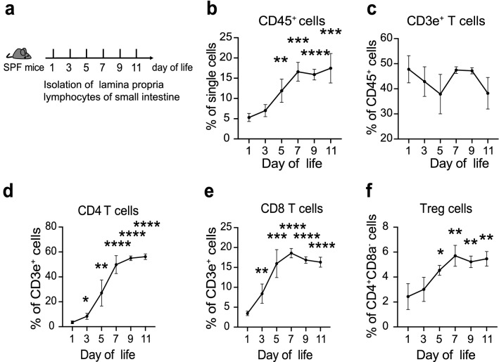
The ontogeny of intestinal immune cells was investigated in normal neonatal mice to seek causes leading to intestinal disease in early life. The composition of immune cells within the lamina propria of small intestine was assessed throughout the first few days of life (Fig. 1a, Additional file 1: Fig. S1). Firstly, we noticed that the proportion of leukocytes (CD45+ cells) increased rapidly in the first 5 days, and kept stable after day 7 (Fig. 1b). However, the percentages of CD3e+ T cells vary dramatically during early life, which indicated the immature state of adaptive immune in the neonatal intestine (Fig. 1c). The average percentages of CD4 or CD8 T cells were below 5% at day 1 (Fig. 1d, e). The frequency of CD4 T cells climbed rapidly from day 3 to day 7, and remained over 50% after day 9. While, the population of CD8 T cells reached a peak of over 15% at day 7 and then decreased progressively (Fig. 1e). The percentages of CD4 and CD8 T cells at day 7 were consistent with the results in lamina propria of human infant intestines [26]. Whereas, the ontogeny trends of CD4 and CD8 T cells different from the study by Dingle et al. [17] showed the average percentage of CD4 T cells remained steady below 15%, and CD8 T cells kept over 75% in the terminal ileum since they measured total cellular content within the intestines in neonatal rats. Meanwhile, we found the percentages of CD4+Foxp3+ Treg cells progressively increased during the first week, then remained stable till to day 11 (Fig. 1f). These results characterized the ontogeny of intestinal adaptive immune cells in the early life of mice.
Fig. 1.
Dynamic changes of T cells in lamina propria of small intestine. a. Diagram of the experimental design. b. The changes tendency of leucocytes (CD45+ cells). c–f. The variations of CD3e+ T cells (c), CD4 T cells (d), CD8 T cells (e), and Treg cells (f) along with time. Three independent experiments were performed, n = 4–5 per group. Data were shown as mean values ± SD. Statistical analyses were performed with Student’s two-tailed unpaired t-test. *Compared with day 1. *p < 0.05; **p < 0.01; ***p < 0.001; ****p < 0.0001
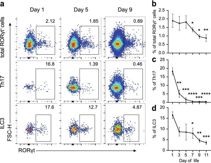
Interestingly, the highest percentage of RAR-related orphan receptor γ, isoform t (RORγt)-expressing cells among total CD45+ cells was observed in the first three days, which decreased to less than 1% after day 9 (Fig. 2a, b). Th17 and ILC3 cells are the two dominant populations that express the transcription factor RORγt in intestinal lamina propria, and they are responsible for various intestinal inflammation diseases [23]. Remarkably, our data showed that the average percentages of Th17 and ILC3 were more than 15% at day 1, and both of them decreased progressively (Fig. 2a, c, d). These findings prompted our hypothesis that the superabundant RORγt+ cells in the first few days of life might be resulting in the susceptibility to necrotizing enterocolitis induction.
Fig. 2.
High percentages of RORγt+ cells during the first few days of life. a. Representative pseudocolor dot plots of total RORγt+ cells, Th17 and ILC3. b–d Dynamic changes of total RORγt+ cells, Th17 and ILC3. Th17 was gated on CD3e+CD4+CD8a−. ILC3 was gated on CD3e−CD8a−B220−LIN−CD90.2+. LIN includes CD11b and CD11c. Three independent experiments were performed, n = 4–5 per group. Data were shown as mean values ± SD. Statistical analyses were performed with Student’s two-tailed unpaired t-test. *Compared with day 1. *p < 0.05; **p < 0.01; ***p < 0.001; ****p < 0.0001
IL-17A+ RORγt+ cells accumulate in intestine of NEC mice
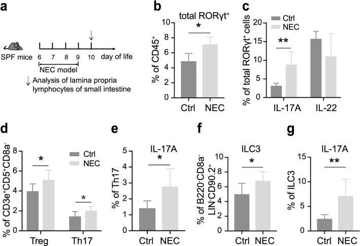
To examine the role of RORγt+ cells in NEC development, animal models were induced in neonatal mice as the method we used previously [27] (Fig. 3a). NEC was identified by the severity of weight loss and tissue impairment (Additional file 2: Fig. S2a, b). We first sought to compare the composition of lymphocytes within the lamina propria of small intestines in NEC and dam-fed control mice. Due to the most commonly affected site of NEC was ileum, we isolated the lymphocytes within lamina propria in small intestine and analyzed them with flow cytometry. Firstly, we observed that similar ratios of CD3e+ and CD4 T cells between the two groups and a reduction of CD8 T cells in NEC mice (Additional file 2: Fig. S2c, d). Not surprisingly, as shown in Fig. 3, the proportions of RORγt+ cells and IL-17A+ RORγt+ cells increased significantly in NEC when compared with controls (Fig. 3b, c). Furthermore, we found that the proportions of Th17 increased in NEC mice, and were accompanied by the increase of Treg (Fig. 3d). Meanwhile, IL-17A, the major inflammatory cytokine of Th17, was also elevated (Fig. 3e). In addition, we found that ILC3, the other dominant population expressed RORγt in intestinal lamina propria, as well as IL-17A+ ILC3 was also elevated when compared with control (Fig. 3f, g). Taken together, these findings suggest that the RORγt+ cells enriched intestinal environment predisposing to the induction of IL-17A lead to the development of NEC.
Fig. 3.
IL-17A+ RORγt+ cells increase in intestine of NEC mice. a. Diagram of the method to induce NEC. b, c Flow cytometric quantification of total RORγt+ cells and related cytokines (IL-17A and IL-22) in the lamina propria of mice with or without NEC. d–g. Flow cytometric quantification of Th17 and ILC3, as well as IL-17A expressed of them. Three independent experiments were performed, n = 4–5 per group. Data were shown as mean values ± SD. Statistical analyses were performed with Student’s two-tailed unpaired t-test. *p < 0.05; **p < 0.01
T cells and RORγt+ cells increased in intestine of NEC patients
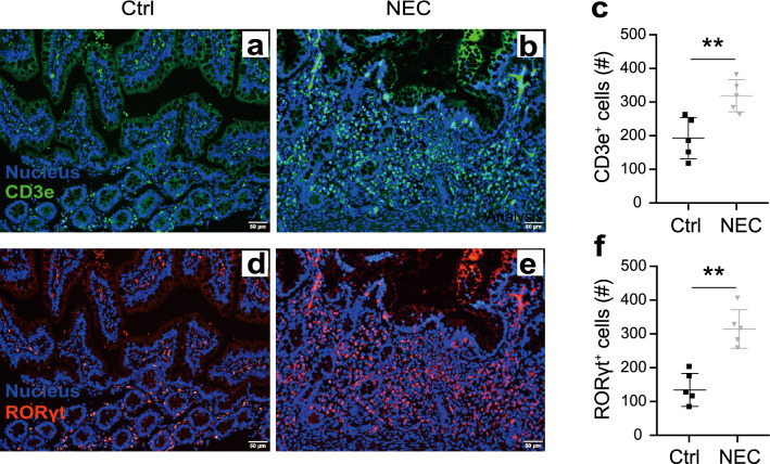
To examine whether the frequencies of RORγt+ cells were perturbed in NEC patients, we detected the composition of lymphocytes within the terminal ileum. As shown by immunofluorescent staining, the number of CD3e+ T cells in NEC patients increased notably when compared with controls (Fig. 4a–c). This is consistent with the previous study [15]. Meanwhile, we found a significant increase of RORγt+ cells in NEC patients as compared with controls (Fig. 4d–f). These data reminded us the influx of RORγt+ cells in the intestine is associated with the development of NEC in humans.
Fig. 4.
Changes of T cells and RORγt+ cells in NEC patients. Representative immunofluorescent staining pictures, and the statistical analyses of the count of CD3e+ T cells (a–c) and RORγt+ cells (d–f). n = 4 per group. Data were shown as mean values ± SD. Statistical analyses were performed with Student’s two-tailed unpaired t-test. **p < 0.01. Scale bars represent 50 μm. Ctrl, tissue from neonatal intestinal atresia patients. The Y-axis means total cell number in a visual field of one patient’s terminal ileum (c, f)
RORγt antagonist (GSK805) ameliorates the severity of NEC by suppressing IL-17A
RORγt is an attractive therapeutic target for the treatment of IL-17-mediated inflammatory disease. Based on our previous results, we considered whether the RORγt antagonist could inhibit IL-17A expressed to prevent the development of NEC in mice. Several small-molecular-weight compounds, including TMP778, SR1001, digoxin, and GSK805 [28–30], have been identified to inhibit the function of RORγt protein and showed efficacy in models of autoimmunity disease. Among them, GSK805 presents more potent at inhibiting Th17 responses in vitro and ameliorating the severity of experimental autoimmune encephalomyelitis (EAE) by oral administration [30].
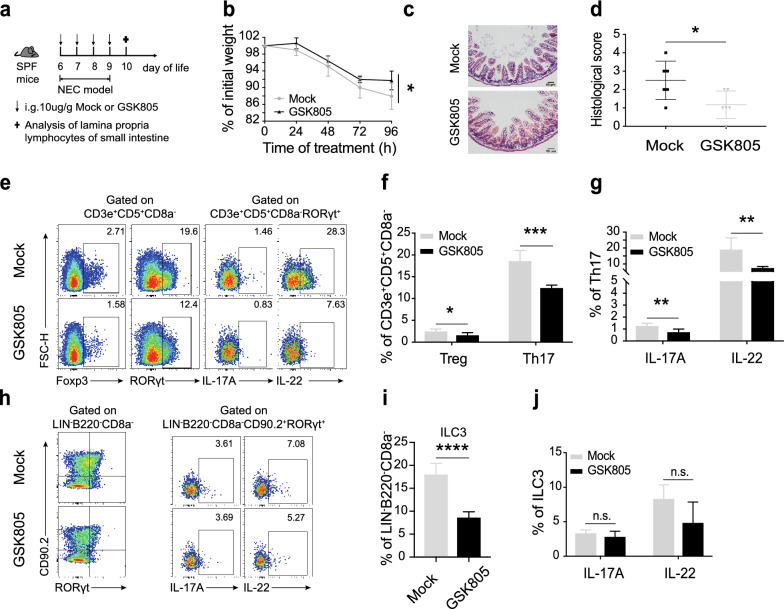
We next examined the in vivo effects of GSK805 on NEC mice. We induced NEC in neonatal mice and treated them with GSK805 orally once a day (Fig. 5a). Compared with Mock (control), GSK805 treatment delayed weight loss and substantially reduced the intestinal tissue impairment demonstrated by hematoxylin and eosin staining and histopathological scores (Fig. 5b–d). Analysis of intestinal samples after 4 days of GSK805 treatment revealed that the drug had no effect on intestinal barrier (Additional file 3: Fig. S3a, b). However, GSK805 effectively reduced the infiltration of immune cells, including both Th17 and Treg cells, and suppressed the expression of IL-17A and IL-22 (Fig. 5e–g). Furthermore, we found that GSK805 significantly inhibits the expression of RORγt in ILCs, resulting in the decrease of ILC3 (Fig. 5h, i). Interestingly, GSK805 had no effect on cytokines expression from ILC3 (Fig. 5h, j), which might be associated with the selective targeting of GSK805 on Th17 and ILC3 [31, 32]. Taken together, these results demonstrate that the RORγt antagonist provides therapeutic potential by inhibiting Th17 responses.
Fig. 5.
GSK805 alleviates intestinal inflammation of NEC mice by inhibiting IL-17A released. a. Scheme of the method of GSK805 treatment. Mice were treated with Mock or GSK805 (10 μg/g) orally. GSK805 was prepared with 1% DMSO in CMC-Na. b. Percentage of initial weight. c. Representative hematoxylin and eosin staining sections of the terminal ileum. d. Histopathological score measuring the severity of tissue lesions in the terminal ileum. e–g. Flow cytometric analysis and quantification of T cell subsets and related cytokines (IL-17A and IL-22) within lamina propria in small intestine. h–j. Flow cytometric analysis and quantification of ILC3 and related cytokines (IL-17A and IL-22). Four independent experiments were performed, n = 4–5 per group. Data were shown as mean values ± SD. Statistical analyses were performed with Student’s two-tailed unpaired t-test. *p < 0.05; **p < 0.01; ***p < 0.001; ****p < 0.0001. Scale bars represent 50 μm
Discussion
With the prominent work of researchers over the last few decades, scientists have made considerable progress in understanding the pathogenesis of NEC. However, there has been very little focus on the state of immune cells within the intestine in newborns before the onset of this disease. Previous studies examining the postnatal ontogeny of lymphocytes in the intestine of human neonates show that CD3+, CD4+, CD8+, and Foxp3+ cells are present in the lamina propria of infants after birth [16]. However, they were unable to find a difference in the proportion or quantities of Treg between term and preterm samples by immunohistochemistry due to technical limitations. Later studies revealed a significant reduction of the proportion of Treg in the ileum of NEC patients [17, 26]. Recent research has characterized the pathoimmunology of NEC and found that the increase of type 3 innate lymphoid cells (ILC3) and deficiency in IL-37 have association with the development of NEC [33]. In the present study, we focused on the state of intestinal immunity before NEC happened and investigated the compositions of immune cells within the small intestine during the early period of neonatal mice. We found that the proportion of RORγt+ cells is very high in the first week of life. Moreover, IL-17A+RORγt+ cells accumulated in the small intestine of NEC mice which might be attributed to the disease. Furthermore, inhibiting the function of RORγt+ cells by GSK805 ameliorated the severity of NEC. These findings provide new insight into the pathomechanistic understanding and treatment of NEC.
The nuclear hormone receptor RORγt, as an inducer of the pro-inflammatory program in lymphocytes, plays a key role in the regulation of immune responses and the maintenance of immune homeostasis. During ontogeny of fetuses, RORγt is firstly expressed in lymphoid tissue inducer (LTi) cells, which determines the development of lymph nodes and Peyer’s patches [21, 34]. Shortly after birth, RORγt+ type 3 innate lymphoid cells (ILC3) and RORγt+ T cells expand within the intestinal lamina propria. RORγt+ T cells include subsets of γδT cells, invariant natural killer T (iNKT) cells, and Th17 cells [24, 35]. All RORγt+ subsets express IL-17, which have been implicated in multiple human chronic inflammatories and autoimmune diseases, including inflammatory bowel disease (IBD) [36], multiple sclerosis [37], rheumatoid arthritis [38], and psoriasis [39]. A recent study has demonstrated that the expression of intestinal IL-17A and IL-17 receptor IL-17RA are elevated during human and mouse NEC [15]. Meanwhile, the other study has shown the risk and outcome of NEC in preterm infants is positively associated with the increase in single nucleotide polymorphisms of IL-17F and IL23R [40]. Here, we firstly revealed that the proportion of RORγt+ cells within intestinal lamina propria was at a high level during the first few days of life. Moreover, IL-17A produced by RORγt+ cells (Th17 and ILC3) increased significantly in NEC mice compared with control. Therefore, it is plausible that the high level of RORγt+ cells within intestines places the newborns at high risk for intestinal inflammation, resulting in a susceptibility to NEC.
Therapeutic targeting of the Th17 cell pathway or blocking of IL-17A has been proved effective in several autoimmune diseases [41, 42], but ineffective in IBD [43–45]. Later researches ascribe the paradoxical effect to the protective functions for IL-17A and IL-22 in the intestine, mediated in part by ILC3 [46–48]. In recent years, inhibition of RORγt by small-molecular-weight compounds offers an alternative method for the treatment of autoimmune disease and intestinal inflammation [28, 29, 31, 49]. Among inhibitors of RORγt, orally available compound GSK805 presents more potent at inhibiting Th17 responses and ameliorating experimental autoimmune encephalomyelitis (EAE) [49]. GSK805 interacts physically with the putative ligand-binding domain of RORγt, but exert less pronounced effects on DNA binding than other inhibitors [49]. Meanwhile, GSK805 has been demonstrated to selectively reduce cytokine production from Th17 but not ILC3, for limiting intestinal inflammation [31]. Accordingly, we investigated the effectiveness of GSK805 in alleviating the severity of NEC. Not surprisingly, GSK805 treatment improved the survival and weight loss in NEC mice and reversed the histological lesion of the intestine. Notably, GSK805 selectively suppressed IL-17A release from Th17 but not ILC3, as discussed above, contributed to its role in ameliorating NEC.
Conclusion
In summary, we characterized the immune state within intestinal lamina propria during the first few days of mouse life. Meanwhile, our results revealed the proportion of RORγt+ cells are at a high level shortly after birth. This might be associated with the development of NEC in newborns. Furthermore, we demonstrated that GSK805, as the selective inhibitor of RORγt, has the potential to mitigate the severity of NEC by inhibiting Th17 function. These results can be very instructive in understanding the pathological mechanism and exploring new effective therapeutic methods for NEC.
Methods
Mice
C57BL/6 neonatal mice were provided by Shanghai Ling Chang Biotech limited company. Mice were maintained under SPF conditions in the Animal Science Centre at the Shanghai Jiao Tong University School of Medicine. Animal experiments were approved by the Animal Care Committee of Children's Hospital of Shanghai and Shanghai Jiao Tong University School of Medicine.
Reagents and antibodies
The drug GSK805 was synthesized and provided by Yonghui Wang’s laboratory. Collagenase VIII (C2139) and DNase I (DN25) were purchased from Sigma-Aldrich. Cell Stimulation Cocktail (plus protein transport inhibitors) (00-4975-93), Foxp3/Transcription Factor Staining Buffer Set (00-5523-00), Permeabilization Buffer (10×) (00-8333), anti-CD3e (45-0031-82), and anti-Foxp3 (25-5773-82) were provided by Thermo Fisher SCIENTIFIC. Anti-CD45 (30-F11), anti-CD4 (553051), anti-CD8a (557668), anti-CD90.2 (563008), anti-B220 (563892), and anti-RORγt (564722) were purchased from BD Bioscience. Anti-CD11b (101224) and anti-CD11c (117322) were purchased from Biolegend.
Experimental design and NEC model
For analyzing the ontogeny of intestinal immune cells in the early life of mice, neonatal mice born at the appointed time were purchased directly from the company. Those mice were sacrificed on the same day for the experiment. For the other experiments, 6- to 7-day-old mice were randomly divided into the NEC + Mock group, NEC + GSK805 group, or control group. Mice in the control group were dam-fed without any other stimulus. NEC model was induced using a method as our previous study [27] modified from Jilling et al. [50]. Briefly, neonatal mice were fed with 50 μl of 33% Esbilac formula (Pet-Ag) by gavage every 4 h for 96 h. Meanwhile, newborns were subjected to hypoxia (99.9% N2 for 90 s) followed by cold stress (4 °C for 10 min) twice a day for 4 days. During the NEC modeling process, mice were treated with Mock or GSK805 (10 μg/g) by intragastric administration once a day for four days. All mice were weighed every morning and euthanized at 96 h for collecting intestinal samples.
Histological analysis
Terminal ileum tissues were fixed with 4% paraformaldehyde, embedded in paraffin, and sectioned, then stained with H&E. NEC was evaluated based on histological changes of the terminal ileum according to a previously described scoring system [51]. The severity of NEC was classified as follows: intact villi were assigned a score of 0, sloughing of cells on villous tips received a score of 1, and mid-villous damage was scored as 2. An NEC score of 3 was recorded when villi were absent, but crypts were still readily detectable, and an NEC score of 4 was assigned in cases of a complete absence of epithelial structures and transmural necrosis. Scores were always determined based on the highest score observed in a specimen.
Isolation of intestinal lamina propria lymphocytes
Intestinal lamina propria lymphocytes in newborns were isolated as our previous method with a modification [52]. In brief, the small intestine excluded duodenum was removed and cut longitudinally. After washing in PBS by a few sharp shakes, the tissue pieces were transferred into solution A (1 mM DTT, 30 mM EDTA, 10 mM HEPES) and incubated in a shaker at 37 °C for 10 min. Then, transferred the tissues into solution B (30 mM EDTA, 10 mM HEPES) and repeated as the previous step. After washing with complete RPMI 1640 medium, the tissues were cut into 1 mm3 piece and digested in RPMI 1640 containing 500 μg/ml collagenase VIII and 100 μg/ml DNase I at 37 °C for 50 min. Due to a relatively low number of lymphocytes in the neonatal intestine, it is not suitable for purifying the cells. Hence, we next washed the single-cell suspension with FACS buffer (0.5% BSA-PBS) and stained with flow antibodies.
Flow cytometry
Intestinal cells were firstly incubated with cell stimulation mixture plus protein transport inhibitor (Thermo) in complete RPMI 1640 at 37 °C for 4 h. After stimulation, cells were stained with antibodies conjugated with the indicated fluorochrome for surface marker analysis. The intracellular expression levels of transcriptional factors and cytokines were detected as the manufacturer’s instructions of Foxp3/Transcription Factor Staining Buffer Set (Thermo). Flow cytometric analyses were performed using a LSRFortessa X-20 (BD Biosciences) and analyzed with FlowJo 10.4 software.
Statistical analysis
Statistical analysis was performed with Student’s two-tailed unpaired t-test and one-way or two-way analysis of variance (ANOVA) followed by Welch’s or Mann–Whitney test. For weight change analyses, two-way ANOVA was used, with treatment as the main effect. All data are expressed as mean values ± SD. p < 0.05 was considered to be significant. Statistical analysis was performed by GraphPad Prism 8.0.
Supplementary Information
Additional file 1: Figure S1. Gate strategy of flow cytometry analysis.The methods to analyze flow data with FlowJo software. LIN presents CD11b and CD11c.
Additional file 2: Figure S2. Confirmation of NEC model and the changes of T cells. a. Percentage of initial weight. b. Representative hematoxylin and eosin staining sections of the terminal ileum. c and d, Changes of CD3e+, CD4, CD8 T cells between Ctrl and NEC mice.
Additional file 3: Figure S3. Effect of GSK805 on intestinal barrier. a. Serum concentrations of FD4 in NEC mice treated with GSK805 or not. b. Representative images of tight junction proteins in small intestinal epithelial cells. FD4, FITC-dextran 4 kDa.
Acknowledgements
The authors appreciate Zhi Li from the pathological department of Shanghai Children’s hospital for his technical assistance. We also thank you for the help during the experiment from Shuiyu Xu, Feng Liu, Ziwei Zhang, Qiuyang Guo, Xiaolan Yan, and Xueyi Jiang, who are undergraduates from Shanghai Jiao Tong University.
Abbreviations
- NEC
Necrotizing enterocolitis
- RORγt
Retinoid-related orphan receptor γt
- ILC
Innate lymphoid cells
- LTi
Lymphoid tissue inducer cells
- TNF
Tumor necrosis factor
- TGF
Transforming growth factor
Authors’ contributions
FW and ZL conceived the project. FW, XZ, and WL designed and conducted the experiments. XZ, WL, RY, LLuo, WW, NS, MY, WX, QS, LLu, and JP assisted to conduct the experiments. YW, NS, and MY provided the drug GSK805 and assisted in the animal experiments. XZ and WL wrote the manuscript, which was modified by FW and ZL. All authors read and approved the final manuscript.
Funding
This study was supported by grants from the National Key Research and Development Program of China (SQ2018YFA090045-01), the National Natural Science Foundation of China (81871194, 81771739, 82071852), the Program for Professor of Special Appointments (Eastern Scholar) at Shanghai Institutions of Higher Learning, the Top Young Talent Program of Shanghai, and the Technology Committee of Shanghai Municipality 18ZR14311300, 18JC1414100 and 20410713800.
Availability of data and materials
Not applicable.
Declarations
Ethics approval and consent to participate
This study was approved by the Ethics Committee of Children's Hospital of Shanghai. Animal experiments were approved by the Animal Care Committee of Children's Hospital of Shanghai and Shanghai Jiao Tong University School of Medicine.
Consent for publication
All authors have approved for publication.
Competing interests
The authors declare that they have no competing interests.
Footnotes
Publisher's Note
Springer Nature remains neutral with regard to jurisdictional claims in published maps and institutional affiliations.
Xiuhao Zhao and Wenhua Liang contributed equally to this work
Contributor Information
Zhibao Lv, Email: zhibaolyu@163.com.
Feng Wang, Email: wangfeng16@sjtu.edu.cn.
References
- 1.Neu J, Walker WA. Necrotizing enterocolitis. N Engl J Med. 2011;364(3):255–264. doi: 10.1056/NEJMra1005408. [DOI] [PMC free article] [PubMed] [Google Scholar]
- 2.Zozaya C, Shah J, Pierro A, Zani A, Synnes A, Lee S, Shah PS, Canadian Neonatal N. the Canadian Neonatal Follow-Up Network I Neurodevelopmental and growth outcomes of extremely preterm infants with necrotizing enterocolitis or spontaneous intestinal perforation. J Pediatr Surg. 2021;56(2):309–316. doi: 10.1016/j.jpedsurg.2020.05.013. [DOI] [PubMed] [Google Scholar]
- 3.Matei A, Montalva L, Goodbaum A, Lauriti G, Zani A. Neurodevelopmental impairment in necrotising enterocolitis survivors: systematic review and meta-analysis. Arch Dis Child Fetal Neonatal Ed. 2020;105(4):432–439. doi: 10.1136/archdischild-2019-317830. [DOI] [PubMed] [Google Scholar]
- 4.Patel RM, Kandefer S, Walsh MC, Bell EF, Carlo WA, Laptook AR, Sanchez PJ, Shankaran S, Van Meurs KP, Ball MB, et al. Causes and timing of death in extremely premature infants from 2000 through 2011. New Engl J Med. 2015;372(4):331–340. doi: 10.1056/NEJMoa1403489. [DOI] [PMC free article] [PubMed] [Google Scholar]
- 5.Calvert W, Sampat K, Jones M, Baillie C, Lamont G, Losty PD. Necrotising enterocolitis—a 15-year outcome report from a UK specialist centre. Acta Paediatr. 2021;110(2):495–502. doi: 10.1111/apa.15510. [DOI] [PubMed] [Google Scholar]
- 6.Nino DF, Sodhi CP, Hackam DJ. Necrotizing enterocolitis: new insights into pathogenesis and mechanisms. Nat Rev Gastro Hepat. 2016;13(10):590–600. doi: 10.1038/nrgastro.2016.119. [DOI] [PMC free article] [PubMed] [Google Scholar]
- 7.Repa A, Thanhaeuser M, Endress D, Weber M, Kreissl A, Binder C, Berger A, Haiden N. Probiotics (Lactobacillus acidophilus and Bifidobacterium bifidum) prevent NEC in VLBW infants fed breast milk but not formula (vol 77, pg 381, 2015) Pediatr Res. 2016;79(1):124–124. doi: 10.1038/pr.2014.192. [DOI] [PubMed] [Google Scholar]
- 8.Frost BL, Modi BP, Jaksic T, Caplan MS. New medical and surgical insights into neonatal necrotizing enterocolitis a review. Jama Pediatr. 2017;171(1):83–88. doi: 10.1001/jamapediatrics.2016.2708. [DOI] [PubMed] [Google Scholar]
- 9.Costeloe K, Hardy P, Juszczak E, Wilks M, Millar MR, Study PPI Bifidobacterium breve BBG-001 in very preterm infants: a randomised controlled phase 3 trial. Lancet. 2016;387(10019):649–660. doi: 10.1016/S0140-6736(15)01027-2. [DOI] [PubMed] [Google Scholar]
- 10.Basha S, Surendran N, Pichichero M. Immune responses in neonates. Expert Rev Clin Immunol. 2014;10(9):1171–1184. doi: 10.1586/1744666X.2014.942288. [DOI] [PMC free article] [PubMed] [Google Scholar]
- 11.Eaton S, Rees CM, Hall NJ. Current research on the epidemiology, pathogenesis, and management of necrotizing enterocolitis. Neonatology. 2017;111(4):423–430. doi: 10.1159/000458462. [DOI] [PubMed] [Google Scholar]
- 12.Sodhi CP, Neal MD, Siggers R, Sho S, Ma CR, Branca MF, Prindle T, Russo AM, Afrazi A, Good M, et al. Intestinal epithelial toll-like receptor 4 regulates goblet cell development and is required for necrotizing enterocolitis in mice. Gastroenterology. 2012;143(3):708–U234. doi: 10.1053/j.gastro.2012.05.053. [DOI] [PMC free article] [PubMed] [Google Scholar]
- 13.Hackam DJ, Good M, Sodhi CP. Mechanisms of gut barrier failure in the pathogenesis of necrotizing enterocolitis: toll-like receptors throw the switch. Semin Pediatr Surg. 2013;22(2):76–82. doi: 10.1053/j.sempedsurg.2013.01.003. [DOI] [PMC free article] [PubMed] [Google Scholar]
- 14.Yazji I, Sodhi CP, Lee EK, Good M, Egan CE, Afrazi A, Neal MD, Jia H, Lin J, Ma C, et al. Endothelial TLR4 activation impairs intestinal microcirculatory perfusion in necrotizing enterocolitis via eNOS-NO-nitrite signaling. Proc Natl Acad Sci USA. 2013;110(23):9451–9456. doi: 10.1073/pnas.1219997110. [DOI] [PMC free article] [PubMed] [Google Scholar]
- 15.Egan CE, Sodhi CP, Good M, Lin J, Jia HP, Yamaguchi Y, Lu P, Ma CR, Branca MF, Weyandt S, et al. Toll-like receptor 4-mediated lymphocyte influx induces neonatal necrotizing enterocolitis. J Clin Investig. 2016;126(2):495–508. doi: 10.1172/JCI83356. [DOI] [PMC free article] [PubMed] [Google Scholar]
- 16.Weitkamp JH, Rudzinski E, Koyama T, Correa H, Matta P, Alberty B, Polk DB. Ontogeny of FOXP3(+) regulatory T cells in the postnatal human small intestinal and large intestinal lamina propria. Pediatr Dev Pathol. 2009;12(6):443–449. doi: 10.2350/08-09-0533.1. [DOI] [PMC free article] [PubMed] [Google Scholar]
- 17.Dingle BM, Liu YY, Fatheree NY, Min J, Rhoads JM, Tran DQ. FoxP3(+) regulatory T cells attenuate experimental necrotizing enterocolitis. PLoS ONE. 2013;8(12):e82963. doi: 10.1371/journal.pone.0082963. [DOI] [PMC free article] [PubMed] [Google Scholar]
- 18.Weitkamp JH, Rosen MJ, Zhao Z, Koyama T, Geem D, Denning TL, Rock MT, Moore DJ, Halpern MD, Matta P, et al. Small intestinal intraepithelial TCRgammadelta+ T lymphocytes are present in the premature intestine but selectively reduced in surgical necrotizing enterocolitis. PLoS ONE. 2014;9(6):e99042. doi: 10.1371/journal.pone.0099042. [DOI] [PMC free article] [PubMed] [Google Scholar]
- 19.Cho SX, Berger PJ, Nold-Petry CA, Nold MF. The immunological landscape in necrotising enterocolitis. Expert Rev Mol Med. 2016;18:e12. doi: 10.1017/erm.2016.13. [DOI] [PMC free article] [PubMed] [Google Scholar]
- 20.Ivanov II, McKenzie BS, Zhou L, Tadokoro CE, Lepelley A, Lafaille JJ, Cua DJ, Littman DR. The orphan nuclear receptor RORgammat directs the differentiation program of proinflammatory IL-17+ T helper cells. Cell. 2006;126(6):1121–1133. doi: 10.1016/j.cell.2006.07.035. [DOI] [PubMed] [Google Scholar]
- 21.Sun Z, Unutmaz D, Zou YR, Sunshine MJ, Pierani A, Brenner-Morton S, Mebius RE, Littman DR. Requirement for RORgamma in thymocyte survival and lymphoid organ development. Science. 2000;288(5475):2369–2373. doi: 10.1126/science.288.5475.2369. [DOI] [PubMed] [Google Scholar]
- 22.Kurebayashi S, Ueda E, Sakaue M, Patel DD, Medvedev A, Zhang F, Jetten AM. Retinoid-related orphan receptor gamma (ROR gamma) is essential for lymphoid organogenesis and controls apoptosis during thymopoiesis. Proc Natl Acad Sci USA. 2000;97(18):10132–10137. doi: 10.1073/pnas.97.18.10132. [DOI] [PMC free article] [PubMed] [Google Scholar]
- 23.Patel DD, Kuchroo VK. Th17 cell pathway in human immunity: lessons from genetics and therapeutic interventions. Immunity. 2015;43(6):1040–1051. doi: 10.1016/j.immuni.2015.12.003. [DOI] [PubMed] [Google Scholar]
- 24.Eberl G. Development and evolution of RORgammat+ cells in a microbe's world. Immunol Rev. 2012;245(1):177–188. doi: 10.1111/j.1600-065X.2011.01071.x. [DOI] [PubMed] [Google Scholar]
- 25.Ueno A, Jeffery L, Kobayashi T, Hibi T, Ghosh S, Jijon H. Th17 plasticity and its relevance to inflammatory bowel disease. J Autoimmun. 2018;87:38–49. doi: 10.1016/j.jaut.2017.12.004. [DOI] [PubMed] [Google Scholar]
- 26.Weitkamp JH, Koyama T, Rock MT, Correa H, Goettel JA, Matta P, Oswald-Richter K, Rosen MJ, Engelhardt BG, Moore DJ, et al. Necrotising enterocolitis is characterised by disrupted immune regulation and diminished mucosal regulatory (FOXP3)/effector (CD4, CD8) T cell ratios. Gut. 2013;62(1):73–82. doi: 10.1136/gutjnl-2011-301551. [DOI] [PMC free article] [PubMed] [Google Scholar]
- 27.Zhao X, Zhou J, Liang W, Sheng Q, Lu L, Chen T, Chen J, Tan K, Lv Z. Probiotics mixture reinforces barrier function to ameliorate necrotizing enterocolitis by regulating PXR-JNK pathway. Cell Biosci. 2021;11(1):20. doi: 10.1186/s13578-021-00530-7. [DOI] [PMC free article] [PubMed] [Google Scholar]
- 28.Huh JR, Leung MW, Huang P, Ryan DA, Krout MR, Malapaka RR, Chow J, Manel N, Ciofani M, Kim SV, et al. Digoxin and its derivatives suppress TH17 cell differentiation by antagonizing RORgammat activity. Nature. 2011;472(7344):486–490. doi: 10.1038/nature09978. [DOI] [PMC free article] [PubMed] [Google Scholar]
- 29.Solt LA, Kumar N, Nuhant P, Wang Y, Lauer JL, Liu J, Istrate MA, Kamenecka TM, Roush WR, Vidovic D, et al. Suppression of TH17 differentiation and autoimmunity by a synthetic ROR ligand. Nature. 2011;472(7344):491–494. doi: 10.1038/nature10075. [DOI] [PMC free article] [PubMed] [Google Scholar]
- 30.Xiao S, Yosef N, Yang J, Wang Y, Zhou L, Zhu C, Wu C, Baloglu E, Schmidt D, Ramesh R, et al. Small-molecule RORgammat antagonists inhibit T helper 17 cell transcriptional network by divergent mechanisms. Immunity. 2014;40(4):477–489. doi: 10.1016/j.immuni.2014.04.004. [DOI] [PMC free article] [PubMed] [Google Scholar]
- 31.Withers DR, Hepworth MR, Wang X, Mackley EC, Halford EE, Dutton EE, Marriott CL, Brucklacher-Waldert V, Veldhoen M, Kelsen J, et al. Transient inhibition of ROR-gammat therapeutically limits intestinal inflammation by reducing TH17 cells and preserving group 3 innate lymphoid cells. Nat Med. 2016;22(3):319–323. doi: 10.1038/nm.4046. [DOI] [PMC free article] [PubMed] [Google Scholar]
- 32.Chang D, Xing Q, Su Y, Zhao X, Xu W, Wang X, Dong C. The conserved non-coding sequences CNS6 and CNS9 control cytokine-induced rorc transcription during T helper 17 cell differentiation. Immunity. 2020;53(3):614–626.e614. doi: 10.1016/j.immuni.2020.07.012. [DOI] [PubMed] [Google Scholar]
- 33.Cho SX, Rudloff I, Lao JC, Pang MA, Goldberg R, Bui CB, McLean CA, Stock M, Klassert TE, Slevogt H, et al. Characterization of the pathoimmunology of necrotizing enterocolitis reveals novel therapeutic opportunities. Nat Commun. 2020;11(1):5794. doi: 10.1038/s41467-020-19400-w. [DOI] [PMC free article] [PubMed] [Google Scholar]
- 34.Eberl G, Littman DR. The role of the nuclear hormone receptor RORgammat in the development of lymph nodes and Peyer's patches. Immunol Rev. 2003;195:81–90. doi: 10.1034/j.1600-065x.2003.00074.x. [DOI] [PubMed] [Google Scholar]
- 35.Eberl G. ROR gamma t, a multitask nuclear receptor at mucosal surfaces. Mucosal Immunol. 2017;10(1):27–34. doi: 10.1038/mi.2016.86. [DOI] [PubMed] [Google Scholar]
- 36.Fujino S, Andoh A, Bamba S, Ogawa A, Hata K, Araki Y, Bamba T, Fujiyama Y. Increased expression of interleukin 17 in inflammatory bowel disease. Gut. 2003;52(1):65–70. doi: 10.1136/gut.52.1.65. [DOI] [PMC free article] [PubMed] [Google Scholar]
- 37.Lock C, Hermans G, Pedotti R, Brendolan A, Schadt E, Garren H, Langer-Gould A, Strober S, Cannella B, Allard J, et al. Gene-microarray analysis of multiple sclerosis lesions yields new targets validated in autoimmune encephalomyelitis. Nat Med. 2002;8(5):500–508. doi: 10.1038/nm0502-500. [DOI] [PubMed] [Google Scholar]
- 38.Kotake S, Udagawa N, Takahashi N, Matsuzaki K, Itoh K, Ishiyama S, Saito S, Inoue K, Kamatani N, Gillespie MT, et al. IL-17 in synovial fluids from patients with rheumatoid arthritis is a potent stimulator of osteoclastogenesis. J Clin Investig. 1999;103(9):1345–1352. doi: 10.1172/JCI5703. [DOI] [PMC free article] [PubMed] [Google Scholar]
- 39.Teunissen MB, Koomen CW, de Waal MR, Wierenga EA, Bos JD. Interleukin-17 and interferon-gamma synergize in the enhancement of proinflammatory cytokine production by human keratinocytes. J Investig Dermatol. 1998;111(4):645–649. doi: 10.1046/j.1523-1747.1998.00347.x. [DOI] [PubMed] [Google Scholar]
- 40.Tian J, Liu Y, Jiang Y, Zhou H, Zhu T, Zhao X, Peng L, Yan C. Association of single nucleotide polymorphisms of IL23R and IL17 with necrotizing enterocolitis in premature infants. Mol Cell Biochem. 2017;430(1–2):201–209. doi: 10.1007/s11010-017-2972-6. [DOI] [PubMed] [Google Scholar]
- 41.Papp KA, Leonardi C, Menter A, Ortonne JP, Krueger JG, Kricorian G, Aras G, Li J, Russell CB, Thompson EH, et al. Brodalumab, an anti-interleukin-17-receptor antibody for psoriasis. N Engl J Med. 2012;366(13):1181–1189. doi: 10.1056/NEJMoa1109017. [DOI] [PubMed] [Google Scholar]
- 42.Genovese MC, Van den Bosch F, Roberson SA, Bojin S, Biagini IM, Ryan P, Sloan-Lancaster J. LY2439821, a humanized anti-interleukin-17 monoclonal antibody, in the treatment of patients with rheumatoid arthritis: A phase I randomized, double-blind, placebo-controlled, proof-of-concept study. Arthritis Rheum. 2010;62(4):929–939. doi: 10.1002/art.27334. [DOI] [PubMed] [Google Scholar]
- 43.Hueber W, Sands BE, Lewitzky S, Vandemeulebroecke M, Reinisch W, Higgins PD, Wehkamp J, Feagan BG, Yao MD, Karczewski M, et al. Secukinumab, a human anti-IL-17A monoclonal antibody, for moderate to severe Crohn's disease: unexpected results of a randomised, double-blind placebo-controlled trial. Gut. 2012;61(12):1693–1700. doi: 10.1136/gutjnl-2011-301668. [DOI] [PMC free article] [PubMed] [Google Scholar]
- 44.Moncada RR, Moron JMV, Manrique HP. The onset of ulcerative colitis during treatment with secukinumab: can anti-IL-17A be a trigger for inflammatory bowel disease? Rev Esp Enferm Dig. 2019;111(9):720. doi: 10.17235/reed.2019.5841/2018. [DOI] [PubMed] [Google Scholar]
- 45.Fauny M, Moulin D, D'Amico F, Netter P, Petitpain N, Arnone D, Jouzeau JY, Loeuille D, Peyrin-Biroulet L. Paradoxical gastrointestinal effects of interleukin-17 blockers. Ann Rheum Dis. 2020;79(9):1132–1138. doi: 10.1136/annrheumdis-2020-217927. [DOI] [PubMed] [Google Scholar]
- 46.Gladiator A, Wangler N, Trautwein-Weidner K, LeibundGut-Landmann S. Cutting edge: IL-17-secreting innate lymphoid cells are essential for host defense against fungal infection. J Immunol. 2013;190(2):521–525. doi: 10.4049/jimmunol.1202924. [DOI] [PubMed] [Google Scholar]
- 47.O'Connor W, Kamanaka M, Booth CJ, Town T, Nakae S, Iwakura Y, Kolls JK, Flavell RA. A protective function for interleukin 17A in T cell-mediated intestinal inflammation. Nat Immunol. 2009;10(6):603–U665. doi: 10.1038/ni.1736. [DOI] [PMC free article] [PubMed] [Google Scholar]
- 48.Sonnenberg GF, Fouser LA, Artis D. Border patrol: regulation of immunity, inflammation and tissue homeostasis at barrier surfaces by IL-22. Nat Immunol. 2011;12(5):383–390. doi: 10.1038/ni.2025. [DOI] [PubMed] [Google Scholar]
- 49.Xiao S, Yosef N, Yang JF, Wang YH, Zhou L, Zhu C, Wu C, Baloglu E, Schmidt D, Ramesh R, et al. Small-molecule ROR gamma t antagonists inhibit T helper 17 cell transcriptional network by divergent mechanisms. Immunity. 2014;40(4):477–489. doi: 10.1016/j.immuni.2014.04.004. [DOI] [PMC free article] [PubMed] [Google Scholar]
- 50.Jilling T, Simon D, Lu J, Meng FJ, Li D, Schy R, Thomson RB, Soliman A, Arditi M, Caplan MS. The roles of bacteria and TLR4 in rat and murine models of necrotizing enterocolitis. J Immunol. 2006;177(5):3273–3282. doi: 10.4049/jimmunol.177.5.3273. [DOI] [PMC free article] [PubMed] [Google Scholar]
- 51.Jilling T, Lu J, Jackson M, Caplan MS. Intestinal epithelial apoptosis initiates gross bowel necrosis in an experimental rat model of neonatal necrotizing enterocolitis. Pediatr Res. 2004;55(4):622–629. doi: 10.1203/01.PDR.0000113463.70435.74. [DOI] [PubMed] [Google Scholar]
- 52.Sun S, Luo L, Liang W, Yin Q, Guo J, Rush AM, Lv Z, Liang Q, Fischbach MA, Sonnenburg JL, et al. Bifidobacterium alters the gut microbiota and modulates the functional metabolism of T regulatory cells in the context of immune checkpoint blockade. Proc Natl Acad Sci USA. 2020;117(44):27509–27515. doi: 10.1073/pnas.1921223117. [DOI] [PMC free article] [PubMed] [Google Scholar]
Associated Data
This section collects any data citations, data availability statements, or supplementary materials included in this article.
Supplementary Materials
Additional file 1: Figure S1. Gate strategy of flow cytometry analysis.The methods to analyze flow data with FlowJo software. LIN presents CD11b and CD11c.
Additional file 2: Figure S2. Confirmation of NEC model and the changes of T cells. a. Percentage of initial weight. b. Representative hematoxylin and eosin staining sections of the terminal ileum. c and d, Changes of CD3e+, CD4, CD8 T cells between Ctrl and NEC mice.
Additional file 3: Figure S3. Effect of GSK805 on intestinal barrier. a. Serum concentrations of FD4 in NEC mice treated with GSK805 or not. b. Representative images of tight junction proteins in small intestinal epithelial cells. FD4, FITC-dextran 4 kDa.
Data Availability Statement
Not applicable.


