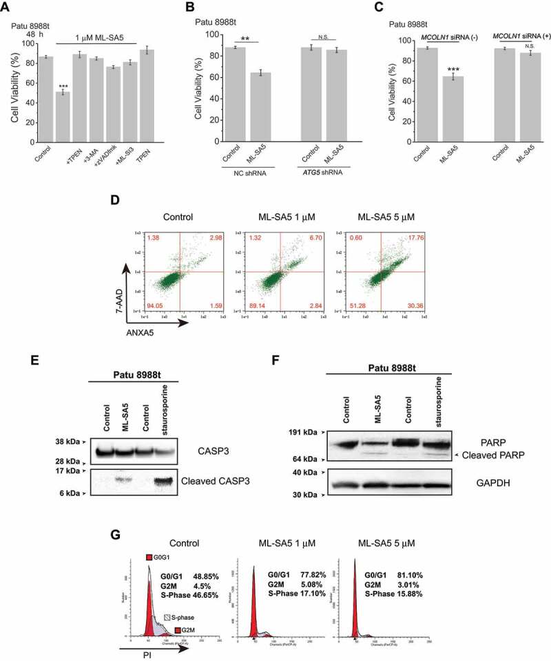
Figure 6.

Following autophagic arrest mediated by ML-SA5, induction of apoptosis and cell cycle arrest triggers cell death in the cancer cells. (A) Patu 8988 t cell death triggered by 1 μM ML-SA5 (48 h) were strongly rescued by co-application of either 3-MA (10 mM, pretreatment for 2 h), or TPEN (1 μM), or zVADfmk (20 μM), or ML-SI3 (20 μM), respectively. n = 10–40. (B) Knockdown of ATG5 genes significantly attenuated cell death triggered by ML-SA5 treatments (1 μM for 48 h) in Patu 8988 t cells. n = 3. (C) ML-SA5 was not lethal to Patu 8988 t cells when MCOLN1 was efficiently knocked down by the MCOLN1 siRNA, evaluated by Trypan blue assay. n = 3–5. (D-F) Induction of apoptosis following ML-SA5 treatments (1 μM) in Patu 8988 t cells was confirmed by detection of ANXA5/annexin V-7-AAD (D), cleaved CASP3 (caspase 3) (E) and cleaved PARP (F). All treatments were for 48 h. (G) Flow cytometry images displaying that G0/G1 phase arrest was triggered upon application of ML-SA5 for 48 h at both 1 μM and 5 μM in Patu 8988 t cells in medium containing 2% FBS, stained by PI. Error bars indicate Mean ± SEMs in panels A, B and C. Significant differences were evaluated using one-way ANOVA followed by Tukey’s test. **P < 0.01; ***P < 0.001
