Abstract
Vogt–Koyanagi–Harada (VKH) disease is a primary autoimmune stromal choroiditis. This review aimed to provide a novel perspective of the disease. We took into account recent developments in the understanding of the disease and crucial progress in investigational modalities of the choroid, which has led to new, simpler diagnostic criteria. We analysed recent novel notions in the literature and new diagnostic tools for VKH. We identified the following updates for VKH disease: (1) A crucial differentiation between the acute initial-onset and the chronic forms of the disease; (2) the integration of new, precise imaging methods to assess choroidal inflammation; (3) the promotion of simplified, more reliable diagnostic criteria for acute initial-onset of the disease, based on the sine qua non presence of diffuse choroiditis, detected with indocyanine green angiography (ICGA) and/or Enhanced Depth Imaging OCT (EDI-OCT); and (4) treatment optimisation through early, vigorous, sustained corticosteroid and nonsteroidal immunosuppression, as the first line of treatment for initial-onset VKH disease, and monitoring subclinical choroidal inflammation during follow-ups. Several studies have shown that most patients could discontinue treatment without an inflammation relapse. ICGA and EDI-OCT represented the methods of choice for precisely monitoring disease evolution. Simplified, precise, new diagnostic criteria allow early diagnosis of VKH. In VKH disease, inflammation exclusively originates in the choroidal stroma. Therefore, in many cases, early, sustained treatment, with dual corticosteroid and nonsteroidal immunosuppressive therapy can result in full “healing”, which obviates chronic, uncontrolled, subclinical choroidal inflammation.
Subject terms: Uveal diseases, Eye manifestations
摘要
小柳-原田病 (Vogt-Koyanagi-Harada, VKH) 是一种原发性自身免疫性间质性脉络膜炎。本综述旨在为疾病的诊断与治疗提供一个新的视角。我们将该疾病发病机理的最新进展以及针对脉络膜成像的关键进展进行综合考虑, 提出了新的、更简单的诊断标准。我们对新近文献中的新概念和的VKH诊断手段进行了分析与综合, 总结了VKH疾病的最新进展如下: (1) 急性初发和慢性疾病之间的关键区别; (2) 整合新的、精确的成像方法以评估脉络膜炎症; (3) 以弥漫性脉络膜炎的存在为非必要诊断条件, 通过吲哚青绿血管造影 (indocyanine green angiography, ICGA) 和/或EDI-OCT (Enhanced Depth Imaging OCT, EDI-OCT) 成像技术, 确立简化的、更可靠的疾病急性起病诊断标准; (4) 通过早期、足量、持续的皮质类固醇和非类固醇免疫抑制剂的优化治疗方案, 作为初发VKH的一线治疗, 且在随访期间监测亚临床脉络膜炎症。不少研究均表明, 大多数患者停止治疗后炎症不会复发。ICGA和EDI-OCT是精确测病情的首选方法。新的精确、简化的诊断标准有利于VKH在早期得到诊断。在VKH中, 炎症仅起源于脉络膜基质。因此在许多情况下, 早期、持续、联合皮质类固醇和非类固醇免疫抑制治疗可实现完全“治愈”, 从而避免慢性、不受控制的亚临床脉络膜炎。
Introduction
Vogt–Koyanagi–Harada (VKH) disease is an inflammatory condition caused by an autoimmune reaction against tyrosinase-like proteins in melanocytes in the uvea, the inner ear, the meninges, and the integumentary system [1–3]. VKH predominantly affects people with high melanin loads, such as Asians, Hispanics, American Indians, and Asian Indians [4, 5]; it occurs less frequently in Caucasians [6].
At the onset of VKH, the main structure involved is the uvea, particularly the choroid. Sometimes, VKH is associated with meningeal and auditory symptoms [4, 7, 8]. Clinically, the process starts as a choroiditis [9], and it evolves to granulomatous panuveitis with exudative retinal detachments. Concomitantly, the auditory system can become involved, and meningitis can occur, with mononuclear pleocytosis in the cerebrospinal fluid [7, 10]. When early and vigorous immunosuppressive treatment is not introduced, uveitis can become chronic, and the integumentary and auditory systems can become involved [11].
The clinical signs differ for acute initial-onset VKH and chronic VKH [12]. Until recently, most studies did not distinguish between initial-onset and chronic disease; thus, most cohorts with VKH disease included mixed acute and chronic cases [13]. In addition, the past diagnostic criteria for VKH disease combined signs of acute and chronic disease. This practice resulted in inadequate definitions, particularly for initial-onset disease [14, 15]. Moreover, those diagnostic criteria are unsatisfactory, because they failed to include new, precise investigational procedures for the choroid that have become available recently. For example, the advent of indocyanine green angiography (ICGA) [16] and choroidal enhanced depth imaging, optical coherence tomography (EDI-OCT) [17] have improved the diagnostic accuracy and the precision of follow-up assessments of VKH disease. The subdivision of VKH disease into its initial-onset and chronic forms and the introduction of new investigational modalities for the choroid have also led to more precise evaluations of prognostic factors and improved therapeutic intervention outcomes in homogenous groups of patients [18].
The purpose of this review was to update the appraisal of VKH disease, propose more adequate diagnostic criteria, and suggest means to optimise management, in light of improved diagnostic and monitoring possibilities and the recent progress in understanding the evolution of VKH disease.
History
In 1906, Alfred Vogt, in Basel, Switzerland, reported the first known case of VKH. That patient had poliosis associated with intraocular inflammation [19]. In 1911, the first Japanese patient with VKH was described by Jujiro Komoto, the first Professor and Chairman of the Department of Ophthalmology of the Imperial University of Tokyo. Three years later, in 1914, Yoshizo Koyanagi reported several more cases of VKH [20, 21]. In 1918 (in Japanese) and in 1926 (in German), Einosuke Harada described a posterior uveitis associated with exudative retinal detachments and accompanied by CSF pleocytosis [22, 23]. Then, the groundbreaking report on VKH was published in 1929 by Koyanagi, in a German ophthalmological journal, which described 16 patients with bilateral chronic iridocyclitis associated with vitiligo, alopecia, poliosis, deafness, and tinnitus [24]. The importance of that article cannot be stressed enough, because it precisely described, with all clinical details, the natural evolution of untreated VKH, due to the lack of available treatments at the time. In 1939, Babel, and in 1949, Bruno and McPherson unified the disorders described by Vogt, Koyanagi, and Harada, and suggested that these apparently disparate entities were a continuum of the same disease process [25, 26]. Since then, this uveomeningoencephalitic syndrome has been known as VKH disease. The pathogenesis of the disease was revealed in the late 1980s, at first mostly in Japanese studies. They showed that the the immune system reacted with melanocyte-associated antigens [27, 28]. In particular, they found that T-cells from patients with VKH were cytotoxic against human melanocytes and melanoma cells [27, 28].
With the development of corticosteroid therapy during the 1950s, for the first time, an inflammation-suppressive treatment (IST) became available for VKH disease. In the late 1950s, several patients had been treated with low doses of corticosteroids during the course of VKH [29]. In 1969, the first report on early high-dose corticosteroid treatment for VKH disease was published. In 1984, pulse intravenous methyprednisolone was first described [30, 31]. From that time on, two evolutionary patterns co-existed. The first pattern was an attenuated disease course, where the disease could potentially resolve, when the IST was vigorous and sufficient. The second pattern was a sustained, chronic evolution, closer to the natural evolution described by Koyanagi, with progressive destruction of ocular pigmented structures in the uvea, which extended to the integumentary and auditory systems [24].
In 1978, Seiji Sugiura’s VKH diagnostic criteria became available to English-speaking readers [32]. Since then, two attempts were made to redefine VKH diagnostic criteria [14, 33]. The early criteria were insufficient, mainly because sensitive investigative procedures for choroiditis were lacking and because the initial-onset and chronic forms of the VKH were not clearly separated.
In the 1990s, with the introduction of ICGA, it became possible to image the choroidal compartment more precisely, which allowed reliable assessments of choroidal inflammation [34]. Because VKH is a primarily choroidal inflammatory disease, ICGA represented a substantial improvement in the appraisal of VKH disease. ICGA provided unparalleled sensitivity in the diagnosis of VKH, and it allowed clinicians to monitor therapy effects and disease progression [16]. One objective of the present review was to advance one step further, by establishing more adequate, simpler diagnostic criteria, based on these sensitive methods for investigating the choroid.
Aetiology and immunopathology
The histopathology of eye involvement in VKH was described by Inomata and Sakamoto. They showed that the choroid was infiltrated by T and B lymphocytes, which accumulated around choroidal melanocytes. This infiltration remained detectable in eyes with “sunset glow fundus”[35]. Although it was established that VKH was an autoimmune disease directed against melanocytes, as shown by Maezawa et al. [27], and by Inomata and Sakamoto [35], the auto-antigen was more precisely identified by Yamaki et al. [36, 37] in an animal model. They immunised Lewis rats with tyrosinase peptides, a melanocyte-associated enzyme involved in the synthesis of melanin. This immunisation produced uveitis, with lymphocytic infiltration in the entire uvea, and subsequent depigmentation of the skin and choroid [36, 37]. They also showed that peripheral blood mononuclear cells from patients with VKH responded to tyrosinase family peptides (TYR, TRP1, and TRP2) [37]. They showed that immunising mice with these peptides induced ocular inflammation, which resembled VKH disease[37]. In 2006, Sugita et al. showed that CD4+ T-cell clones from the eyes of patients with VKH produced inflammatory cytokines in the presence of tyrosinase peptides [3]. Additionally, several studies have indicated that the human leucocyte antigen (HLA)-DR4 haplotypes, HLA-DRB1*0405 and DRB1*0410, were robustly related to VKH susceptibility [38].
One hypothesis of a possible trigger mechanism for VKH disease proposed that CD4+ T cells sensitised to cytomegalovirus peptides might cross-react with tyrosinase peptides, due to a certain degree of homology (molecular mimicry) [39].
Alongside T lymphocytes, B cells play a central role in the development and propagation of autoimmune diseases. In addition to producing autoantibodies, B cells may capture antigens in their B-cell receptors and contribute to autoimmunity by presenting autoantigens to pathogenic T cells. In turn, those T cells produce proinflammatory cytokines [40–42] Moreover, B lymphocytes were identified in the choroidal inflammatory cell infiltrate in VKH disease [35, 43]. Consequently, B-cell depletion, with the anti-CD20 monoclonal antibody, rituximab, is an informed choice of treatment for patients with refractory, chronic recurrent uveitis, associated with VKH disease [44].
Clinicopathology
Acute initial-onset VKH disease
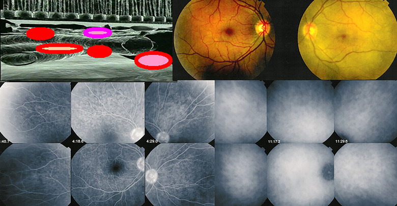
Before the clinical signs of VKH become apparent, patients often present prodromal symptoms, such as headache, nausea, fever, vertigo, tinnitus, meningismus, scalp hypersensitivity, or orbital pain [45, 46]. The prodromal stage of VKH disease typically lasts a few hours or days, but sometimes weeks. This stage corresponds to subclinical ocular choroiditis or meningeal inflammation, which can only be identified at this stage by performing ICGA [16] (Fig. 1) or a CSF analysis [47]. By the same token, it has been shown that, in preclinical VKH disease, only the internal limiting membrane folds could be detected initially. However, when ICGA was performed 24 h later, massive ICGA lesions were observed [48]. With EDI-OCT, marked choroidal thickening could be detected as evidence of diffuse choroidal inflammation [17].
Fig. 1. The prodromal stage of VKH disease.
(Top left) Cartoon shows that, during the prodromal stage of VKH disease, subclinical choroidal inflammation silently develops in the choroidal stroma. This subclinical choroidal involvement can only be detected with ICGA, and possibly, with EDI-OCT. (Top right) Photographs show the fundi of both eyes. The right image shows that the fundus is discoloured yellow, due to massive choroidal infiltration. The left image shows a normal fundus; thus, this patient was diagnosed with “unilateral” VKH disease. (Bottom left) Six images of FA show no lesions, but (bottom right) six images of ICGA clearly show numerous hypofluorescent dark dots (HDDs), which indicate choroidal granulomas in the apparently uninvolved eye.
After the prodromal phase, the disease becomes clinically apparent. Choroidal inflammation secondarily affects adjacent structures, including the optic disc, the retina, and subsequently, the ciliary body, and sometimes the anterior chamber. During the acute phase, a full-thickness granuloma can form that prevents normal choroidal flow [49] Thus, the filling of choroidal arteries is delayed, followed by a delay in choriocapillaris filling. This mechanically induced delay in blood supply is thought to induce acute ischaemic damage to the retinal pigment epithelium, which could contribute to exudative retinal detachment.
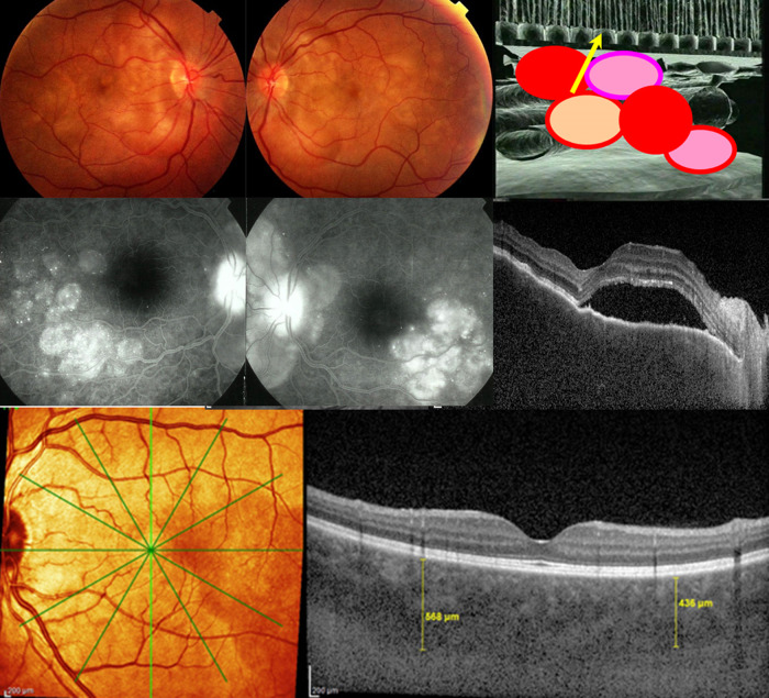
In this second, uveitic/exudative stage, the disease manifests as a bilateral, mostly granulomatous, panuveitis with predominantly posterior involvement. In most cases, it is characterised by exudative retinal detachment [50–52]. The involvement is always bilateral, and it includes papillitis, serous detachments of the retina, and mild to moderate vitritis. In the absence of therapy, most of the time, it extends anteriorly, in the form of granulomatous uveitis, with small to mid-sized granulomatous keratic precipitates [41, 50–52] (Fig. 2). When the inflammation is severe, the process can involve the ciliary body, which produces supraciliary fluid. This fluid accumulates at the origin of a ciliary detachment and causes myopisation, and sometimes, angle-closure glaucoma [52].
Fig. 2. Signs of initial-onset, acute exudative VKH disease.
a Fundoscopy images show bilateral retinal exudative detachments that resulted from (b, cartoon) choroidal inflammation spill-over into the retina and optical disc. c FA and d OCT images show exudative retinal detachments. e EDI-OCT images show choroidal thickening in excess of 400 µm (yellow vertical lines), in early disease.
It is of utmost importance to be aware that, in acute initial-onset VKH disease, the initial inflammatory events take place in the uvea, starting in the choroid, or more precisely, in the choroidal stroma. The stroma is the exclusive origin of all inflammation; hence, it is called primary stromal choroiditis [53]. Other structures, like the retina and optic disc, only become involved secondary to choroidal inflammation. That is, these other structures become inflamed as a consequence of choroiditis; they do not initiate inflammation by themselves [54]. (Fig. 2)
Therefore, the approach to VKH disease management is based on the fact that the single and exclusive source of all intraocular inflammation is the choroidal stroma. This feature makes VKH disease unique, compared to other choroiditis entities, such as sarcoidosis, which involves both the choroid and retina at random [55], or birdshot retinochoroiditis, which can display dual, parallel, and independent involvement of the choroid and retina [56]. Diagnostic criteria and investigations of early-onset VKH disease will be discussed in the next section.
Chronic VKH disease
When appropriate therapy is not introduced diligently, within “the therapeutic window of opportunity”, after initial-onset VKH is diagnosed [18], the disease will evolve to chronic VKH disease. Chronic VKH disease must be distinguished from initial-onset disease, in terms of the clinical signs, evolution, response to treatment, and complications [13].
The chronic course of VKH disease can exhibit a subclinical smouldering evolution, recurrent bouts of inflammation, or a combination of both these patterns [57]. The clinical signs, at the ocular level, comprise chronic granulomatous anterior uveitis; sunset glow fundus (SGF), defined as progressive fundus depigmentation; peripheral atrophic foci, which represent scars of Dalen-Fuchs nodules; and pigment migration. Beyond the eye, the clinical signs comprise integumentary changes, like vitiligo, poliosis, and alopecia, and auditory involvement, in the form of dysacusis, tinnitus, or hearing loss [8]. Logically, chronic VKH disease affects mainly the anterior segment, because melanocytes persist in this part of the uvea after the earlier phases of the disease have largely destroyed melanocytes in the choroid [58, 59]. However, a sensitive analysis of the choroid, performed with ICGA, has clearly shown that concomitant subclinical inflammation is active in the choroid during chronic clinical anterior recurrences [60]. Indeed, currently, ICGA provides information on the whole fundus. In contrast, with the OCT instruments currently available, EDI-OCT can provide only posterior pole imaging.
In bilateral chronic recurrent anterior granulomatous or panuveitis, in addition to compatible clinical findings, such as SGF, ICGA is a relevant complementary investigational tool for confirming the diagnosis. Although exudative retinal detachments do not typically occur during recurrences or chronic disease, ICGA is useful, because it shows typical hypofluorescent dark dots (HDDs) evenly distributed over the entire fundus, which probably indicates ongoing immune cell infiltration around the remaining choroidal melanocytes [60–62]. In addition, a choroidal thickness measurement, with EDI-OCT or another choroidal OCT method, such as Swept Source OCT, provides useful information on posterior involvement in chronic disease [17, 63, 64]. However, chronic disease includes a mixture of choroidal atrophy and choroidal thickening, due to new areas of inflammatory activity; therefore, the EDI-OCT method is less reliable than ICGA for assessing and monitoring activity in this post-acute situation. Indeed, in a recent study, ICGA was much more sensitive than EDI-OCT in detecting inflammatory choroidal foci in patients with chronic VKH that displayed SGF [65].
Clearly, chronic disease will require prolonged therapy, because several, mostly (but not only) anterior recurrences, will occur during the evolution of the disease. Moreover, chronic disease often leads to complications. Abu El-Asrar et al. showed that chronic VKH disease was significantly associated with severe anterior segment inflammation, ocular complications, and a poor visual outcome [51]. They compared follow-up assessments of treated patients between an initial-onset group and a chronic VKH group, and we found that, respectively, 16% versus 66.3% had cataracts, 13% versus 32% had glaucoma, and 0% versus 17.5% had subretinal neovascular membranes [51]. Moreover, chronic evolution and SGF were shown to be associated with worse retinal sensitivity [66]. Unlike the mostly treatable complications, like cataract, glaucoma, and subretinal neovascular membranes, subretinal fibrosis is a more deleterious complication that occurs rarely in chronic VKH. In one series of 101 patients, subretinal fibrosis occurred in 6% of patients [67]. However, its occurrence depended on the severity of disease and the diligence and strength of therapy. One risk factor for subretinal fibrosis is the development of bullous retinal detachments [68].
Update on diagnostic criteria of initial-onset VKH
More than 20 years have elapsed since a workshop was held on revised diagnostic criteria for VKH disease (October 19–21,1999, University of California Conference Centre, Lake Arrowhead, California) [14]. Discussions were very lively, and some participants were uncomfortable with the final result, because initial-onset and chronic disease were not considered separately; thus, the consensus group tried to accommodate both stages in one set of criteria. The criteria distinguished three categories: complete, incomplete, and probable VKH disease, based on the presence of acute or chronic ocular signs, the neurological/auditory findings, and the integumentary changes.
However, little by little, it became clear that VKH should be subdivided into initial-onset and chronic disease forms, including, for the latter, all cases that lacked early, vigorous treatment [13]. For example, the inclusion of both acute and chronic ocular and extraocular signs in the criteria caused the complete VKH form to become rarely observed in all subsequent studies that adopted this classification, because acute and chronic signs coexist only exceptionally. Moreover, in the revised criteria, diffuse choroiditis was considered the sine qua non feature of early ocular disease. However, evidence of diffuse choroiditis was based on the clinical finding of secondary retinal detachment, fluorescein angiography (FA) findings of leaks and pooling, which are only indirect signs of diffuse choroiditis, or a rough, ultrasonographic demonstration of choroidal thickening. Thus, another shortcoming of the criteria was the failure to include ICGA, which is the best method for characterising choroidal inflammation. Moreover, EDI-OCT has recently become available. EDI-OCT is another precious investigational tool for assessing choroidal inflammation. Consequently, it appeared justified to propose more adequate diagnostic criteria, and in particular, to elaborate separate criteria for initial-onset VKH and chronic VKH.
In that regard, a remarkable study was performed by Yang et al. [69] to distinguish the initial-onset from the late phase of VKH disease [69]. However, they proposed criteria that did not include ICGA, even though it is the most sensitive modality for detecting choroidal lesions, HDDs, and other signs of initial-onset VKH that were present in 100% of patients in numerous previous studies [70–73]. Indeed, Yang et al. [69] reported that ICGA detected HDDs in 91.3% of their patients.
Although ICGA is not practiced in some parts of the world, it should not be discarded, because the criteria are intended to be universal. Instead, ICGA should be included in conjunction with EDI-OCT, a surrogate for ICGA, when ICGA is not available.
Another interesting imaging modality that has also been used in assessing VKH disease is optical coherence tomography angiography (OCTA). In one study, OCTA showed choriocapillaris voids that correlated with ICGA signs [74]. That correlation could probably be explained by the fact that, in VKH disease, HDDs are full-thickness stromal lesions, and they exert pressure on the choriocapillaris. However, VKH disease is principally a choroidal stromal disease; consequently, we cannot anticipate that OCTA will constitute a routinely relevant modality, and it cannot serve as a diagnostic tool.
In the diagnostic criteria proposed by our group (Table 1), diffuse choroiditis remains a sine qua non diagnostic criterion. Therefore, precise choroidal imaging is crucial, and it was included for the early, accurate diagnosis of initial-onset VKH disease.
Table 1.
The proposed new diagnostic criteria for initial-onset VKH disease.
| 1. No ocular trauma or surgery preceding disease onseta |
| 2. Bilateral involvement (verified with ICGA and/or EDI-OCT)a |
| 3. Exclusion of other infectious, inflammatory, or masquerading entities, particularly other stromal choroiditis entities (e.g., tuberculosis, sarcoidosis, or syphilis)a |
| 4. Diffuse choroiditis, evidenced by ICGA and/or EDI-OCTa |
| 5. Signs and symptoms having persisted for less than 4 weeksa |
| 6. Absence of clinical findings compatible with chronic disease (i.e., SGF or integumentary signs, like vitiligo, alopecia, or poliosis)a |
| 7. Exudative retinal detachment (evidenced by pooling and pinpoint leaks on FA and ICGA) (a very helpful criterion when present) |
| 8. Disc hyperfluorescence (a helpful criterion) |
| 9. Neurological/auditory findings (meningismus, tinnitus, acute hearing loss) (a helpful criterion) |
aEssential required criteria.
The study by Yang et al. [69] also proposed diagnostic criteria for late-stage VKH disease, which are straightforward. However, the criteria did not include an appraisal of patients assessed beyond 4 weeks of the initial manifestation or patients that had not received adequate treatment. Although the latter patient group does not yet present the characteristic signs of late-stage disease, this group should be included in the diagnosis of chronic disease.
Due to the advent of ICGA and EDI-OCT, the diagnostic criteria presented here for initial-onset VKH disease have become much simpler and handier than the original criteria. These new imaging modalities (ICGA and EDI-OCT) are highly sensitive for analysing choroidal inflammation, which is the initial site of inflammatory activity.
Management of initial-onset VKH disease (the essential role of ICGA)
The key to successfully managing acute initial-onset VKH disease is prompt, vigorous, sustained therapy. The expected goal to be achieved is no less than a cure of the disease [75]. Several prerequisites must be fulfilled for a favourable outcome, including: (1) early diagnosis, (2) early treatment, (3) combined treatment of steroidal and nonsteroidal immunosuppression [75, 76], and (4) close follow-up monitoring to detect subclinical choroidal disease and subclinical choroidal reactivation [62, 77].
Early diagnosis
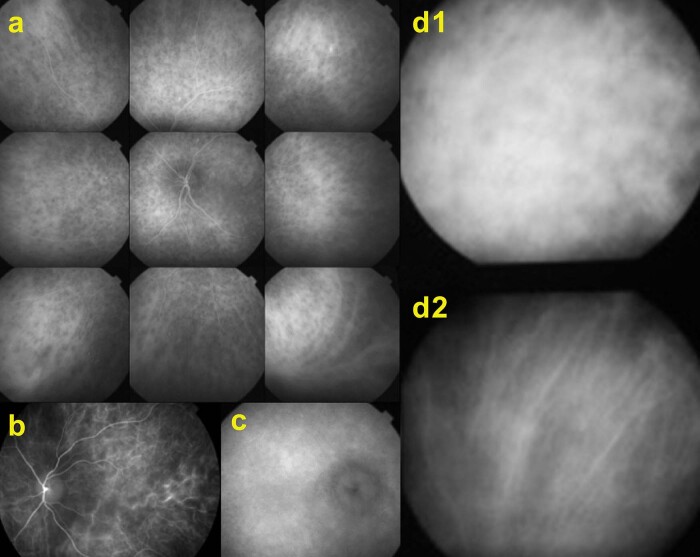
Within 4 weeks of disease onset, an early diagnosis is crucial. Thanks to the recently available tools for imaging the choroid, an early diagnosis can be expected, even when the presentation is not full-blown and typical serous retinal detachments are absent, or not yet present. Investigational modalities, such as ICGA and EDI-OCT, are highly precise in establishing the “disease defining sign” of diffuse choroiditis, which contributes substantially to reaching an early diagnosis. For diagnosing initial-onset VKH, ICGA displayed 100% sensitivity for identifying signs in multiple studies [70–73] and over 90% sensitivity in other studies [69]. In these studies, the semiology of ICGA in initial-onset VKH disease has been precisely established. Four main ICGA signs have been defined, including early hyperfluorescent choroidal vessels, HDDs, fuzzy choroidal vessels, and disc hyperfluorescence. (Fig. 3) EDI-OCT had a slightly lower sensitivity for identifying diffuse choroiditis, but its sensitivity was above 90% [69]. The major drawback of currently available instruments is that they do not show peripheral involvement; however, new generations of these instruments may overcome this disadvantage.
Fig. 3. Four main ICGA signs of acute initial-onset VKH disease.
a Images show numerous, evenly-sized hypofluorescent dark dots (HDDs), regularly distributed over the entire fundus, the most demonstrative and quantifiable ICGA sign of VKH. b Early hyperfluorescent vessels and c a hyperfluorescent disc (normally hypofluorescent on ICGA) indicates severe inflammation. (d1) Fuzzy, indistinct choroidal vessels represent the fourth ICGA sign. (d2) After 3 days of intravenous methylprednisolone, the courses and structures of choroidal vessels are again distinctly visible.
In chronic VKH disease, EDI-OCT is less reliable than ICGA in detecting choroiditis, because the choroidal thickness is globally reduced. Therefore, EDI-OCT does not always detect disease reactivation [65, 78].
Early treatment
The main principle in treating early-onset VKH disease is to suppress the initial intraocular inflammation in the acute posterior uveitic/exudative stage, with early, aggressive IST. This early treatment will shorten the duration of the disease, may prevent progression to the chronic stage, and may also reduce the incidence of extraocular manifestations. Many studies have shown that early treatment is essential for the successful management of initial-onset VKH [75, 79–81]. The so-called “window of therapeutic opportunity”, in immune-mediated diseases, was defined as the time interval, after the initial-onset of disease, during which adequate treatment will substantially modify the disease outcome, and possibly, even lead to a cure. For VKH, this window probably lies between 2 and 4 weeks of onset, and closer to two weeks, when the disease is more severe [18].
Combined steroidal and nonsteroidal immunosuppression
When VKH is not properly treated, intraocular inflammation will proceed to chronic recurrent granulomatous anterior uveitis with typical SGF [50, 51]. Moreover, several studies have reported that, despite proper early treatment with corticosteroid monotherapy, chronic smouldering and/or recurrent granulomatous inflammation and SGF developed, with peripapillary atrophy and small, depigmented, atrophic lesions at the level of the retinal pigment epithelium [50, 82].
Sakata et al. reported that patients with VKH had a high rate of clinical recurrence after receiving early treatment with high-dose corticosteroids. The treatment was given within 30 days of disease onset, and later, it was slowly tapered off. In that cohort, 79% of patients progressed to chronic recurrent disease, and 38% developed subretinal fibrosis [83]. Similarly, Chee et al. observed that, among patients that received high-dose corticosteroid therapy within 2 weeks of onset, one-third progressed to chronic recurrent disease [82]. Keino et al. demonstrated that, despite high-dose corticosteroid therapy administered at the initial VKH onset, 17.5% of patients developed chronic ocular inflammation [84].
Compared to initial-onset, acute VKH disease treated with appropriate combined therapy, chronic recurrent VKH disease is associated with significantly more severe anterior segment inflammation, worse visual acuity, and a worse mean retinal sensitivity score [66, 76, 85] Chronic recurrent granulomatous inflammation in the anterior segment with SGF is also more refractory to treatment and more prone to complications, compared to initial-onset, acute VKH disease [50, 51, 76].
These reports clearly indicated that systemic corticosteroid therapy, even when given promptly in initial-onset VKH disease, did not seem to prevent chronic evolution in a large proportion of cases. Therefore, we performed a systematic literature search to compare the rates of chronic evolution and SGF between patients that received early corticosteroid monotherapy and patients treated with dual steroidal and nonsteroidal immunosuppression, immediately at the onset of VKH disease [75].
We identified 20 studies on treatments for early-onset VKH disease. In 16 studies (802 patients), corticosteroid monotherapy was applied, and in four studies, 172 patients received dual steroidal and nonsteroidal immunosuppression. In the corticosteroid monotherapy group, 44% of patients displayed chronic evolution, and 59% displayed SGF. In the dual steroidal plus nonsteroidal immunosuppression group, these proportions were substantially lower, with rates of 2.3% for chronic evolution and 17.5% for SGF [75]. These proportions changed slightly, according to geographic areas, but the difference between treatments remained highly significant [86]. These data left no remaining doubt that combining steroidal and nonsteroidal immunosuppression is the management of choice for initial-onset VKH disease.
Currently, two questions remain open: (1) What additional immunosuppressant agent should be used? And, (2) How long should therapy be given? The latter question is discussed in the next section.
The four studies [12, 69, 77, 85] on combined treatment in our literature review [75, 86] administered diverse immunosuppressive agents. In a study by Abu El-Asrar et al., which included 38 patients, the nonsteroidal immunosuppressant was mycophenolate mofetil. They reported no cases of chronic evolution or SGF [85]. Moreover, those patients discontinued treatment without a relapse of inflammation [85]. Those findings suggested that, similar to rheumatoid arthritis, there is a therapeutic window of opportunity for the highly successful treatment of uveitis associated with VKH disease. This window occurs during the early initial-onset, acute uveitic phase, likely because the underlying disease process has not fully matured in that phase [18]. In the study by Yang et al., in addition to corticosteroids, 105 patients received immunosuppressive agents, including cyclosporine, cyclophosphamide, methotrexate, or azathioprine. They reported no case of chronic evolution, and 22.9% of patients developed mild SGF [69, 87]. In a study by Lodhi et al., concomitant azathioprine was administered as an immunosuppressive agent, and they observed rates of 17% for chronic evolution and 25% for SGF [12]. Bouchenaki and Herbort administered azathioprine, mycophenolate mofetil, cyclosporine, adalimumab, and interferon-α to two patients that received two immunosuppressants in addition to corticosteroids [77]. They noted no chronic evolution or SGF.
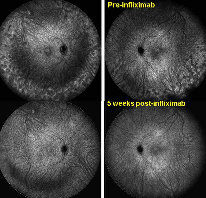
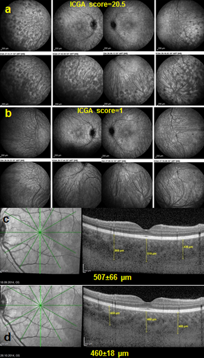
In those four studies, several different immunosuppressive agents were administered, and all had similarly good results. This finding suggested that the choice of immunosuppressant does not matter as much as the choice of giving corticosteroids combined with a nonsteroidal immunosuppressant as the fist-line therapy. However, for each individual case, the efficacy of the chosen treatment regimen should be monitored with ICGA to determine whether HDDs have regressed or persisted, despite treatment (Fig. 4 and 5).
Fig. 4. Extreme sensitivity and global information on whole fundus involvement, assessed with ICGA.
(Top) ICGA images show VKH disease responsiveness to initial high-dose corticosteroids, then mycophenolic acid (Myfortic®) and cyclosporine. Note peripheral recurrence, characterised by numerous HDDs. (Bottom) After introducing infliximab, complete resolution of choroiditis occurred within 5 weeks. This finding established infliximab as the therapy of choice in this patient with VKH that showed treatment responsiveness. Posterior pole involvement was minimal, and choroidal OCT did not reflect a spectacular improvement in choroiditis. ICGA-assisted monitoring of choroidal inflammation is crucial for prompt determinations of the inefficacy or efficacy of immunosuppressive treatment for each individual patient.
Fig. 5. Indocyanine green angiography is more sensitive and reactive than EDI-OCT for precise follow-ups in VKH disease.
Images are from the same patient as shown in Fig. 4a Here in a, the ICGA score of 20.5/40 indicates that the choroiditis is not responding to mycophenolic acid and cyclosporine. After 6 months of treatment, infliximab was added (Remicade®, an anti-TNF-agent, 5 mg/kg per infusion). b Five weeks later (after three infusions), the HDDs completely disappeared, and the ICGA score decreased from 20.5 ± 4.9 to 1 (p < 0.03). c, d The choroidal thickness (yellow vertical lines) decreased slightly, but not significantly, from a mean of c 507 ± 66 to a mean of d 460 ± 18 µm (p = 0.31).
Several trials have demonstrated the efficacy of cyclosporine [88], azathioprine [89], mycophenolate mofetil [90], methotrexate [90], adalimumab [91], and rituximab in VKH disease [44, 92]. During the GOIW congress, in June 2019, in Sapporo, Japan, a workshop was held to launch a clinical trial that aimed to determine the optimal choice of immunosuppressant for combining with corticosteroids. However, that trial was not launched, because a strong tendency towards corticosteroid monotherapy prevailed in the assembly, and finally, the issue was not addressed.
In uveitis, and in immunogenic inflammatory diseases as a whole, the present trend is to use less corticosteroids. This tendency had a double advantage for VKH, because, in addition to the IST effect, the ocular barrier could be restored, and with that, we could expect a potential cure of the disease. Currently, we have at our disposal of a large array of immunosuppressive agents which we can manipulate more appropriately [93, 94]. When the severity of side effects was compared between corticosteroids and conventional immunosuppressants, such as mycophenolate mofetil, azathioprine, or cyclosporine, the immunosuppressants were clearly more favourable, and biologic agents showed even more favourability [44, 91, 92]. The low prevalence of side-effects and the corticosteroid-sparing effect of nonsteroidal immunosuppressants provide benefits that largely exceed the inconvenience of their use. Indeed, close to half of patients with initial-onset VKH could be spared from chronic evolution. Moreover, the benefits are even higher, when considering the prevalence of SGF, an indicator of ongoing smouldering choroiditis. SGF developed in only 17.5% of patients treated with combined therapy, compared to 60% of patients treated with steroidal therapy alone [75].
Close monitoring of subclinical choroiditis for determining treatment duration
VKH is a primary stromal choroiditis because the inflammation exclusively originates from the choroid. Once subclinical choroiditis has been eradicated, all intraocular inflammation is eliminated. Therefore, it is crucial to use the performing investigational modalities at our disposal to detect and monitor choroidal inflammatory lesions, even when they are subclinical. It has been shown that ICGA could detect early subclinical disease and subclinical recurrences, which can occur when tapering off the therapy [77]. ICGA could also detect occult, concomitant choroidal inflammation during anterior segment recurrence, when there was no apparent posterior uveal activity [60]. Therefore, ICGA appears to be the modality of choice for detecting and following VKH disease, because it provides information on the whole fundus, and on the crucial core structure of the disease process, the choroid, which is not otherwise available. EDI-OCT, which can detect choroidal thickening, is another quality imaging procedure that can be used to follow choroidal involvement in initial-onset VKH disease [17, 63, 95, 96]. EDI-OCT complements ICGA in investigations of diffuse choroiditis, and it can be used instead of ICGA, when ICGA is unavailable. However, EDI-OCT is less reliable and less precise than ICGA for detecting minute changes that require an adjustment to therapy [78] (Fig. 5). Thanks to ICGA, and to a lesser extent, EDI-OCT, it is currently possible to follow inflammatory choroidal lesions and their evolution very precisely. This is the fourth prerequisite for the optimal management of VKH disease: monitoring subclinical choroidal inflammation, until the absence of occult choroiditis is verified.
It is well known that, in most cases, controlling a clinically apparent disease is not sufficient for a cure. ICGA-monitoring during VKH treatment has shown that, once the clinical signs and functional parameters have normalised (i.e., the fundus picture, OCT, FA, and visual acuity), choroidal inflammation remains and continues to evolve [61, 62, 77]. The persistence of choroidal subclinical disease explains why, in most patients that receive “standard” corticosteroid monotherapy, VKH continues to evolve towards SGF [97]. In practice, when these elements are taken into account, initial-onset VKH disease should be treated in two main phases: (1) treating the early uveitic/exudative acute stage, and (2) maintaining sufficient therapy, including first-line nonsteroidal immunosuppressive agents, until all choroidal inflammation has resolved; thus, tapering the therapy should be assisted, preferably with ICGA monitoring, or when ICGA is not available, with EDI-OCT monitoring. Moreover, because ICGA is an invasive, costly modality, a combination of ICGA and EDI-OCT can be used, or EDI-OCT can be used alone, depending on the setting and resources.
The severity of acute VKH disease requires treatment with high-dose corticosteroids [98]. A 3-day course of intravenous methylprednisolone (500–000 mg/day) is often recommended, followed by high-dose oral prednisone (1.0 mg/kg) for 4–6 weeks. Although the need for intravenous corticosteroid administration during the first 3 days of treatment has not been proven definitively; however, common sense has it that, in case of hyperacute uveitis, a rapid resolution of inflammation is desirable [99]. As indicated earlier, the addition of a first-line, nonsteroidal immunosuppressant is crucial in the early phase of the disease.
After the clinical resolution of VKH disease, the task is to verify the resolution of all subclinical choroidal inflammation with ICGA (or EDI-OCT) and to succeed in tapering the therapy without subclinical recurrences. This process sometimes leads to a temporary increase in therapy, which may be repeated, until the final tapering of therapy results in a choroiditis-free resolution of the disease. In our experience, this treatment lasted 27.3 months ± 38.2 months (range 9–114), which was too prolonged to use corticosteroids alone [77]. The advantage of this relentless therapy approach was that a high proportion of patients could be ″healed″, with no recurrent activity, within a mean follow-up period 26 ± 14.8 months without therapy, and a low proportion of patients developed SGF [77]. Thus, to cure this autoimmune condition, we recommend an ICGA-monitored regression of choroidal disease in the post-acute phase of VKH disease and a long-term tapering of inflammation-suppressive therapy, until no recurrence of choroidal disease is observed (zero tolerance of choroiditis).
Meaningful ICGA-monitoring presupposes that ICGA can be performed every 5–6 weeks during the first 4–5 months of treatment, and then every 2–3 months, during the long-term tapering-off period. However, there are cost considerations, and EDI-OCT might achieve comparable results [77]. Performing an ICGA every 6 months during the follow-up cannot be called ICGA-monitoring, because a low frequency of ICGAs does not allow treatment modifications in a timely fashion. Therefore, it represents an ICGA-documented follow-up. Nevertheless, early and sustained treatment, with ICGA or EDI-OCT monitoring, can modify the VKH phenotype.
A practical approach that we have used increasingly is early, high-dose systemic steroids, combined with a rapid-acting immunosuppressive drug, such as cyclosporine, together with a well-tolerated immunosuppressant, such as mycophenolic acid (Myfortic®), which requires several weeks to develop full activity. This approach can accelerate corticosteroid tapering, and it requires a limited period (4–5 months) of cyclosporine use. When this therapeutic regimen fails, disease activity can be controlled with a biologic therapy, for example, anti-TNF agents or the B-cell inhibitor, rituximab [44, 91, 92]. Adalimumab has been approved for treating non-infectious posterior uveitis or panuveitis; thus, it should be considered a first-line biologic agent for patients with VKH. Moreover, several case reports have shown that infliximab was effective for treating VKH [100].
We can expect to cure this autoimmune condition more readily than other autoimmune diseases, because VKH takes place in a secluded organ. Thus, when the blood-ocular barriers are promptly restored with early, vigorous, sustained therapy, chronic aggression cannot occur.
Conclusion
This review showed that the current diagnostic criteria for VKH must be reconsidered, in light of the new investigational capacities, particularly because the existing criteria have not separated acute from chronic signs, which leads to confusion. Here, new criteria were proposed for initial-onset VKH disease; similarly, new criteria will have to be proposed for chronic VKH.
VKH disease belongs to the group of primary stromal choroiditis entities, which are characterised by inflammation that originates exclusively from within the choroid. Inflammatory damage to adjacent structures only occurs when inflammatory factors spill over to those compartments. Because inflammation initiates only in the choroid, this structure should be targeted with therapy. The choroidal compartment, unlike the retina, is easily accessible to systemic therapy. Increasing evidence has shown that, when treatment is conducted with zero tolerance to choroiditis recurrence, detected with ICGA or EDI-OCT, a substantial proportion of patients can be healed before depigmentation occurs, and SGF would no longer be inevitable.
Supplementary information
Author contributions
CPH Jr, ITT and AA: concept of the manuscript and redaction of parts of the manuscript. CPH Jr, ITT, AA, AG, MT, CF, AH, CU, and IP elaboration of the diagnostic. criteria, writing of parts of the MS, reading and correction of the manuscript. CPH Jr, IP: figures and legends and references
Compliance with ethical standards
Conflict of interest
The authors declare no competing interests.
Ethical approval
All procedures performed in studies involving human participants were in accordance with the 1964 Helsinki declaration and its later amendments or comparable ethical standards. For this type of study, formal consent is not required.
Footnotes
Publisher’s note Springer Nature remains neutral with regard to jurisdictional claims in published maps and institutional affiliations.
These authors contributed equally: Carl P. Herbort Jr, Ilknur Tugal-Tutkun, Ahmed Abu El-Asrar
Supplementary information
The online version contains supplementary material available at 10.1038/s41433-021-01573-3.
References
- 1.Gocho K, Kondo I, Yamaki K. Identification of autoreactive T cells in Vogt-Koyanagi-Harada disease. Investig Ophthalmol Vis Sci. 2001;42:2004–9. [PubMed] [Google Scholar]
- 2.Damico FM, Cunha-Neto E, Goldberg AC, Iwai LK, Marin ML, Hammer J, et al. T-cell recognition and cytokine profile induced by melanocyte epitopes in patients with HLA-DRB1*0405-positive and -negative Vogt-Koyanagi-Harada uveitis. Investig Ophthalmol Vis Sci. 2005;46:2465–71. doi: 10.1167/iovs.04-1273. [DOI] [PubMed] [Google Scholar]
- 3.Sugita S, Takase H, Taguchi C, Imai Y, Kamoi K, Kawaguchi T, et al. Ocular infiltrating CD4+ T cells from patients with Vogt-Koyanagi-Harada disease recognize human melanocyte antigens. Investig Ophthalmol Vis Sci. 2006;47:2547–54. doi: 10.1167/iovs.05-1547. [DOI] [PubMed] [Google Scholar]
- 4.Lavezzo MM, Sakata VM, Morita C, Rodriguez EE, Abdallah SF, da Silva FT, et al. Vogt-Koyanagi-Harada disease: review of a rare autoimmune disease targeting antigens of melanocytes. Orphanet J Rare Dis. 2016;11:29. doi: 10.1186/s13023-016-0412-4. [DOI] [PMC free article] [PubMed] [Google Scholar]
- 5.Yokoyama MM, Matsui Y, Yamashiroya HM, O’Donnell MJ, Tseng CH, Snyder DA, et al. Humoral and cellular immunity studies in patients with Vogt-Koyanagi-Harada syndrome and pars planitis. Investig Ophthalmol Vis Sci. 1981;20:364–70. [PubMed] [Google Scholar]
- 6.Tran VT, Auer C, Guex-Crosier Y, Pittet N, Herbort CP. Epidemiological characteristics of uveitis in Switzerland. Int Ophthalmol. 1994;18:293–8. doi: 10.1007/BF00917833. [DOI] [PubMed] [Google Scholar]
- 7.Nakamura S, Nakazawa M, Yoshioka M, Nagano I, Nakamura H, Onodera J, et al. Melanin-laden macrophages in cerebrospinal fluid in Vogt-Koyanagi-Harada syndrome. Arch Ophthalmol. 1996;114:1184–8. doi: 10.1001/archopht.1996.01100140384003. [DOI] [PubMed] [Google Scholar]
- 8.Noguchi Y, Nishio A, Takase H, Miyanaga M, Takahashi H, Mochizuki M, et al. Audiovestibular findings in patients with Vogt-Koyanagi-Harada disease. Acta Otolaryngol. 2014;134:339–44. doi: 10.3109/00016489.2013.868604. [DOI] [PubMed] [Google Scholar]
- 9.Hirooka K, Saito W, Namba K, Mizuuchi K, Iwata D, Hashimoto Y, et al. Significant role of the choroidal outer layer during recovery from choroidal thickening in Vogt-Koyanagi-Harada disease patients treated with systemic corticosteroids. BMC Ophthalmol. 2015;15:181. doi: 10.1186/s12886-015-0171-3. [DOI] [PMC free article] [PubMed] [Google Scholar]
- 10.Fang W, Yang P. Vogt-Koyanagi-Harada syndrome. Curr Eye Res. 2008;33:517–23. doi: 10.1080/02713680802233968. [DOI] [PubMed] [Google Scholar]
- 11.Herbort CP, Jr, Asrar AMAbuEL, Yamamoto JH, Pavesio CE, Gupta V, Khairallah M, et al. Reappraisal of the management of Vogt-Koyanagi-Harada disease: sunset glow fundus is no more a fatality. Int Ophthalmol. 2017;37:1383–95. doi: 10.1007/s10792-016-0395-0. [DOI] [PMC free article] [PubMed] [Google Scholar]
- 12.Lodhi SAK, Reddy JML, Peram V. Clinical spectrum and management options in Vogt-Koyanagi-Harada disease. Clin Ophthalmol. 2017;11:1399–406. doi: 10.2147/OPTH.S134977. [DOI] [PMC free article] [PubMed] [Google Scholar]
- 13.Urzua CA, Herbort CP, Jr, Valenzuela RA, Abu E-Asrar AM, Arellanes-Garcia L, Schlaen A, et al. Initial-onset acute and chronic stages are two distinctive courses of Vogt-Koyanagi-Harada disease. J Ophthalmic Inflamm Infect. 2020;14:23. doi: 10.1186/s12348-020-00214-2. [DOI] [PMC free article] [PubMed] [Google Scholar]
- 14.Read RW, Holland GN, Rao NA, Tabbara KF, Ohno S, Arellanes-Garcia L, et al. Revised diagnostic criteria for Vogt-Koyanagi-Harada disease: report of an international committee on nomenclature. Am J Ophthalmol. 2001;131:647–52. doi: 10.1016/s0002-9394(01)00925-4. [DOI] [PubMed] [Google Scholar]
- 15.Hedayatfar A, Khochtali S, Khairallah M, Takeuchi M, El-Asrar AA, Herbort CP., Jr “Revised diagnostic criteria” for Vogt-Koyanagi-Harada disease fail to improve disease management. J Curr Ophthalmol. 2018;31:1–7. doi: 10.1016/j.joco.2018.10.011. [DOI] [PMC free article] [PubMed] [Google Scholar]
- 16.Bouchenaki N, Herbort CP. The contribution of indocyanine green angiography to the appraisal and management of Vogt-Koyanagi-Harada disease. Ophthalmology. 2001;108:54–64. doi: 10.1016/s0161-6420(00)00428-0. [DOI] [PubMed] [Google Scholar]
- 17.Nakai K, Gomi F, Ikuno Y, Yasuno Y, Nouchi T, Ohguro N, et al. Choroidal observations in Vogt-Koyanagi-Harada disease using high-penetration optical coherence tomography. Graefes Arch Clin Exp Ophthalmol. 2012;250:1089–95. doi: 10.1007/s00417-011-1910-7. [DOI] [PubMed] [Google Scholar]
- 18.Herbort CarlP, Jr, Abu El Asrar AhmedM, Takeuchi Masuru, Pavésio CarlosE, Couto Cristobal, Hedayatfa Alireza, et al. Catching the therapeutic window of opportunity in early initial-onset Vogt-Koyanagi-Harada uveitis can cure the disease. Int Ophthalmol. 2019;39:1419–25. doi: 10.1007/s10792-018-0949-4. [DOI] [PubMed] [Google Scholar]
- 19.Vogt A, Frühzeitiges ergrauen der Zilien und Bermerkungen über den sogenannten plötzlichen Eintritt dieser Veränderung. Klin Monbl Augenheilkd, 1906;44:228–42.
- 20.Herbort CP, Mochizuki M. Vogt-Koyanagi-Harada disease: inquiry into the genesis of a disease name in the historical context of Switzerland and Japan. Int Ophthalmol. 2007;27:67–79. doi: 10.1007/s10792-007-9083-4. [DOI] [PubMed] [Google Scholar]
- 21.Koyanagi Y. Nippon Ganka Gakkai Zasshi, 1914;18:1188.
- 22.Harada E. Jikken Ganka Zasshi 1918;2:199.
- 23.Harada E. Beitrag zur klinischen Kenntnis von nichteitriger Choroiditis (choroiditis diffusa acta) Acta Soc Ophthalmol Jpn. 1926;30:356–78. [Google Scholar]
- 24.Koyanagi Y. Dysakusis, alopecia und poliosis bei schwerer uveitis nicht traumatischen ursprungs. Klin Monbl Augenheilkd. 1929;82:228–42.
- 25.Babel J. Syndrome de Vogt-Koyanagi (Uveite bilateral, poliosis, alopecie, vitiligo et dysacousie). Schweiz Med Wochenschr. 1932;44:1136–40.
- 26.Bruno MM, McPherson SD., Jr Harada’s disease. Am J Ophthalmol. 1949;32:513–22. doi: 10.1016/0002-9394(49)90876-4. [DOI] [PubMed] [Google Scholar]
- 27.Maezawa N, Yano A. Two distinct cytotoxic T lymphocyte subpopulations in patients with Vogt-Koyanagi-Harda disease that recognize human melanoma cells. MicrobiolImmunol. 1984;28:219–31. doi: 10.1111/j.1348-0421.1984.tb00673.x. [DOI] [PubMed] [Google Scholar]
- 28.Norose K, Yano A, Aosai F, Segawa K. Immunologic analysis of cerebrospinal fluid lymphocytes in Vogt-Koyanagi-Harada disease. Investig Ophthalmol Vis Sci. 1990;31:1210–6. [PubMed] [Google Scholar]
- 29.Reed H, Lindsay A, Silversides JL, Speakman J, Monckton G, Rees DL. The uveo-encephalitic syndrome or Vogt-Koyanagi-Harada disease. Can MedAssoc J. 1958;79:451–9. [PMC free article] [PubMed] [Google Scholar]
- 30.Masuda K, Tanishima T. Early treatment of Harada’s disease. Jpn J Clin Ophthalmol. 1969;23:553. [Google Scholar]
- 31.Kotake S, Ohno S. Pulse methyl prednisolone therapy in the treatment of Vogt-Koyanagi-Harada disease. Jpn J Clin Ophthalmol. 1984;38:1053–8. [Google Scholar]
- 32.Sugiura S. Vogt-Koyanagi-Harada Disease. Jpn J Ophthalmol. 1978;22:9–35. [Google Scholar]
- 33.Snyder DA, Tessler HH. Vogt-Koyanagi-Harada syndrome. Am J Ophthalmol. 1980;90:69–75. [PubMed] [Google Scholar]
- 34.Herbort CP, Guex-Crosier Y, LeHoang P. Schematic interpretation of indocyanine green angiography. Ophthalmology. 1994;2:169–76. doi: 10.1016/S0161-6420(98)93024-X. [DOI] [PubMed] [Google Scholar]
- 35.Inomata H, Sakamoto T. Immunohistochemical studies of Vogt-Koyanagi-Harada disease with sunset sky fundus. Curr Eye Res. 1990;9:35–40. doi: 10.3109/02713689008999417. [DOI] [PubMed] [Google Scholar]
- 36.Yamaki K, Kondo I, Nakamura H, Miyano M, Konno S, Sakuragi S. Ocular and axtraocular inflammation induced by immunazation of tyrosinase related protein 1 and 2 in Lewis rats. Exp Eye Res. 2000;71:361–69. doi: 10.1006/exer.2000.0893. [DOI] [PubMed] [Google Scholar]
- 37.Yamaki K, Gocho K, Hayakawa K, Kondo I, Sakuragi S. Tyrosinase family proteins are antigens specific to Vogt-Koyanagi-Harada. J Immunol. 2000;165:7323–9. doi: 10.4049/jimmunol.165.12.7323. [DOI] [PubMed] [Google Scholar]
- 38.Shindo Y, Inoko H, Yamamoto T, Ohno S. HLA-DRB1 typing of Vogt-Koyanagi-Harada disease by PCR-RFLP and the strong association with DRB1*0405 and DRB1*0410. Br J Ophthalmol. 1994;78:223–6. doi: 10.1136/bjo.78.3.223. [DOI] [PMC free article] [PubMed] [Google Scholar]
- 39.Mochizuki M. Regional immunity of the eye. Acta Ophthalmol. 2010;88:292–99. doi: 10.1111/j.1755-3768.2009.01757.x. [DOI] [PubMed] [Google Scholar]
- 40.Musette P, Bouaziz JD. B cell modulation strategies in autoimmune diseases: new concepts. Front Immunol. 2018;9:622. doi: 10.3389/fimmu.2018.00622. [DOI] [PMC free article] [PubMed] [Google Scholar]
- 41.Abu El-Asrar AM, Bergman N, Al-Obeidan SA, et al. Differential CXC and CX3C chemokine expression profiles in aqueous humor of patients with specific endogenous uveitic entities. Investig Opthalmol Vis Sci. 2018;59:2222–8. doi: 10.1167/iovs.17-23225. [DOI] [PubMed] [Google Scholar]
- 42.Abu El-Asrar AM, Berghmans N, Al-Obeidan SA, et al. Local cytokine expression profiling in patients with specific autoimmune uveitic entities. Ocul Immunol Inflamm. 2020;28:453–62. doi: 10.1080/09273948.2019.1604974. [DOI] [PubMed] [Google Scholar]
- 43.Chan CC, Palestine AG, Kuwabara T, Nussenblatt RB. Immunopathologic study of Vogt-Koyanagi-Harada syndrome. Am J Ophthalmol. 1988;105:607–11. doi: 10.1016/0002-9394(88)90052-9. [DOI] [PubMed] [Google Scholar]
- 44.Abu El-Asrar AM, Dheyab A, Khatib D, Struyf S, Van Damme J, Opdenakker G. Efficacy of B cell depletion therapy with rituximab in refractory chronic recurrent uveitis associated with Vogt-Koyanagi-Harada disease. Ocul Immunol Inflamm. 2020;29:1–8. doi: 10.1080/09273948.2020.1820531. [DOI] [PubMed] [Google Scholar]
- 45.Khairallah M. Headache as an initial manifestation of Vogt-Koyanagi-Harada disease. Saudi J Ophthalmol. 2014;28:239–42. doi: 10.1016/j.sjopt.2013.10.003. [DOI] [PMC free article] [PubMed] [Google Scholar]
- 46.Kosma KK, Kararizou E, Markou I, Eforakopoulou E, Kararizos G, Mitsonis C, et al. Headache as a first manifestation of Vogt-Koyanagi-Harada disease. Med Princ Pr. 2008;17:253–4. doi: 10.1159/000117802. [DOI] [PubMed] [Google Scholar]
- 47.Kamondi A, Szegedi A, Papp A, Sees A, Szirmmai I. Vogt-Koyanagi-Harada disease presenting initially as aseptic meningoencephalitis. Eur J Neurol. 2000;7:719–22. doi: 10.1046/j.1468-1331.2000.00156.x. [DOI] [PubMed] [Google Scholar]
- 48.Almalki K, Alsulaiman SM, Abouammoh A. Internal limiting membrane folds as a presenting sign in acute initial-onset Vogt-Koyanagi-Harada disease: a case report. Ocul Immunol Inflamm 2020; 10.1080/09273948.2020.1828489. [DOI] [PubMed]
- 49.Fardeau C, Tran TH, Gharbi B, Cassoux N, Bodaghi B, LeHoang P. Retinal fluorescein and indocyanine green angiography and optical coherence tomography in successive stages of Vogt-Koyanagi-Harada disease. Int Ophthalmol. 2007;27:163–72. doi: 10.1007/s10792-006-9024-7. [DOI] [PubMed] [Google Scholar]
- 50.Yang P, Ren Y, Li B, Fang W, Meng Q, Kijlstra A. Clinical characteristics of Vogt-Koyanagi-Harada syndrome in Chinese patients. Ophthalmology. 2007;114:606–14. doi: 10.1016/j.ophtha.2006.07.040. [DOI] [PubMed] [Google Scholar]
- 51.Abu El-Asrar AM, Al Tamimi M, Hemachandran S, Al-Mezaine HS, Al-Muammar A, Kangave D. Prognostic factors for clinical outcomes in patients with Vogt-Koyanagi-Harada disease treated with high-dose corticosteroids. Acta Ophthalmol. 2013;91:e486–e493. doi: 10.1111/aos.12127. [DOI] [PubMed] [Google Scholar]
- 52.Mantovani A, Resta A, Herbort CP, Abu El Asrar A, Kawaguchi T, Mochizuki M, et al. Work-up, diagnosis and management of acute Vogt-Koyanagi-Harada disease: a case of acute myopisation with granulomatous uveitis. Int Ophthalmol. 2007;27:105–15. doi: 10.1007/s10792-007-9052-y. [DOI] [PubMed] [Google Scholar]
- 53.Bouchenaki N, Herbort CP. Stromal choroiditis. In: Pleyer U, Mondino B, editors Essentials in Ophthalmology: Uveitis and Immunological Disorders. Berlin, Heidelberg, New York: Springer; 2004 pp 234–53.
- 54.Bouchenaki N, Cimino L, Auer C, Tran VT, Herbort CP. Assessment and classification of choroidal vasculitis in posterior uveitis using indocyanine green angiography. Klin Monbl Augenheilkd. 2002;219:243–9. doi: 10.1055/s-2002-30661. [DOI] [PubMed] [Google Scholar]
- 55.El Ameen A, Herbort CP., Jr Comparison of retinal and choroidal involvement in sarcoidosis related chorioretinitis using fluorescein and indocyanine green angiography. J Ophthalmic Vis Res. 2018;13:426–32. doi: 10.4103/jovr.jovr_201_17. [DOI] [PMC free article] [PubMed] [Google Scholar]
- 56.Elahi S, Herbort CP., Jr Vogt-Koyanagi-Harada disease and birdshot retinochoroiditis, similarities and differences: a glimpse into the clinicopathology of stromal choroiditis, a perspective and a review. Klin Monbl Augenheilkd. 2019;236:492–510. doi: 10.1055/a-0829-6763. [DOI] [PubMed] [Google Scholar]
- 57.Attia S, Khochtali S, Kahloum R, Zouali S, Khairallah M. Vogt-Koyanagi-Harada disease. Expert Rev Ophthalmol. 2012;7:565–85. [Google Scholar]
- 58.Hu DN, Simon JD, Sarna T. Role of ocular melanin in ophthalmic physiology and pathology. Photochem Photobio. 2008;84:639–44. doi: 10.1111/j.1751-1097.2008.00316.x. [DOI] [PubMed] [Google Scholar]
- 59.Kaczurowski MI. The pigment elements of the human iris and ciliary body. Am J Opthalmol. 1965;59:299–308. doi: 10.1016/0002-9394(65)94795-1. [DOI] [PubMed] [Google Scholar]
- 60.Bascal K, Wen DS, Chee SP. Concomitant choroidal inflammation during anterior segment recurrence in Vogt-Koyanagi-Harada disease. Am J Ophthalmol. 2008;145:480–6. doi: 10.1016/j.ajo.2007.10.012. [DOI] [PubMed] [Google Scholar]
- 61.da Silva FT, Hirata CE, Sakata VM, Olivalves E, Preti R, Pimentel SLG, et al. Indocyanine green angiography findings in patients with long-standing Vogt-Koyanagi-Harad disease: a cross-sectional study. BMC Ophthalmol. 2012;12:40. doi: 10.1186/1471-2415-12-40. [DOI] [PMC free article] [PubMed] [Google Scholar]
- 62.Kawaguchi T, Horie S, Bouchenaki N, Ohno-Matsui K, Mochizuki M, Herbort CP. Suboptimal therapy controls clinically apparent disease but not subclinical progression of Vogt-Koyanagi-Harada disease. Int Ophthalmol. 2010;30:41–50. doi: 10.1007/s10792-008-9288-1. [DOI] [PubMed] [Google Scholar]
- 63.da Silva FT, Sakata VM, Nakashima A, Hirata CE, Olivalves E, Takahashi WY, et al. Enhanced depth imaging optical coherence tomography in long-standing Vogt-Koyanagi-Harada disease. Br J Ophthalmol. 2013;97:70–4. doi: 10.1136/bjophthalmol-2012-302089. [DOI] [PubMed] [Google Scholar]
- 64.Han YS, Shin KS, Lee WH, Kim JY. Changes in central macular thickness and retinal nerve fiber layer thickness in eyes with Vogt-Koyanagi-Harada disease: a 2-year follow-up study. Ophthalmologica. 2018;239:143–50. doi: 10.1159/000481863. [DOI] [PubMed] [Google Scholar]
- 65.Murata T, Sako N, Takayama K, Harimoto K, Kanda K, Herbort CP, Jr, et al. Identification of underlying inflammation in Vogt-Koyanagi-Harada disease with sunset glow fundus by multiple analyses. J Ophthalmol. 2019;2019:3853794. doi: 10.1155/2019/3853794. [DOI] [PMC free article] [PubMed] [Google Scholar]
- 66.Abu El-Asrar AM, Al Mudhaiyan T, Al Najashi AA, Hemachandran S, Hariz R, Mousa A, et al. Chronic recurrent Vogt-Koyanagi-Harada disease and development of “sunset glow fundus predict worse retinal sensitivity. Ocul Immunol Inflamm. 2017;25:475–85. doi: 10.3109/09273948.2016.1139730. [DOI] [PubMed] [Google Scholar]
- 67.Read RW, Rechodouni A, Butani N, Johnston R, LaBree D, Smith RE, et al. Complications and prognostic factors in Vogt-Koyanagi-Harada disease. Am J Ophthalmol. 2001;131:599–606. doi: 10.1016/s0002-9394(01)00937-0. [DOI] [PubMed] [Google Scholar]
- 68.Golzarri MF, Cheja-Kalb R, Concha-Del-Rio LE, Gonzales-Salinas R, Arellanes-Garcia L Risk factors for subretinal fibrosis in patients with Vogt-Koyanagi-Harada syndrome. Ocul Immunol Inflamm 2020;1–5. 10.1080/09273948.2020.1825750. [DOI] [PubMed]
- 69.Yang P, Zhong Y, Du L, Chi W, Chen L, Zhang R, et al. Development and Evaluation of Diagnostic Criteria for Vogt-Koyanagi-Harada Disease. JAMA Ophthalmol. 2018;136:1025–31. doi: 10.1001/jamaophthalmol.2018.2664. [DOI] [PMC free article] [PubMed] [Google Scholar]
- 70.Kim P, Sun HJ, Ham DI. Ultra-wide-field angiography findings in acute Vogt-Koyanagi-Harad disease. Br J Ophthalmol. 2019;103:942–8. doi: 10.1136/bjophthalmol-2018-312569. [DOI] [PubMed] [Google Scholar]
- 71.Balci O, Jeannin B, Herbort CP. Contribution of dual fluorescein and indocyanine green angiography to the appraisal of posterior involvement in birdshot choriretinitis and Vogt-Koyanagi-Harada disease. Int Ophthalmol. 2018;38:527–39. doi: 10.1007/s10792-017-0487-5. [DOI] [PubMed] [Google Scholar]
- 72.Abouammoh MA, Gupta V, Hemachandran S, Herbort CP, Abu El-Asrar AM. Indocyanine green angiographic findings in initial-onset acute Vogt-Koyanagi-Harada disese. Acta Ophthalmol. 2016;94:573–8. doi: 10.1111/aos.12974. [DOI] [PubMed] [Google Scholar]
- 73.Miyanaga M, Kawaguchi T, Miyata K, Horie S, Mochizuki M, Herbort CP. Indocyanine green angiography findings in initial acute pretreatment Vogt-Koyanagi-Harada disease in Japanese patients. Jpn J Ophthalmol. 2010;54:377–82. doi: 10.1007/s10384-010-0853-6. [DOI] [PubMed] [Google Scholar]
- 74.Aggarwal k, Agarwal A, Mahajan S, Invernizzi A, SKR Mandadi, et al. OCTA Study Group. The role of Optical Coherence Tomography Angiography in the diagnosis and management of acute Vogt-Koyanagi-Harada diseases. Ocul Immunol Inflamm. 2018;26:142–53. doi: 10.1080/09273948.2016.1195001. [DOI] [PubMed] [Google Scholar]
- 75.Papasavvas I, Tugal-Tutkun I, Herbort CP., Jr Vogt-Koyanagi-Harada disease is a curable autoimmune disease: early diagnosis and dual steroidal and non-steroidal immunosuppression are crucial prerequisites. J Curr Ophthalmol. 2020;32:310–14. doi: 10.4103/JOCO.JOCO_190_20. [DOI] [PMC free article] [PubMed] [Google Scholar]
- 76.Abu El-Asrar AM, Hemachandran S, Al-Mezaine HS, Kangave D, Al-Muammar AM. The outcomes of mycophenolate mofetil therapy combined with systemic corticosteroids in acute uveitis associated with Vogt-Koyanagi-Harada disease. Acta Ophthalmol. 2012;90:e603–e608. doi: 10.1111/j.1755-3768.2012.02498.x. [DOI] [PubMed] [Google Scholar]
- 77.Bouchenaki N, Herbort CP. Indocyanine green angiography guided management of Vogt-Koyanagi-Harada disease. J Ophthalmic Vis Res. 2011;6:241–8. [PMC free article] [PubMed] [Google Scholar]
- 78.Balci O, Gasc A, Jeannin B, Herbort CP., Jr (2016) Enhanced depth imaging is less suited than indocyanine green angiography for close monitoring of primary stromal choroiditis: a pilot study. Int Ophthalmol. 2016;37:737–48. doi: 10.1007/s10792-016-0303-7. [DOI] [PubMed] [Google Scholar]
- 79.Bouchenaki N, Morisod L, Herbort CP. Vogt-Koyanagi-Harada syndrome: Importance of rapid diagnosis and therapeutic intervention. Klin Monbl Augenheilkd. 2000;216:290–4. doi: 10.1055/s-2000-10987. [DOI] [PubMed] [Google Scholar]
- 80.Giordano VE, Schlaen A, Guzman-Sanchez MJ, Couto C. Spectrum and visual outcomes of Vogt-Koyanagi-Harada disease in Argentina. Int J Ophthalmol. 2017;10:98–102. doi: 10.18240/ijo.2017.01.16. [DOI] [PMC free article] [PubMed] [Google Scholar]
- 81.Park UC, Cho IH, Lee EK, Yu HG. The effect on choroidal changes of the route of systemic corticosteroids in Acute Vogt-Koyanagi-Harada disease. Graefes Arch Clin Exp Ophthalmol. 2017;255:1203–11. doi: 10.1007/s00417-017-3654-5. [DOI] [PubMed] [Google Scholar]
- 82.Chee SP, Jap A, Bascal K. Spectrum of Vogt-Koyanagi-Harada disease in Singapore. Int Ophthalmol. 2007;27:137–42. doi: 10.1007/s10792-006-9009-6. [DOI] [PubMed] [Google Scholar]
- 83.Sakata VM, da Silva FT, Hirata CE, Marin ML, Rodrigues H, Kalil J, et al. High-rate of clinical recurrence in patients with Vogt-Koyanagi-Harada disease treated with early high-dose corticosteroids. Graefes Arch Clin Exp Ophthalmol. 2015;253:785–90. doi: 10.1007/s00417-014-2904-z. [DOI] [PubMed] [Google Scholar]
- 84.Keino H, Goto H, Usui M. Sunset glow fundus in Vogt-Koyanagi-Harada disease with or without chronic ocular inflammation. Graefe’s Arch Clin Exp Ophthalmol. 2002;240:878–82. doi: 10.1007/s00417-002-0538-z. [DOI] [PubMed] [Google Scholar]
- 85.Abu EL-Asrar AM, Dosari M, Hemachandran S, Hikandi PW, Al-Muamar A. Mycophenolate mofetil combined with systemic corticosteroids prevents progression to chronic recurrent inflammation and development of “sunset glow fundus” in initial-onset acute uveitis associated with Vogt-Koyanagi-Harada disease. Acta Ophthalmol. 2017;95:85–90. doi: 10.1111/aos.13189. [DOI] [PubMed] [Google Scholar]
- 86.Herbort CP, Jr, Tugal-Tutkun I, Khairallah M, Abu EL-Asrar AM, Pavésio CE, Soheilian M. Vogt-Koyanagi-Harada disease: recurrence rates after initial-onset disease differ according to treatment modalities and geographic area. Int Ophthalmol. 2020;40:2423–33. doi: 10.1007/s10792-020-01417-1. [DOI] [PubMed] [Google Scholar]
- 87.Yang P, Ye Z, Zhou Q, Qi J, Liang L, Wu L. et alNovel treatment regimen of Vogt-Koyanagi-Harada disease with reduced dose of corticosteroids combined with immunosuppressive agents. Curr Eye Res. 2018;43:254–61. doi: 10.1080/02713683.2017.1383444. [DOI] [PubMed] [Google Scholar]
- 88.Haruta M, Yoshioka M, Fukutomi A, Minami T, Mashimo H, Shimojo H, et al. The effect of low-dose cyclosporine (100 mg once daily) for chronic Vogt-Koyanagi-Harada disease. Nippon Ganka Gakkai Zasshi. 2017;121:474–9. [PubMed] [Google Scholar]
- 89.Yu HG, Kim SJ. The use of low-dose azathioprine in patients with Vogt-Koyanagi-Harada disease. Ocul Immunol Inflamm. 2007;15:381–7. doi: 10.1080/09273940701624312. [DOI] [PubMed] [Google Scholar]
- 90.Shen E, Rathinam SR, Babu M, Kanakath A, Thundikandy R, Lee SM, et al. Outcomes of Vogt-Koyanagi-Harada disease: a subanalysis from a randomizes clinical trial of antimetabolite therapies. Am J Ophthalmol. 2016;168:279–86. doi: 10.1016/j.ajo.2016.06.004. [DOI] [PMC free article] [PubMed] [Google Scholar]
- 91.Couto C, Schlaen A, Frick M, Khoury M, Lopez M, Hurtado E, et al. Adalumimab treatment in patients with Vogt-Koyanagi-Harada disease. Ocul Immunol Inflamm. 2018;26:485–9. doi: 10.1080/09273948.2016.1236969. [DOI] [PubMed] [Google Scholar]
- 92.Dolz-Marco R, Gallego-Pinazo R, Diaz-Llopis M. Rituximab in refractory Vogt-Koyanagi-Harada disease. J Ophthalmic Inflamm Infect. 2011;1:177–80. doi: 10.1007/s12348-011-0027-9. [DOI] [PMC free article] [PubMed] [Google Scholar]
- 93.Kempen JH, Daniel E, Dunn JP, Foster CS, Gangaputra S, Hanish A, et al. Overall and cancer related mortality among patients with ocular inflammation treated with immunosuppressive drugs: retrospective cohort study. BMJ. 2009;339:b2480. doi: 10.1136/bmj.b2480. [DOI] [PMC free article] [PubMed] [Google Scholar]
- 94.Multicenter Uveitis Steroid Treatment (MUST) Trial Research Group. Kempen JH, Altaweel MM, Holbrook JT, Jabs DA, Louis TA, Sugar EA, et al. Randomized comparison of systemic anti-inflammatory therapy versus fluocinolone acetonide implant for intermediate, posterior, and panuveitis: the multicenter uveitis steroid treatment trial. Ophthalmology. 2011;118:1916–26. doi: 10.1016/j.ophtha.2011.07.027. [DOI] [PMC free article] [PubMed] [Google Scholar]
- 95.Tagawa Y, Namba K, Mizuuchi K, Takemoto Y, Iwata D, Uno T, et al. Choroidal thickening prior to anterior recurrence in patients with Vogt-Koyanagi-Harada disease. Br J Ophthalmol. 2016;100:473–7. doi: 10.1136/bjophthalmol-2014-306439. [DOI] [PubMed] [Google Scholar]
- 96.Jap A, Chee SP. The role of enhanced depth imaging optical coherence tomography in chronic Vogt-Koyanagi-Harada disease. Br J Ophthalmol. 2017;101:186–9. doi: 10.1136/bjophthalmol-2015-308091. [DOI] [PubMed] [Google Scholar]
- 97.Keino H, Goto H, Mori H, Iwasaki T, Usui M. Association between severity of inflammation in CNS and development of sunset glow fundus in Vogt-Koyanagi-Harada disease. Am J Ophthalmol. 2006;141:1140–42.. doi: 10.1016/j.ajo.2006.01.017. [DOI] [PubMed] [Google Scholar]
- 98.Miyanaga M, Kawaguchi T, Shimizu K, Miyata K, Mochizuku M. Influence of early cerebrospinal fluid-guided diagnosis and early high-dose corticosteroid therapy on ocular outcomes of Vogt-Koyanagi-Harada disease. Int Ophthalmol. 2007;27:183–8. doi: 10.1007/s10792-007-9076-3. [DOI] [PubMed] [Google Scholar]
- 99.Read RW, Yu F, Accorinti Bodaghi B, Chee SP, Fardeau C, Goto H, et al. Evaluation of the effect on outcomes of the route of administration of corticosteroids in acute Vogt-Koyanagi-Harada disease. Am J Ophthalmol. 2006;142:119–24. doi: 10.1016/j.ajo.2006.02.049. [DOI] [PubMed] [Google Scholar]
- 100.Wang Y, Gaudio PA. Infliximab therapy for 2 patients with Vogt-Koyanagi-Harada syndrome. Ocul Immunol Inflamm. 2008;16:167–71. doi: 10.1080/09273940802204527. [DOI] [PubMed] [Google Scholar]
Associated Data
This section collects any data citations, data availability statements, or supplementary materials included in this article.


