Abstract
Screw-retained implant restorations have an advantage of predictable retention, retrievability, and lack of potentially retained subgingival cement. However, a few disadvantages exist such as need for precise placement of the implant for optimal and esthetic location of the screw access hole and obtaining passive fit. Malo bridge with customization of abutment can establish a precise patient's gingival architecture. It is the most esthetically advanced form of fixed prosthodontic rehabilitation for complete and partially edentulous patients. This prosthesis is combined with three-dimensional (3D)–printed computer-aided design and computer-aided manufacturing technology to gain the precise fit and added esthetics. It also has advantages such as elimination of screw access openings, makes it possible to remove and repair the fractured porcelain of the individual crown without removing the whole structure, excellent precision, avoids casting errors, light weight, reduced complexity of laboratory procedures, high definition of morphology, and time-consuming. This case report presents replacement of partially edentulous maxilla using 3D-printed Malo bridge.
Keywords: Customized abutment, fixed prosthetic rehabilitation, Malo concept, partially edentulous maxilla, screw retained, three-dimensional printed
Introduction
Dental implants are considered to be an important contribution to dentistry as they have revolutionized the way by which missing teeth are replaced with a high success rate. The key for successful outcome is to have an optimum selection for designing of the implant position and angulations based on the clinical situation, but every clinical scenario will not be the same to achieve an ideal implant positioning; hence, the need of customization of abutment comes into point. The advantage of customized abutment is to establish a precise patient's gingival architecture.[1] Generally, customization can be done through customized abutment or milling; however, again, the choice for precision fit is a big debate. A three-dimensional (3D) printing has been held as a disruptive technology, which will change the manufacturing. Direct metal laser sintering (DMLS) is a type of 3D printing technology available as a novel technology for customization of implant abutment with accurate fit and passivity. In excessive crown height space, the major risk factor is mechanical complications of implant-supported rehabilitations such as screw loosening or porcelain fractures.[2,3] Therefore, in these compromised situations, a digitally approached 3D-printed Malo bridge with customized abutment will be the best treatment of choice. This concept has individual crown that can be removed and repaired without the need to remove the entire structure.[4,5] This is believed to have advantages of precision, avoids casting errors, and time-consuming. Hence, this case report aimed to highlight and describe on step-by-step anterior esthetic rehabilitation of a young patient, who had missed his anterior teeth due of trauma.
Case Report
A 23-year-old male patient reported to Krishnadevaraya College of Dental Sciences and Hospital, with the chief complaint of missing upper front teeth. On examination, it was observed that the teeth that missed were right-sided maxillary central, lateral incisor, and canine showing compromised horizontal bone width, with the vertical height being slightly deficient. The fracture of the tooth crown was noticed in the first premolar. Periodontal health of the all other teeth was good. The patient was healthy with no known medical issues. Cone-beam computed tomography evaluation revealed sufficient bone for implant placement, so it was decided that the patient was a suitable candidate to receive implant-supported prostheses.
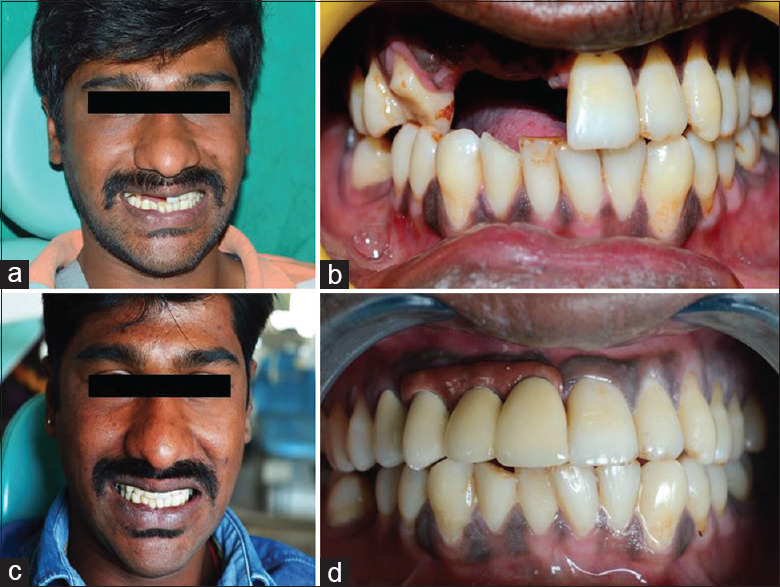
Implant (MIS 3.75 mm × 11.5 mm - standard platform) was placed using manual acrylic surgical guide obtained through wax up. Standard protocol of implant placement was followed with 40N torque to exhibit excellent primary stability. After 6 months of healing period, osseointegration was evaluated, and second-stage surgery was performed. Placement of the implant was not ideal as the bone architecture was compromised; henceforth, it was decided to make a customized abutment with screw-retained substructure framework with cement-retained individual single-unit crown, which gives the concept of Malo bridge. After 3 weeks, implant-level impression was made with polyvinyl siloxane-3M impression material. Implant laboratory analog (MIS) was attached to the impression coping (MIS-Closed tray), and implant cast was fabricated that incorporated a soft tissue gingival mask (Gi-MASK coltene) with a Type IV die stone (Kalrock). On examination of master cast with wax up, opening access hole was at the labial surface of the connector region, and also, there was need for soft tissue replacement for better esthetics to maintain pink and white ratio. Hence, the customization of abutment with Malo bridge pattern was planned digitally, and the cast was scanned with scan body using laboratory scanner. The customized abutment with substructure of Malo bridge was designed in software (exocad) and it was printed in DMLS 3D printing technology [Figure 1]. This prosthesis consists of two parts: the first being substructure with customized abutment and the next part being superstructure, i.e., individual crowns. After printing the substructure, pink composite (Adoro-Ivoclar Vivadent) was layered to mimic soft tissue [Figure 2]. Pink color gingival component was matched using photograph and customized shade tab. Framework was tried in patient mouth and checked for passive and precise fit of the prosthesis. After trial, ceramic-layered DMLS crowns were fabricated based on previously corrected provisional restoration [Figure 2]. The abutment screw was then tightened in 30N torque followed by which cotton pellet and gutta-percha were packed to prevent the rotation of abutment screw and to seal the access hole. An individual ceramic-layered crown was cemented on 3D-printed metal substructure using self-adhesive universal resin cement (Relyx Unicem, 3M) [Figure 3]. The inner surface of the prosthesis was given modified ridge lap pattern to avoid food lodgment. The fractured premolar was endodontically treated and was restored with full veneer crown. Water Pik was recommended to the patient to maintain oral hygiene.
Figure 1.

(a) Computer-aided design of substructure. (b) Design on the virtual model. (c) Three-dimensional–printed metal substructure with customized printed abutment. (d) Metal framework trial
Figure 2.

(a) Indirect composite layering on framework. (b) Individual crowns on the framework. (c) Framework trial. (d) Try-in of Malo bridge component
Figure 3.

(a and b) Preoperative photographs. (c and d) Postoperative Photographs
Discussion
Metal ceramic fixed prosthesis for implant restoration is rarely recommended for atrophic jaw.[6] The huge size, excessive weight, and thermal expansion during the application of the porcelain layer may interfere with precision–fit due to repeated firing cycles.[7] Another choice is hybrid prosthesis which has a more chance of acrylic debonding and breakage.[8,9] Hence, the best choice is the individual ceramic-layered crowns cemented on a 3D-printed metal substructure, which addresses to greater extent in those limitations. Hence, Malo bridge design was chosen as it was nonideal placement of the implant due to vertical bone loss. On the other hand, this type of prosthesis eliminates the screw access openings in the occlusal surface of the crowns and also makes it possible to remove and repair the fractured porcelain of the individual crown without removing the whole structure. In addition, sealing of the gingival component resembles the esthetics of the anatomical gingival sulcus and allows removal of the excess cement without damaging the pink esthetic. DMLS is one of the 3D printing technologies that directly can create a metal component from its 3D computer model. The implant abutment and hex connection were printed through this technology to eliminate all casting errors and to achieve precise passive fit of nonengaging abutment.
Conclusion
In Malo concept, each crown is separate, so it can be removed and fixed again without hampering the whole fixed bridge. The natural look is excellent, the recurrent cost is less, and it is considered to be one of the best methods, as it saves time in future correction. Through 3D printing technology, the fabrication of Malo bridge, a boon for novel approach, has great success outcome in upcoming era of digital prosthesis in implantology for its high precision fit and passivity.
Declaration of patient consent
The authors certify that they have obtained all appropriate patient consent forms. In the form the patient(s) has/have given his/her/their consent for his/her/their images and other clinical information to be reported in the journal. The patients understand that their names and initials will not be published and due efforts will be made to conceal their identity, but anonymity cannot be guaranteed.
Financial support and sponsorship
Nil.
Conflicts of interest
There are no conflicts of interest.
References
- 1.Amit R, Atul B. Preservation of optimal gingival architecture through customised healing abutment in immediate implant placement: A clinical report. Int J Adv Res. 2015;3:156–60. [Google Scholar]
- 2.Nissan J, Ghelfan O, Gross O, Priel I, Gross M, Chaushu G. The effect of crown/implant ratio and crown height space on stress distribution in unsplinted implant supporting restorations. J Oral Maxillofac Surg. 2011;69:1934–9. doi: 10.1016/j.joms.2011.01.036. [DOI] [PubMed] [Google Scholar]
- 3.Birdi H, Schulte J, Kovacs A, Weed M, Chuang SK. Crown-to-implant ratios of short-length implants. J Oral Implantol. 2010;36:425–33. doi: 10.1563/AAID-JOI-D-09-00071. [DOI] [PubMed] [Google Scholar]
- 4.Maló P, de Araújo Nobre M, Borges J, Almeida R. Retrievable metal ceramic implant-supported fixed prostheses with milled titanium frameworks and all-ceramic crowns: Retrospective clinical study with up to 10 years of follow-up. J Prosthodont. 2012;21:256–64. doi: 10.1111/j.1532-849X.2011.00824.x. [DOI] [PubMed] [Google Scholar]
- 5.Maló P, de Sousa ST, De Araújo Nobre M, Moura Guedes C, Almeida R, Roma Torres A, et al. Individual lithium disilicate crowns in a full-arch, implant-supported rehabilitation: A clinical report. J Prosthodont. 2014;23:495–500. doi: 10.1111/jopr.12137. [DOI] [PubMed] [Google Scholar]
- 6.Ameri N, Alikhasi M, Rezayani V. Full mouth rehabilitation with retrievable metal-ceramic implant-supported fixed prostheses for a young patient with atrophic jaws: A clinical report. Clin Case Rep. 2017;5:1531–5. doi: 10.1002/ccr3.1112. [DOI] [PMC free article] [PubMed] [Google Scholar]
- 7.Piermatti J. Using CAD-CAM technology for the full-mouth, fixed, retrievable implant restoration: A clinical report. J Oral Implantol. 2007;33:23–7. doi: 10.1563/0-831.1. [DOI] [PubMed] [Google Scholar]
- 8.Goodacre CJ, Bernal G, Rungcharassaeng K, Kan JY. Clinical complications with implants and implant prostheses. J Prosthet Dent. 2003;90:121–32. doi: 10.1016/S0022-3913(03)00212-9. [DOI] [PubMed] [Google Scholar]
- 9.Julia RO, Nieves AM, Cosme GE. Prevalence of complications after the oral rehabilitation with implant-supported hybrid prostheses. Med Oral Patol Oral Cir Bucal. 2012;17:116–21. doi: 10.4317/medoral.17099. [DOI] [PMC free article] [PubMed] [Google Scholar]


