Abstract
Objective:
The purpose of this study was to evaluate the effect of pretreatment with of matrix metalloproteinase inhibitors on the shear bond strength (SBS) of Adper Single Bond 2 total etch adhesive to the primary teeth dentin following 6 months of storage in artificial saliva.
Materials and Methods:
One hundred and twenty primary anterior teeth extracted for orthodontic reasons were selected. After etching, dentin blocks from each tooth were pretreated for 60 s with: (i) phosphate-buffered saline (0.01 M, pH 7.2) as the control group, (ii) 17% ethylenediaminetetraacetic acid (EDTA), (iii) 2% doxycycline (DO) solution, and (iv) 2% chlorhexidine (CHX) solution, with subsequent application of an etch-and-rinse adhesive system (Adper Single Bond 2). The composite was placed in clear Teflon cylinders. The SBS values were determined immediately and following 6 months of aging with a universal testing machine. Failure mode was evaluated using the stereomicroscope and scanning electron microscope. Data were analyzed by the SPSS software using the one-way analysis of variance and post hoc tests (P = 0.05).
Results:
At baseline, no significant difference was observed between control, EDTA, CHX, and DO groups (P = 0.554). Following 6 months of aging, the SBS of the CHX group was significantly higher than the control group (P = 0.013). However, the SBS of the control, EDTA, and DO groups was not statistically different (P < 0.05).
Conclusions:
Following 6 months of aging, among different groups of the study, only CHX significantly preserved the SBS of composite resin to primary teeth dentin using Adper Single Bond 2 adhesive.
Keywords: Composite, matrix metalloproteinases inhibitor, primary teeth, shear bond strength
Introduction
Long-term studies have shown a decrease in the bond strength of resin-bonded dentin over time.[1] Deterioration of the hybrid layer is considered as the primary reason for compromising the resin-dentin bond durability.[1,2] Apart from the extrinsic factors, such as water or oral fluid sorption and polymer swelling, some intrinsic host-derived enzymes like matrix metalloproteinases (MMPs) are also responsible for disintegration of the hybrid layer.[3]
MMPs are a group of zinc and calcium-dependent enzymes, which are trapped within the mineralized dentin matrix during the tooth development process. Simplified etch and rinse adhesives and less destructive versions of self-etch adhesives used during dental procedures, and the caries process itself can contribute to the release and activation of these endogenous MMPs and lead to dentin-adhesive bond failure.[3,4]
Moreover, lower grades of resin monomer infiltration within the acid-etched dentin using etch-and-rinse adhesives may result in the formation of incompletely infiltrated zones and denuded collagen fibrils at the bottom of the hybrid layer.[5] Dentin MMPs can degrade these unprotected collagen fibrils.[6] From a clinical standpoint, MMP inhibitors such as chlorhexidine (CHX) can play an imperative role in the longevity of the resin bond to dentin.[2]
CHX chelates and sequestrates cations such as calcium and zinc which are required for the activation of the MMPs, thus inhibiting collagenase and gelatinase activity in dentin matrices.[7] Recent in vivo and in vitro studies have demonstrated that the application of CHX has a broad-spectrum MMP-inhibitory effect and considerably preserves the unity of the hybrid layer formed by etch-and-rinse adhesives.[3]
Moreover, tetracyclines (TCs) and their semisynthetic forms (minocycline and doxycycline [DO]) that are commonly used in the treatment of periodontitis, show inhibitory effects on the collagenase and gelatinase enzymes like MMPs.[4] Based on the findings of a recent research, pretreatment of acid-etched dentin with aqueous solutions of semisynthetic TCs (minocycline and DO) improves immediate bonding performance.[8]
Another extrinsic agent with MMP inhibitory capacity is ethylenediaminetetraacetic acid (EDTA). The application of EDTA as a chelating compound has been shown to inactivate the endogenous MMP action in human dentin.[4] Singh et al. showed that EDTA had an MMP inhibitory effect, which could enhance the durability of the resin-dentin bond.[9]
The beneficial effects of dentin surface pretreatment with MMP inhibitors would possibly become apparent over time, as the dentin bond strength is not immediately impaired.[10]
To date, the efficacy of these MMP inhibitors to prevent the loss of dentin bond strength over time has not been determined in the primary dentition. Thus, the present study intended to evaluate the effect of primary teeth dentin pretreatment with inhibitors of MMP enzymes on the shear bond strength (SBS) of Adper Single Bond 2 adhesive, immediately and following 6 months of aging, with microscopic evaluation of the bond failure mode.
Materials and Methods
Initial specimen preparation
The study was approved by the local ethics committee. One hundred and twenty primary anterior teeth (from boys and girls, with ages between 8 and 10 years) extracted for orthodontic reasons (from June/2019 to August/2019) were selected for the study (September/2019). Written informed consent was obtained from the parents or guardians at the time of tooth extraction. The parents were informed about the purpose of the study, privacy preservation, and data anonymity. After visual inspection, the selected teeth were confirmed to be devoid of discoloration, carious lesions, or any other defect. The enamel was removed to create a flat dentinal surface, and the root was cut at the cementoenamel junction. A dentin block (6.0 mm × 6.0 mm × 2.0 mm) was obtained from each tooth. Lack of enamel residue was confirmed using a stereomicroscope (12 × SZ51/61, Olympus, Tokyo, Japan). The dentin blocks were polished with a #600-grit (Water Proof Silicon Carbide Paper, Struers, Erkrath, Germany) wet silicon carbide abrasive paper. Subsequently, the samples were rinsed thoroughly with water.
Etching and bonding procedures and treatment groups
The samples were conditioned with 37% phosphoric acid gel (Scotchbond Etchant, 3M ESPE) for 20 s and washed with water for 15 s. Then, the dentin samples were pretreated with three MMP inhibitors as the following groups, with subsequent application of an etch-and-rinse adhesive system (Adper Single Bond 2).
Group I: control group (n = 30): 0.01M phosphate-buffered saline, pH 7.2 (Sigma-Aldrich, USA) was used. Dentinal surfaces were dried with an absorbent and a stream of air. Subsequently, the resin adhesive layer was applied
Group II: EDTA (Coltene/Whaledent AG, Altstatten, Switzerland) was applied for 60 s[8,10] on the dentin blocks with a micro-brush. Dentinal surfaces were washed and then dried with an absorbent and a stream of air. Subsequently, the resin adhesive layer was applied
Group III-DO (n = 30): 2% DO solution (Sigma-Aldrich, USA) was applied for 60 s on the dentin blocks with a micro-brush. Dentinal surfaces were dried with an absorbent and a stream of air. Subsequently, the resin adhesive layer was applied
Group IV-CHX (n = 30): 2% CHX gluconate solution (Consepsis, Ultradent, USA) was applied for 60 s on the dentin blocks using a micro-brush. Dentinal surfaces were dried with an absorbent and a stream of air. Subsequently, the resin adhesive layer was applied.
Acid-etching, pretreatment, and adhesive application was completed according to the manufacturer's instructions [Table 1]. Subsequently, the composite resin was placed. In all four groups, a clear Teflon trade cylinder (Tygon tubes, ET, Shandong China) measuring 2.65 mm in diameter and 3 mm in length was secured to the lapped tooth surface and served as a mold into which the composite (Filtek 3M, USA) was inserted. The composite was cured (Woodpecker, China) for total time of 40 s from three different sides (20 s from the top and two 10 s from the sides). The specimens were stored in artificial saliva at 37°C. The SBS values were determined immediately for half of the samples in each group (n = 15) and also following 6 months of aging in artificial saliva for another half (n = 15) with a universal testing machine (Zwick/Roll Z020, Zwick GmbH and Co, Germany). The test was performed by securing the specimens in a mounting jig, and a sharp straight-edge chisel attached to the cross-head was used to apply a shearing force of 0.5 mm/min until failure.
Table 1.
Acid etching and adhesive application procedures
| Acid etching | Adhesive application | |
|---|---|---|
| Material | Acid etch (Scotchbond Etchant, 3M ESPE) | Adper Single Bond 2 (3M ESPE, USA) |
| Composition | Acid phosphoric 37% | Bis-GMA, HEMA, water, dimethacrylates, ethanol, photoinitiator system, methacrylate functional copolymer of polyacrylic and polyitaconic acids, 10% by weight of 5 nm-diameter spherical silica nanoparticles |
| Application technique | Apply etchant for 20 s Rinse for 15 s Gently air-dry (10 s at 20 cm) |
Apply one coat of adhesive with gentle agitation Gently air-dry (10 s at 20 cm) Apply second coat of adhesive Gently air-dry (10 s at 20 cm) Light curing for 20 s |
Bis-GMA: Bisphenol A glycerolate dimethacrylate; HEMA: Hydroxyethyl methacrylate
Mode of fracture failure
All specimens were examined under a stereomicroscope (BS-3060C, BestScope, China) to determine modes of failure, which were categorized as follows:
Type I: adhesive failure in the tooth-composite interface
Type II: cohesive failure in the composite or dentin structure
Type III: mixed adhesive and cohesive failure.
Preparation for visualization using field-emission scanning microscope
Two cut sections of sheared dentinal surfaces from each group were examined using magnifications up to ×1130 for analysis, with emphasis on areas of adhesive or cohesive failure. The specimens were mounted on aluminum stubs with conductive silver liquid, sputter-coated with gold, and examined under a field-emission scanning electron microscope (SEM) (TE-SCAN, VEGA3, USA) for verification of the type of failure.
Statistical analysis
SPSS version 20 (SPSS Inc., IL, USA) was used to assess the collected data. Data were analyzed using the one-way analysis of variance (ANOVA) analysis and least significant difference (LSD) Post hoc test. P < 0.05 was considered statistically significant. The mean results of baseline SBS tests and aging tests were compared by the paired t-test.
Results
The mean SBS values and their respective standard deviations of different groups of the study for immediate tests (0 month) and following 6 months of aging tests are presented in Table 2. The highest numerical SBS values were observed when CHX was applied in both periods of times. DO group recorded the second highest bond strength values.
Table 2.
Comparison of mean shear bond strength between 0 and 6 month in groups of the control, chlorhexidine, ethylenediaminetetraacetic acid, doxycycline and comparison of 4 group (the control, chlorhexidine, ethylenediaminetetraacetic acid, doxycycline) in baseline and 6 months
| Variable | SBS base (0 month) |
SBS after 6 months |
Paired t | P | ||
|---|---|---|---|---|---|---|
| Mean | SD | Mean | SD | |||
| Control | 19.20 | 2.55 | 16.44 | 2.48 | 89.28 | ≤0.001* |
| EDTA | 19.50 | 3.44 | 17.45 | 3.48 | 78.40 | ≤0.001* |
| DO | 19.80 | 3.89 | 18.30 | 3.80 | 13.84 | ≤0.001* |
| CHX | 21.60 | 5.73 | 21.18 | 5.72 | -1.33 | 0.214 |
| F | 0.708 | 2.56 | ||||
| P | 0.554† | 0.070† | ||||
*Paired sample t-test; †One-way ANOVA test ≤0.05 is significant. *EDTA: Ethylenediaminetetraacetic acid; DO: Doxycycline; CHX: Chlorhexidine; SBS: Shear bond strength; SD: Standard deviation
At baseline, one-way ANOVA analysis did not show any significant difference between control, EDTA, CHX, and DO groups (P = 0.554) [Table 1].
The results of one-way ANOVA following 6 months of intervention in four groups showed that there was not a significant difference between control, EDTA, CHX, and DO groups (P = 0.070). However, LSD Post hoc test was used to compare the differences between these groups.
The results showed that the mean of postintervention test in the CHX group was significantly higher than the control group (P = 0.013). According to the findings, there were no statistically significant differences between the mean SBS values of the control, EDTA, and DO groups (P > 0.05) [Table 3].
Table 3.
Least significant difference Post hoc analysis results
| Group | Mean difference | P |
|---|---|---|
| Control versus CHX | −4.74 | 0.013* |
| Control versus EDTA | −1.01 | 0.571 |
| Control versus DO | −1.86 | 0.323 |
| CHX versus EDTA | 3.73 | 0.416 |
| CHX versus DO | 2.88 | 0.130 |
| EDTA verrsus DO | −0.85 | 0.643 |
P≤0.05 is significant. EDTA: Ethylenediaminetetraacetic acid; DO: Doxycycline; CHX: Chlorhexidine
Finally, comparing the mean results of baseline SBS tests and aging tests by paired t-test showed statistically significant differences (P ≤ 0.001) except for the CHX group. Bond strength values were preserved when CHX was used (P = 0.214) [Table 2].
Microscopic analysis
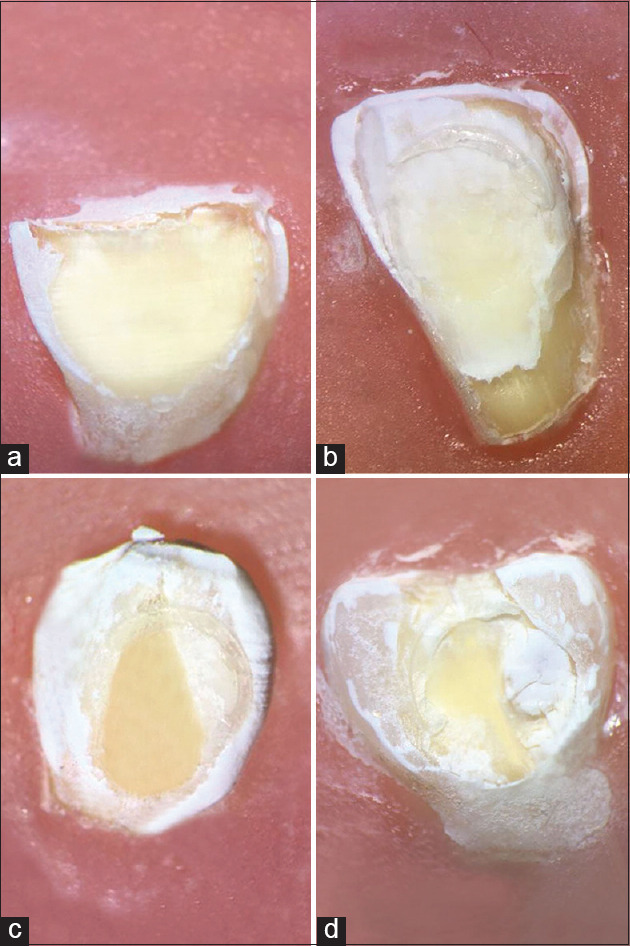
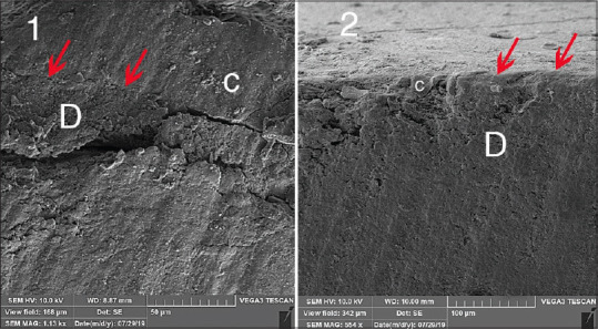
The prevalence of the failure modes confirmed by stereomicroscope is shown in Table 4. The most commonly occurring failure modes were mixed failures. Figure 1 shows the images related to the stereomicroscope evaluation for all groups of the study after 6 months of aging. The results of SEM analysis were also consistent with the mentioned data analysis. Figure 2 illustrates representative images from visualization of cut sections of sheared dentinal surfaces by SEM In images I (CHX group) and II (control group), which are related to the groups with highest and lowest SBS values (mixed failure and adhesive failure are evident, respectively.)
Table 4.
The prevalence of the failure modes
| Group | n | Immediate failure mode |
Aging failure mode |
|||||
|---|---|---|---|---|---|---|---|---|
| I | II | III | I | II | III | |||
| I | Control | 15 | 11 | 0 | 4 | 10 | 0 | 5 |
| II | EDTA | 15 | 5 | 5 | 5 | 4 | 6 | 5 |
| III | DO | 15 | 4 | 5 | 6 | 4 | 3 | 8 |
| IV | CHX | 15 | 3 | 2 | 10 | 3 | 3 | 9 |
EDTA: Ethylenediaminetetraacetic acid; DO: Doxycycline; CHX: Chlorhexidine
Figure 1.

Stereomicroscope evaluation for all study groups. (a) Control group, (b) Ethylenediaminetetraacetic acid group, (c) doxycycline group, (d) chlorhexidine group
Figure 2.

Representative scanning electron microscope images of the cut sections of sheared dentinal surfaces. 1: Chlorhexidine group and 2: Control group (C: Composite, D: Dentin)
Discussion
Numerous factors affect the bond strength of adhesive agents. Mechanical stresses induced by chewing forces, variations in intraoral pH and temperature, water sorption, resin shrinkage, and enzymatic action of MMPs affect the bond integrity to varying extents.[11]
Despite the development of simplified adhesive systems in recent years, etch-and-rinse adhesives are still considered as the gold standard in terms of durability and bond strength.[8] The acidic monomers in etch-and-rinse adhesives expose the collagen fibrils of the organic matrix during bonding procedures.
Hybrid layer formation requires the infiltration of adhesive resin monomers to the exposed collagen network. However, a layer of exposed collagen resulting from incomplete resin infiltration may remain at the bottom of the hybrid layer. These collagen fibrils are susceptible to degradation by MMP host-derived enzymes activated through acid contact and water uptake.[12]
Pretreatment with MMP inhibitors is considered as a valid alternative to prolong the resin-dentin bond stability by overcoming this self-degradation process.[9]
On the other hand, deciduous teeth have less mineral and more organic matrix than permanent teeth. The advantages of using MMP inhibitors would be evident in deciduous teeth as their hybrid layer is more susceptible to degradation over time.[12]
In the present study, the effect of three different MMP inhibitors on the SBS of resin composite to primary teeth dentin was assessed at baseline and following 6 months of storage of samples in the artificial saliva.
The results of the present study showed improved dentin bond strength with the application of CHX on etched dentin at baseline which was preserved following 6 months of aging. No significant reductions in bond strength value were observed when CHX was used (P = 0.214). Other studies also observed that applying a 2% CHX digluconate solution before composite placement successfully preserved the bond strength for 6 months when etch-and-rinse adhesive systems were used.[13,14,15] Studies by Manfro et al. and Breschi et al. reported that bond strengths in CHX-treated specimens were significantly higher than the nontreated groups after 12 months of aging.[16,17] The results of the present study corroborated these findings.
The use of CHX preserved the bond interface, possibly due to its inhibitory effect on MMPs found in etched dentin.[6] This observation was in agreement with the results of the studies by Carrilho et al. and Zheng et al.[18,19]
CHX maintains a strong affinity to the dental tissue by binding to the phosphate groups of mineralized dentin crystallites and negative carboxyl groups of the collagen matrix. The long-term effectiveness of CHX can be explained by its high substantivity, regardless of the concentration. CHX can oversaturate protease binding sites and remain bound to collagen fibrils for later release in higher concentrations.[7]
Furthermore, CHX water-based solutions act as rehydrating agents by maintaining the humidity needed to keep the collagen network of dried dentin in an expanded condition.[2]
Ricci et al. confirmed that the use of CHX reduced the rate of resin-dentin bond deterioration within the first few months of restoration.[20] An in vivo study on primary teeth showed extensive nanoleakage and degradation of the hybrid layer even in teeth with clinically intact cavosurface margins after only 6 months of clinical service. Conversely, when CHX was applied to etched dentin, the hybrid layer deterioration was significantly reduced.[21]
Contradictory results have been reported in a few studies on the primary dentition. Findings of a study by Lenzi et al. showed that CHX did not influence the immediate bond strength to sound or caries-affected dentin of primary and permanent teeth.[22] The results of the study by Viera et al. in this context showed the adverse effect of CHX on bond strength of single bond adhesive to the primary teeth dentin.[23] The differences observed could be attributed to the different materials and techniques used or the pretreatment of caries-affected dentin.
The study by Thompson et al. asserted that 17% EDTA significantly inhibits endogenous MMP activity of human dentin within 1–2 min.[24]
However, in the present study, the application of EDTA did not show significant differences in SBS in comparison with the control group. Furthermore, EDTA is water soluble, and it might be rinsed off the EDTA-treated dentin. Hence, it may have been unable to sustain MMP inhibition for as long as 6 months.[7]
Based on our findings, the highest numerical SBS values of the composite to primary teeth dentin were observed for the CHX followed by DO groups after 6 months, as was reported in the study by Li et al.[8] Loguercio et al. showed that the use of 2% minocycline and 2% CHX as pretreatment of acid-etched dentin retarded the degradation of the resin-dentin interface over 24 months.[25]
However, in some studies, DO negatively affected the bonding of etch-and-rinse adhesives.[26,27] The increased depth of demineralization was not compatible with the extent of resin monomer infiltration in the study by Elkassas and El Zohairy.[26] In the study by Stanislawczuk et al.,[27] the phase separation observed for DO after mixing with bonding agents resulted in lower SBS values observed.
Evaluating the sheared surfaces by stereomicroscope and SEM, the present study revealed that the failure modes were mostly adhesive for the control group. An increased percentage of mixed failures with MMP inhibitors was observed, which could be attributed to the increased SBS values.
Although the performance of CHX was superior in our study, it should be mentioned that the molecules of this inhibitor are water-soluble and may gradually leach out from the adhesive interface, especially when it comes in contact with an external environment (for example, through marginal gaps). Therefore, further long-term laboratory and clinical studies (longer than 6 months) are suggested to confirm our findings.
Conclusions
Following 6 months of aging, among different groups of the study, only CHX significantly preserved the SBS of composite resin to primary teeth dentin using Adper Single Bond 2 adhesive. Future clinical trials are recommended to verify the effect of CHX and other MMP inhibitors on long-term bonding durability in primary dentition.
Financial support and sponsorship
Nil.
Conflicts of interest
There are no conflicts of interest.
Acknowledgments
The authors thank the Vice-Chancellery of Shiraz University of Medical Sciences for supporting this research (Grant# 20969). The authors also thank Dr. Vossoughi from the Center for Research Improvement of the School of Dentistry for statistical analysis and Editage Service for improving the use of English in the manuscript.
References
- 1.Mohammed Hassan A, Ali Goda A, Baroudi K. The effect of different disinfecting agents on bond strength of resin composites. Int J Dent. 2014;2014:231235. doi: 10.1155/2014/231235. [DOI] [PMC free article] [PubMed] [Google Scholar]
- 2.Soares CJ, Pereira CA, Pereira JC, Santana FR, do Prado CJ. Effect of chlorhexidine application on microtensile bond strength to dentin. Oper Dent. 2008;33:183–8. doi: 10.2341/07-69. [DOI] [PubMed] [Google Scholar]
- 3.Zhou J, Tan J, Yang X, Cheng C, Wang X, Chen L. Effect of chlorhexidine application in a self-etching adhesive on the immediate resin-dentin bond strength. J Adhes Dent. 2010;12:27–31. doi: 10.3290/j.jad.a17543. [DOI] [PubMed] [Google Scholar]
- 4.Longhi M, Cerroni L, Condò SG, Ariano V, Pasquantonio G. The effects of host derived metalloproteinases on dentin bond and the role of MMPs inhibitors on dentin matrix degradation. Oral Implantol (Rome) 2014;7:71–9. [PMC free article] [PubMed] [Google Scholar]
- 5.Stanislawczuk R, Amaral RC, Zander-Grande C, Gagler D, Reis A, Loguercio AD. Chlorhexidine-containing acid conditioner preserves the longevity of resin-dentin bonds. Oper Dent. 2009;34:481–90. doi: 10.2341/08-016-L. [DOI] [PubMed] [Google Scholar]
- 6.Bin-Shuwaish MS. Effects and effectiveness of cavity disinfectants in operative dentistry: A literature review. J Contemp Dent Pract. 2016;17:867–79. doi: 10.5005/jp-journals-10024-1946. [DOI] [PubMed] [Google Scholar]
- 7.Matos AB, Trevelin LT, Silva BT, Francisconi-Dos-Rios LF, Siriani LK, Cardoso MV. Bonding efficiency and durability: Current possibilities. Braz Oral Res. 2017;31:e57. doi: 10.1590/1807-3107BOR-2017.vol31.0057. [DOI] [PubMed] [Google Scholar]
- 8.Li H, Li T, Li X, Zhang Z, Li P, Li Z. Morphological effects of MMPs inhibitors on the dentin bonding. Int J Clin Exp Med. 2015;8:10793–803. [PMC free article] [PubMed] [Google Scholar]
- 9.Singh S, Nagpal R, Tyagi SP, Manuja N. Effect of EDTA Conditioning and Carbodiimide Pretreatment on the Bonding Performance of All-in-One Self-Etch Adhesives. International Journal of Dentistry. 2015;2015:141890. doi: 10.1155/2015/141890. DOI:10.1155/2015/141890. [DOI] [PMC free article] [PubMed] [Google Scholar]
- 10.Stape TH, Menezes MS, Barreto BC, Aguiar FH, Martins LR, Quagliatto PS. Influence of matrix metalloproteinase synthetic inhibitors on dentin microtensile bond strength of resin cements. Oper Dent. 2012;37:386–96. doi: 10.2341/11-256-L. [DOI] [PubMed] [Google Scholar]
- 11.Dionysopoulos D. Effect of digluconate chlorhexidine on bond strength between dental adhesive systems and dentin: A systematic review. J Conserv Dent. 2016;19:11–6. doi: 10.4103/0972-0707.173185. [DOI] [PMC free article] [PubMed] [Google Scholar]
- 12.Leitune VC, Portella FF, Bohn PV, Collares FM, Samuel SM. Influence of chlorhexidine application on longitudinal adhesive bond strength in deciduous teeth. Braz Oral Res. 2011;25:388–92. doi: 10.1590/s1806-83242011000500003. [DOI] [PubMed] [Google Scholar]
- 13.Gunaydin Z, Yazici AR, Cehreli ZC. In vivo and in vitro effects of chlorhexidine pretreatment on immediate and aged dentin bond strengths. Oper Dent. 2016;41:258–67. doi: 10.2341/14-231-C. [DOI] [PubMed] [Google Scholar]
- 14.Francisconi-dos-Rios LF, Casas-Apayco LC, Calabria MP, Francisconi PA, Borges AF, Wang L. Role of chlorhexidine in bond strength to artificially eroded dentin over time. J Adhes Dent. 2015;17:133–9. doi: 10.3290/j.jad.a34059. [DOI] [PubMed] [Google Scholar]
- 15.Pilo R, Cardash HS, Oz-Ari B, Ben-Amar A. Effect of preliminary treatment of the dentin surface on the shear bond strength of resin composite to dentin. Oper Dent. 2001;26:569–75. [PubMed] [Google Scholar]
- 16.Manfro AR, Reis A, Loguercio AD, Imparato JC, Raggio DP. Effect of different concentrations of chlorhexidine on bond strength of primary dentin. Pediatr Dent. 2012;34:e11–5. [PubMed] [Google Scholar]
- 17.Breschi L, Cammelli F, Visintini E, Mazzoni A, Vita F, Carrilho M, et al. Influence of chlorhexidine concentration on the durability of etch-and-rinse dentin bonds: A 12-month in vitro study. J Adhes Dent. 2009;11:191–8. [PMC free article] [PubMed] [Google Scholar]
- 18.Carrilho MR, Geraldeli S, Tay F, de Goes MF, Carvalho RM, Tjäderhane L, et al. In vivo preservation of the hybrid layer by chlorhexidine. J Dent Res. 2007;86:529–33. doi: 10.1177/154405910708600608. [DOI] [PubMed] [Google Scholar]
- 19.Zheng P, Zaruba M, Attin T, Wiegand A. Effect of different matrix metalloproteinase inhibitors on microtensile bond strength of an etch-and-rinse and a self-etching adhesive to dentin. Oper Dent. 2015;40:80–6. doi: 10.2341/13-162-L. [DOI] [PubMed] [Google Scholar]
- 20.Ricci HA, Sanabe ME, de Souza Costa CA, Pashley DH, Hebling J. Chlorhexidine increases the longevity of in vivo resin-dentin bonds. Eur J Oral Sci. 2010;118:411–6. doi: 10.1111/j.1600-0722.2010.00754.x. [DOI] [PubMed] [Google Scholar]
- 21.Hebling J, Pashley DH, Tjäderhane L, Tay FR. Chlorhexidine arrests subclinical degradation of dentin hybrid layers in vivo. J Dent Res. 2005;84:741–6. doi: 10.1177/154405910508400811. [DOI] [PubMed] [Google Scholar]
- 22.Lenzi TL, Tedesco TK, Soares FZ, Loguercio AD, Rocha Rde O. Chlorhexidine does not increase immediate bond strength of etch-and-rinse adhesive to caries-affected dentin of primary and permanent teeth. Braz Dent J. 2012;23:438–42. doi: 10.1590/s0103-64402012000400022. [DOI] [PubMed] [Google Scholar]
- 23.Vieira Rde S, da Silva IA., Jr Bond strength to primary tooth dentin following disinfection with a chlorhexidine solution: An in vitro study. Pediatr Dent. 2003;25:49–52. [PubMed] [Google Scholar]
- 24.Thompson JM, Agee K, Sidow SJ, McNally K, Lindsey K, Borke J, et al. Inhibition of endogenous dentin matrix metalloproteinases by ethylenediaminetetraacetic acid. J Endod. 2012;38:62–5. doi: 10.1016/j.joen.2011.09.005. [DOI] [PMC free article] [PubMed] [Google Scholar]
- 25.Loguercio AD, Stanislawczuk R, Malaquias P, Gutierrez MF, Bauer J, Reis A. Effect of minocycline on the durability of dentin bonding produced with etch-and-rinse adhesives. Oper Dent. 2016;41:511–9. doi: 10.2341/15-023-L. [DOI] [PubMed] [Google Scholar]
- 26.Elkassas DW, El Zohairy A. The effect of cavity disinfectants on the micro-shear bond strength of dentin adhesives. Eur J Dent. 2014;8:184. doi: 10.4103/1305-7456.130596. [DOI] [PMC free article] [PubMed] [Google Scholar]
- 27.Stanislawczuk R, da Costa JA, Polli LG, Reis A, Loguercio AD. Effect of tetracycline on the bond performance of etch-and-rinse adhesives to dentin. Braz Oral Res. 2011;25:459–65. doi: 10.1590/s1806-83242011000500014. [DOI] [PubMed] [Google Scholar]


