Abstract
Aim:
To evaluate the in-vitro microleakage of traditional micro hybrid composite resin and 0.2% chitosan-incorporated composite resin when restored in Class V cavities using total etch versus self-etch adhesives after storing in artificial saliva for 24 h.
Materials and Methodology:
Sixty permanent maxillary premolars collected and Class V cavities were prepared on buccal surface of each tooth (dimensions: mesio-distally 3 mm, occluso cervically 2 mm, and depth of 1.5 mm) and restored with Group 1: micro hybrid (30 teeth) and Group 2: chitosan-incorporated composite (30 teeth), which was further subdivided into: (a) 15 teeth using total-etch adhesives. (b) 15 teeth using self-etch adhesives. Next dye extraction test was carried out using spectrophotometer.
Results:
Comparison within groups: In Group 1: Self-etch demonstrated less microleakage (0.0129) compared with total etch (0.0183). The difference was statistically significant, and in Group 2: No statistically significant difference was found in mean microleakage scores after using either self-etch (0.0118) or total etch adhesives (0.0120).
Conclusion:
It can be concluded that chitosan-incorporated composite seems to have improved mechanical properties with a stable bond when used with either self-etch or total etch adhesives in addition to being antibacterial. It may be clinically useful in restoring Class V cavities in patients with high caries risk. However, further in vitro and in-vivo studies need to be carried out.
Keywords: Chitosan, dye extraction test, micro hybrid composite, microleakage, self-etch adhesives, total etch adhesives
Introduction
Effective preventive protocol and enhanced dental care have decreased the incidence of dental caries over a period of time.[1] However, with the increase in geriatric population, root caries and cervical defects have become more prevalent,[2] whether the etiology is caries, erosion, tooth wear, or excessive tooth brushing.[3]
Selecting a restorative material for such lesion is challenging as the etiology is multi-factorial coupled with difficulties in isolation and bonding to root dentin. Hence, one prefers to select a material that is less technique sensitive, has low modulus of elasticity (MOE) to allow the restoration to flex with the tooth on masticatory load, and has less propensity for plaque accumulation.[3] Micro hybrid composite resins with filler size of 0.7–3.6 μm is considered the material of choice in Class V lesions due to its low MOE and the forces generated by polymerization shrinkage are less compared to that of conventional composites.[4]
However, various in vitro and in vivo studies showed greater number of bacteria and plaque accumulation on the surface of composite resin in addition to unsatisfactory antibacterial properties. When compared with other restorative materials, there is evidence of growth of plaque adjacent to the restoration margins, which may lead to secondary caries in vivo and limit the longevity of RBCs (resin-based composites).[5,6]
Hence, attempts have been made to incorporate antibacterial agents into resin component to inactivate bacteria and prevent recurrent caries and decrease plaque accumulation.[5]
Although various antibacterial agents such as chlorhexidine,[5] calcium,[6] antibiotics,[7] magnesium, zinc oxide,[5] and silver ions[7] have been tried, studies showed that resin containing soluble antibacterial agents leach out of mass within few days, resulting in its short-term effectiveness. Another problem was its adverse influence on the mechanical properties. Furthermore, the antibacterial agents may have toxic effect if the release is not properly controlled.[8,9] Hence, there is always a quest for newer natural biomaterial.
A potential biomaterial is chitosan which is naturally acquired polysaccharide that is prepared by deacetylation of chitin, obtained from crab and shrimp shells. It is considered nontoxic, biocompatible, biodegradable, and antibacterial.[10] Moreover it is suggested as a bio-adhesive polymer that provides extended retention in oral cavity. In dental field, chitosan has been used in studies for the prevention of dental caries as it provides bactericidal and/or bacteriostatic characteristics.[11] Chitosan when incorporated in resin composite showed increased biocompatibility and decreased adsorption capacity of bacteria without changing the flexural strength and mechanical properties of the composite.[8] Chitosan, when used in lower concentration, has shown improvement in its adhesion when used with Bisphenol A or HEMA group; this is because at such low concentration Chitosan releases high volume of free amino group.[12] Adhesive system also plays a significant role (total etch or self-etch) for durable composite restoration in providing an impervious seal, but it depends on the procedure of application and mechanism of adhesion. Many researches summarized that total etch systems are technique sensitive where isolation and accessibility are difficult especially in Class V cavities and where dentin is the major substrate. In such situation, self-etch system, due to decrease in number of steps for bonding, would be a preferred option. There is a significant difference in the extent of microleakage evidenced between tooth–restoration interface by using different bonding agents, and its compatibility with the resin contributes to the bondability of the restoration.[13]
Several studies previously have evaluated the properties of chitosan-incorporated composite using total etch adhesive[14] and because no data have been reported so far on the seal-ability of this new combination of resin using self-etch and total etch adhesives and the comparison of microleakage using both, we have undertaken this in vitro study to evaluate the microleakage of chitosan-incorporated composite resin to dentin when used as restorative material in Class v cavities using total etch versus self-etch adhesives.
Aims and objectives
To evaluate the in vitro microleakage of traditional micro hybrid composite resin and 0.2% chitosan-incorporated composite resin when restored in class V cavities using total etch versus self-etch adhesives after storing in artificial saliva for 24 h.
Materials and Methodology
Source of data: Sixty noncarious human permanent maxillary premolars extracted for various reasons were collected.
Inclusion criteria
Intact, noncaries, permanent maxillary premolars with complete root formation were included in the study.
Exclusion criteria
Carious teeth
Previous restoration
Preexisting fractures or cracks
Previous endodontic treatment
Non-carious lesions (attrition, fluorosis)
Tooth with severe root curvatures.
Infection control protocols for extracted teeth were collected for educational purposes:
Collection, storage, sterilization, and handling of extracted teeth were used in the study following OSHA and CDC recommendations and guidelines.
Materials used:
High-speed aerator (NSK Japan)
Straight hand-piece (NSK Japan)
Diamond and carbide bur (Komet, Gebr. Brasseler)
35% phosphoric acid (3M ESPE, St. Paul, MN, USA)
Total etch adhesive: Adper™ Single Bond 2 (3M ESPE)
Self-etch adhesive: Adper™ SE Plus (3M ESPE)
Micro hybrid composite resin (Brilliant NG coltene whaledent)
Light-curing unit (VIP, Bisco Inc., Schaumburg, IL, USA)
Sof-Lex abrasive discs (3M ESPE)
Nail varnish
Artificial saliva
Thermocycling unit
2% methylene blue
UV photo spectrometer
0.2% Chitosan gel (EVEREST BIOTECH)
Centrifuge apparatus.
Sixty freshly extracted maxillary premolars were collected. Calculus and debris were cleaned with a rubber cup and slurry of pumice, disinfected in 0.5% chloramine, and the teeth were stored in artificial saliva at 35°C for <1 month before the restorative procedure. Standardized Class V cavities were prepared on the buccal surface of each tooth with aerator and round diamond bur under air-water cooling. The burs were replaced after every four preparations. The cavities prepared with the following dimensions:
Mesio-distal width: 3 mm, occlusocervical length: 2 mm depth: 1.5 mm.
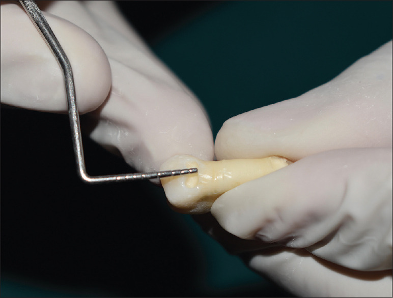
The cavity margins were standardized following the study done by Tavangar et al.[15] The margins were kept in dentin and above the cemento-enamel junction. A William's graduated periodontal probe was used to gauge the dimensions of the cavity [Figure 1]. Subsequently, the teeth were randomly assigned into two experimental groups of thirty teeth in each group which were further divided into two subgroups (n = 15):
Figure 1.

Dimension verified with Perio probe
-
Group 1 – Restored with unmodified composite (control).
- 15 teeth with total etch adhesives
- 15 teeth with self-etch adhesives.
-
Group 2 – Restored with chitosan-composite
- 15 teeth with total etch adhesives
- 15 teeth with self-etch adhesives.
The total etch group specimen were etched with 37% phosphoric acid and left for 15 s and rinsed and Adper Single Bond (fifth generation) was applied according to manufacturer's instruction and light cured for 40 s and restored with micro hybrid composite.
In the self-etch group, Adhesive Adper SE Plus (sixth generation) was applied according to manufacturer's instruction and light cured for 40 s then restored with micro hybrid composite and chitosan-composites, respectively. Polishing of the restoration was done using Sof-Lex abrasive disks. Each specimen was stored in artificial saliva and placed in an incubator (37°C ± 2°C) at 100% humidity for 24 h. The test specimens were then thermo-cycled in water baths held at 5°C and 55°C with a dwell time of 60 s each for 10,000 cycles prior to testing. The teeth were prevented from dehydration by immersing in artificial saliva at room temperature after restoration.
Preparation of chitosan was done by using chitosan powder (0.2%) and resin composite flakes. In a 50-mL glass beaker, chitosan was incorporated into resin composite and homogenously mixed in a dark room with a glass rod. Incorporation was carried out according to a study done by Mirani SA, et al.[16]
Methodology for dye extraction test
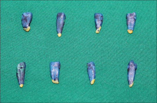
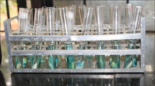
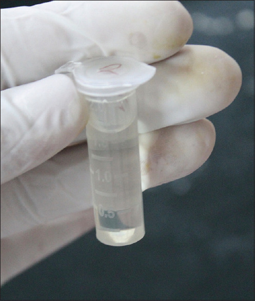
After the restoration, the apical portion of the teeth was sealed with sticky wax. Samples with 1 mm of restoration margins were covered with two layers of nail varnish and immersed in 2% methylene blue solution for 24 h. After 24 h, nail varnish was removed by polishing disks. Then, the samples were put in vials containing 65 wt% nitric acid for 3 days to let methylene blue within restoration dentin interface dilute in nitric acid [Figure 2]. There was 1000 μl acid volume in each vial [Figure 3]. The vials were centrifuged at 14,000 rpm for 5 min and 100 μl of the supernatant from each was transferred to a plate [Figure 4]. The dye absorption was measured by an automatic spectrophotometer at 550 nm using concentrated nitric acid as blank. The results of spectrophotometer indicate the light absorption of methylene blue at the resin–dentin interface which actually showed the microleakage in the restoration.[17]
Figure 2.

Samples after 24 hours storage in Methylene Blue
Figure 3.

Teeth samples dissolved in Nitric Acid for 3 days
Figure 4.

Tooth solution after Centrifugation
Results
Statistical analysis
Independent Student's t-test was used to compare the mean dye penetration (in nm) between Group 1 and Group 2 using total etch adhesive and self-etch adhesive, respectively.
Student's paired t-test was used to compare the mean dye penetration (in nm) between both total-etch groups and both self-etch groups.
Level of significance: (P value) was set at P < 0.05.
Computation: Table 1 demonstrates the results from Independent Student's t-test used and Student's Paired t-test used and the P value.
Table 1.
Comparison of mean dye penetration (nm) between Group 1 and 2 with self-etch and total-etch using independent Student’s t-test
| Group | Time | n | Mean | SD | SEM | Mean difference | t | P |
|---|---|---|---|---|---|---|---|---|
| Group 1 | Total etch | 15 | 0.01834 | 0.0018103 | 0.00047 | 0.00540 | 8.940 | <0.00001* |
| Self-etch | 15 | 0.01294 | 0.0021952 | 0.00057 | ||||
| Group 2 | Total etch | 15 | 0.01204 | 0.0028112 | 0.00073 | 0.00022 | 0.210 | 0.418 |
| Self-etch | 15 | 0.01182 | 0.0031157 | 0.00080 |
*Statistically significant. Group 1: Composite (control); Group 2: Chitosan + composite. SD: Standard deviation; SEM: Standard error of mean
Comparison of mean dye penetration (in nm) between Group 1 and Group 2 after using total etch and self-etch adhesives
The total-etch samples when tested demonstrated that Group 1 exhibited a mean microleakage score of 0.0183 nm and Group 2 a mean microleakage of 0.0120 nm. There was a statistically significant difference between the microleakage scores.
The self-etch samples demonstrated a mean leakage for Group 1 as 0.0129 nm and Group 2 as 0.0118 nm. No statistically significant difference was found between them.
Comparison within the groups:
Group 1: total etch demonstrated a mean leakage of 0.0183 nm and self-etch demonstrated a mean leakage score of 0.0129 nm. There was a statistically significant difference found
Group 2: total etch demonstrated a mean leakage 0.0120 and self-etch groups demonstrated a mean leakage score of 0.0118 nm; the difference was not found statistically significant.
Discussion
Location of Class V lesions make selection of a restorative material hard task as there is always a constant application of masticatory load which has destructive effect of tooth flexure on the cervical region. Micro hybrid composite is considered the material of choice due to its low MOE which allows the restoration to flex with the tooth.[4] They exhibit reduced polymerization shrinkage and offer improved strength. Hence, micro hybrid composite was used in our study.
On analyzing the results of our study, it was found that the unmodified composite group showed a mean microleakage of 0.0129 nm using self-etch and increased microleakage of 0.0183 nm using total etch [Table 1]. The results were in accordance with studies conducted by Nair et al., Moosavi et al., and Kambale et al. and many other researches previously.[18,19,20] Studies have shown that there is presence of tightly clustered spheres that fills the gaps of smaller sized spheres in micro hybrid composites.[21] It has been proved that the low acidity (pH >2.5) leaves dentinal tubules intact with smear plug in self-etch adhesives. This partial demineralization has reported to have an advantage because of the possibility of chemical interaction between some functional monomers (such as MDP and 4-META) and remaining hydroxyapatite crystals along the collagen fibrils. This chemical bonding is suggested to improve the bond durability of the restoration.[13]
The active monomers have acidic properties which dissolve smear layer and demineralize underlying dentin. This depicts that the resultant morphological aspect of the bonded interface is largely dependent on characteristics of the dentin to which the adhesive is being applied and on the aggressiveness of the acidic monomers. Thus, the success of self-etch adhesives in our study could be largely related to the substrate being dentin and the ability to etch and infiltrate simultaneously, thus preventing the discrepancies between demineralization and infiltration.[22]
Studies have shown that water is necessary to maintain collagen fibril expansion in etch and rinse for resin infiltration, but on the contrary, it plays an antagonistic role in hybrid layer formation. This reduces the mechanical properties of the interface and reduces the durability of the bonded surface. These uneven stress distributions along the components of the hybridized zone might have caused enzymatic degradation of collagen fibrils that were left exposed and the hydrolysis of the poorly formed adhesive polymer. It is a proven fact that there is matrix and/or filler deterioration due to mechanical and/or environmental loads, interfacial debonding, micro cracking, and/or filler particle fracture.[13] All these factors may have contributed to increased microleakage seen in unmodified micro hybrid composite group using total etch adhesive after storage in artificial saliva.
In the chitosan + composite group: Comparing the total etch samples, chitosan-composite group showed lower microleakage value (0.0120 nm) compared to unmodified composite group (0.0118 nm), and the difference was found to be statistically significant [Table 1]. This indicates that incorporation of chitosan has not adversely affected the bonding properties of composite to dentin.
On comparing microleakage within self-etch groups: The microleakage of unmodified composite group (0.0129 nm) was not found to be statistically significant compared to chitosan-composite group (0.0118 nm), suggested an improve bond stability of the restoration using self-etch adhesive [Table 2].
Table 2.
Comparison of mean dye penetration (nm) between self-etch and total-etch in Group 1 and Group 2 using student’s paired t-test
| Group | Time | n | Mean | SD | SEM | Mean difference | t | P |
|---|---|---|---|---|---|---|---|---|
| Total etch | Group 1 | 15 | 0.01834 | 0.0018 | 0.00047 | 0.0063 | 6.004 | 0.00002* |
| Group 2 | 15 | 0.01204 | 0.0028 | 0.00073 | ||||
| Self-etch | Group 1 | 15 | 0.01294 | 0.0022 | 0.00057 | 0.0011 | 1.066 | 0.15223 |
| Group 2 | 15 | 0.01182 | 0.0031 | 0.00080 |
*Statistically significant. SD: Standard deviation; SEM: Standard error of mean
Within chitosan-composite groups: Comparing between total etch and self-etch adhesives, there was no significant increase in microleakage scores (from 0.0120 to 0.0118 nm), indicating that there was hardly any bond degradation occurring with either of the bonding agent (self-etch/total etch) used even after storing in artificial saliva for 24 h.
The reduction in microleakage values evident in the chitosan-composite group may be attributed to the fact that: Chitosan incorporated in the composite resin may act as a space occupier since the amine groups makes it very reactive along with–OH group as revealed in the Fourier transform infrared spectroscopy (FTIR) analysis according to a study done by Satheesh et al. Further, chitosan (a biomedical cationic amino polysaccharide) is said to act as an inert filler having–NH2 chain and the presence of high nitrogen content (6.89%) resulted in improved adhesion between the constituents of composite resin, less leaching of resin monomer into the liquid prevented hydrolytic bond degradation, decreasing the volumetric shrinkage compared to regular composites there by reducing microleakage.[23]
Furthermore, it has been speculated by Satheesh et al. in their study that at lower chitosan loading (2.5 wt% and below), relatively uniform dispersion of the additive can be achieved. The thermal stability of the system increases with chitosan loading while mechanical tensile strength is not compromised. According to them, the utilization of HMDA hardener and the introduction of excessive free amine groups through chitosan chains may have improved the adhesion between constituent components.[23]
Several authors have also proved that − 0.12% and 0.25% (w/w) chitosan does not adversely affect adhesive properties of the bonding system. Because chitosan is a cycloaliphatic amine, the amine groups present in chitosan could increase in thermal stability which may be associated with the increase in cross linking of the epoxy with chitosan.[24] A similar mechanism could have been responsible for the reduction of microleakage in our study.
It appears that the chitosan-modified composite is an exciting combination, beneficial especially in class v cavities. Hence, it may be considered an alternative to traditional micro hybrid composite as it combines the benefits of antibacterial properties and better marginal seal.
However, further long-term in vitro and in vivo studies with regard to its antibacterial effectiveness, water sorption, solubility, and long-term stability together with refinement in formulation technique is necessary before it can be recommended for routine clinical usage.
Conclusion
Within the limitation of the present in vitro study, the following conclusions can be drawn:
When total etch samples when tested, Group 2 exhibited a significantly reduced microleakage of 0.0120 nm than Group 1 with a mean microleakage of 0.0183 nm. This indicates that addition of chitosan didn't interfere with the bonding of composite resin to dentin
The self-etch group demonstrated a mean leakage of 0.0129 nm with Group 1 and 0.0118 nm with Group 2. There was no statistically significant difference between the mean microleakage scores which signifies that the self-etch adhesives sealed the margins satisfactorily among both the type of composites used
Comparing within Group 1, self-etch group demonstrated less microleakage compared to total etch. The difference was statistically significant in this group suggesting one step self-etch agents have better bond-ability than total etch adhesives.
In Group 2, no significant difference was found in the microleakage score after using either self-etch or total etch adhesives, suggesting that no bond degradation had taken place after incorporation of chitosan in composite resin.
Clinical implications
Based on the results of our study and that found in the literature, it is evident that chitosan-incorporated composite in addition to being antibacterial, seemed to have improved mechanical properties and bond stability compared to unmodified micro hybrid composite when used with either self-etch or total etch adhesives.
Considering the above advantageous properties of this material, their use may be clinically helpful in restoring class v cavities in patients with high caries risk.
However, further in vitro and in vivo studies need to be carried out to evaluate this noble restorative material for its long-term durability, bond-ability, color stability, solubility, and most importantly retention of its antibacterial property over a longer period of time.
References
- 1.Lagerweij MD, van Loveren C. Declining caries trends: Are we satisfied? Curr Oral Health Rep. 2015;2:212–7. doi: 10.1007/s40496-015-0064-9. [DOI] [PMC free article] [PubMed] [Google Scholar]
- 2.Namita RR. Adolescent rampant caries. Contemp Clin Dent. 2012;3(Suppl 1):S122. doi: 10.4103/0976-237X.95122. [DOI] [PMC free article] [PubMed] [Google Scholar]
- 3.Perez Cdos R, Gonzalez MR, Prado NA, de Miranda MS, Macêdo Mde A, Fernandes BM. Restoration of noncarious cervical lesions: When, why, and how. Int J Dent. 2012;2012:687058. doi: 10.1155/2012/687058. [DOI] [PMC free article] [PubMed] [Google Scholar]
- 4.Srirekha A, Bashetty K. A comparative analysis of restorative materials used in abfraction lesions in tooth with and without occlusal restoration: Three-dimensional finite element analysis. J Conserv Dent. 2013;16:157–61. doi: 10.4103/0972-0707.108200. [DOI] [PMC free article] [PubMed] [Google Scholar]
- 5.Aydin Sevinç B, Hanley L. Antibacterial activity of dental composites containing zinc oxide nanoparticles. J Biomed Mater Res B Appl Biomater. 2010;94:22–31. doi: 10.1002/jbm.b.31620. [DOI] [PMC free article] [PubMed] [Google Scholar]
- 6.Sousa RP, Zanin IC, Lima JP, Vasconcelos SM, Melo MA, Beltrão HC, et al. In situ effects of restorative materials on dental biofilm and enamel demineralisation. J Dent. 2009;37:44–51. doi: 10.1016/j.jdent.2008.08.009. [DOI] [PubMed] [Google Scholar]
- 7.Mirani SA, Sangi L, Kumar N, Ali D. Investigating the antibacterial effect of chitosan in dental resin composites: A pilot study. Pak Oral Dent J. 2015;35:304–6. [Google Scholar]
- 8.Kim JS, Shin DH. Inhibitory effect on Streptococcus mutans and mechanical properties of the chitosan containing composite resin. Restor Dent Endod. 2013;38:36–42. doi: 10.5395/rde.2013.38.1.36. [DOI] [PMC free article] [PubMed] [Google Scholar]
- 9.Imazato S. Antibacterial properties of resin composites and dentin bonding systems. Dent Mater. 2003;19:449–57. doi: 10.1016/s0109-5641(02)00102-1. [DOI] [PubMed] [Google Scholar]
- 10.Casadidio C, Peregrina DV, Gigliobianco MR, Deng S, Censi R, Di Martino P. Chitin and chitosans: Characteristics, eco-friendly processes, and applications in cosmetic science. Mar Drugs. 2019;17:369. doi: 10.3390/md17060369. [DOI] [PMC free article] [PubMed] [Google Scholar]
- 11.Husain S, Al-Samadani KH, Najeeb S, Zafar MS, Khurshid Z, Zohaib S, et al. Chitosan biomaterials for current and potential dental applications. Materials (Basel) 2017;10:602. doi: 10.3390/ma10060602. [DOI] [PMC free article] [PubMed] [Google Scholar]
- 12.Satheesh B, Tshai K. Y., Warrior N. A. “Effect of Chitosan Loading on the Morphological, Thermal, and Mechanical Properties of Diglycidyl Ether of Bisphenol A/Hexamethylenediamine Epoxy System”. Journal of Composites. 2014;vol. 2014:8. Article ID 250290. [Google Scholar]
- 13.Gupta A, Tavane P, Gupta PK, Tejolatha B, Lakhani AA, Tiwari R, et al. Evaluation of microleakage with total etch, self etch and universal adhesive systems in class V restorations: An in vitro study. J Clin Diagn Res. 2017;11:ZC53–6. doi: 10.7860/JCDR/2017/24907.9680. [DOI] [PMC free article] [PubMed] [Google Scholar]
- 14.de Carvalho Nunes RA, do Amaral FL, França FM, Turssi CP, Basting RT. Chitosan in different concentrations added to a two-step etch-and-rinse adhesive system: Influence on bond strength to dentin. Braz Dent Sci. 2017;20:55–62. [Google Scholar]
- 15.Tavangar M, Zohri Z, Sheikhnezhad H, Shahbeig S. Comparison of microleakage of class V cavities restored with the embrace wetbond class V composite resin and conventional opallis composite resin. J Contemp Dent Pract. 2017;18:867–73. doi: 10.5005/jp-journals-10024-2141. [DOI] [PubMed] [Google Scholar]
- 16.Mirani SA, Sangi L, Kumar N, Ali D. Investigating the antibacterial effect of Chitosan in dental resin composites: A pilot study. Pak Oral Dent J. 2015;35:304–6. [Google Scholar]
- 17.Moosavi H, Yazdi FM, Moghadam FV, Soltani S. Comparison of resin composite restorations microleakage: An in vitro study. Open J Stomatol. 2013;3:209. [Google Scholar]
- 18.Nair M, Paul J, Kumar S, Chakravarthy Y, Krishna V, Shivaprasad Comparative evaluation of the bonding efficacy of sixth and seventh generation bonding agents: An in vitro study. J Conserv Dent. 2014;17:27–30. doi: 10.4103/0972-0707.124119. [DOI] [PMC free article] [PubMed] [Google Scholar]
- 19.Moosavi H, Yazdi F, Moghadam F, Soltani S. Comparison of resin composite restorations microleakage: An in-vitro study. Open Journal of Stomatology. 2013;3:209–214. [Google Scholar]
- 20.Kambale S, Hedge V, Munavalli A, Ramesh S, Bandekar DS. Effect of single step adhesives on the marginal permeability of class v resin composites – An in vitro study. IOSR JDMS. 2014;13:44–9. [Google Scholar]
- 21.Sharma RD, Sharma J, Rani A. Comparative evaluation of marginal adaptation between nanocomposites and microhybrid composites exposed to two light cure units. Indian J Dent Res. 2011;22:495. doi: 10.4103/0970-9290.87082. [DOI] [PubMed] [Google Scholar]
- 22.Giannini M, Makishi P, Ayres AP, Vermelho PM, Fronza BM, Nikaido T, et al. Self-etch adhesive systems: A literature review. Braz Dent J. 2015;26:3–10. doi: 10.1590/0103-6440201302442. [DOI] [PubMed] [Google Scholar]
- 23.Satheesh B, Tshai KY, Warrior NA. Effect of chitosan loading on the morphological, thermal, and mechanical properties of diglycidyl ether of bisphenol A/hexamethylenediamine epoxy system. J Compos. 2014;31:273–79. [Google Scholar]
- 24.Elsaka SE. Antibacterial activity and adhesive properties of a chitosan-containing dental adhesive. Quintessence Int. 2012;43:603–13. [PubMed] [Google Scholar]


