Abstract
Pollution and diseases such as the coronavirus pandemic (COVID-19) are major issues that may be solved partly by nanotechnology. Here we review the synthesis of ZrO2 nanoparticles and their nanocomposites using compounds from bacteria, fungi, microalgae, and plants. For instance, bacteria, microalgae, and fungi secret bioactive metabolites such as fucoidans, digestive enzymes, and proteins, while plant tissues are rich in reducing sugars, polyphenols, flavonoids, saponins, and amino acids. These compounds allow reducing, capping, chelating, and stabilizing during the transformation of Zr4+ into ZrO2 nanoparticles. Green ZrO2 nanoparticles display unique properties such as a nanoscale size of 5–50 nm, diverse morphologies, e.g. nanospheres, nanorods and nanochains, and wide bandgap energy of 3.7–5.5 eV. Their high stability and biocompatibility are suitable biomedical and environmental applications, such as pathogen and cancer inactivation, and pollutant removal. Emerging applications of green ZrO2-based nanocomposites include water treatment, catalytic reduction, nanoelectronic devices, and anti-biofilms.
Keywords: ZrO2 nanoparticles, Green synthesis, Biomedical applications, Environmental remediation, ZrO2-based nanocomposites
Introduction
Environmental pollution and climate change are presenting as two of the most globally critical issues in the context of the coronavirus pandemic (COVID-19) and circular economy (Muhammad et al. 2020; Ufnalska and Lichtfouse 2021). The advent of nanoscience and nanotechnology can solve these problems; thereby, holding the key to economic recovery, and environmental mitigation (Weiss et al. 2020; Tang et al. 2021). Nanomaterials play a central role in nanotechnology, raising the demands for sustainable development (Srivastava et al. 2021). The chemical synthesis of nanomaterials chiefly does not satisfy the strict requirements for this trend, and also mismatches with the principles of green chemistry. Currently, the green approaches for the synthesis of nanomaterials are of remarkable significance due to their eco-friendliness as well as cost-effectiveness (Tran et al. 2020a). This also pays the way for multiple applications of nanomaterials in the environmental mitigation, biomedical, and catalysis fields.
Zirconium (Zr) is classified as a transition metal element (d-block) in the titanium family (group IV) with the atomic number 40. Zr does not basically capture neutrons, offering a potential metallic cladding for the fuel rods in the nuclear reactors (Cazado et al. 2021). Zirconium dioxide (ZrO2) or zirconia is one of the highly stable oxides, created by thermalizing zirconium compounds (Hassan and Jalil 2022). Depending on the various synthesis routes, ZrO2 can present in the crystalline phases involving monoclinic, tetragonal, and cubic (Zhang et al. 2018). Bulk ZrO2 has a wide bandgap energy, typically ranging from 5.0 to 7.0 eV. Because of the ultrahigh stability and very low toxicity, ZrO2 has exhibited an intensive range of practical technologies for heat-resistant ceramic superalloys (Wang et al. 2021), dental restorations (Chen et al. 2021), fuel cells (Rambabu et al. 2020), and heterogeneous catalysis (Jiang et al. 2020). Such promising utilizations make ZrO2 an ideal nanomaterial, promoting the green strategy for synthesizing ZrO2 nanoparticles.
In general, there are two major approaches to synthesize the ZrO2 nanoparticles, involving top-down and bottom-up (Jadoun et al. 2021). The former implies the conversion of bulk material into thinner crystallites by the physical route. This means that it needs the participation of enormous mechanical energy sources such as milling, and ionic sputtering (Shrimal et al. 2020). As a result, top-down strategy brings many inevitable drawbacks of causing secondary impressions or intermediates, altering the physicochemical property and surface chemistry of as-synthesized nanoparticles (Indiarto et al. 2021). More importantly, nano-sized particles are mostly unattainable by top-down approach. Meanwhile, the latter implies the formation of particles by creating building blocks from ultra-small particles such as atoms or molecules, and then assembling them together. By this way, the nanostructured particles can be intentionally attainable under the control of fabrication conditions (Rana et al. 2020). The bottom-up strategy involves the physical (e.g., vapor decomposition, plasma irradiation, and ultrasonication), chemical (e.g., sol–gel, co-precipitation, chemical reduction, and hydrothermal), and biological (e.g., plant, fungi, algae, and bacteria) methods. However, the physical bottom-up method requires a high investment cost for operating instruments and consumes a large amount of heating or electric energies (Bolade et al. 2020). The chemical bottom-up method necessarily adopts toxic chemicals for its protocols, resulting in adverse impacts on the environment. Therefore, it may be greatly difficult for physical and chemical methods to attain a critical green strategy.
The biological bottom-up method is the most adequate for the green synthesis of ZrO2 nanoparticles because it offers an effective, tunable, and eco-friendly approach (Shafey 2020). The biogenic synthesis uses low-cost and locally available sources such as plants or other biocompatible sources such as fungi, algae, and bacteria for ZrO2 fabrication (Rana et al. 2020; Nguyen et al. 2021d). The biomolecules extracted from the biological sources play a vital role as extremely efficient bioreducing, biocapping, and biostabilizing agents, bringing excellent ZrO2 production yields (Bandeira et al. 2020). Such biological substrates can safely replace almost highly expensive and toxic chemicals or energy-consuming physical instruments (Agarwal et al. 2017). Biological method for synthesis of ZrO2 nanoparticles also match well with principle of sustainable and green chemistry (Yadi et al. 2018). Because of diminishing the potential risks from chemical and physical methods, generating no hazardous intermediates, and secondary pollutions, biogenic synthesis of ZrO2 nanoparticles can be therefore considered as the green synthesis (Jadoun et al. 2021).
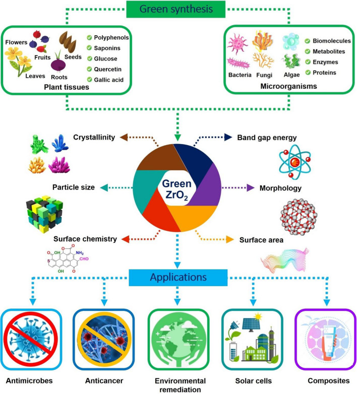
To our knowledge, there is only a limited number of relevant works (about twenty research articles) reported the green synthesis of ZrO2 nanoparticles using various biological sources such as bacteria, fungi, and plant extract. Moreover, there is currently no comprehensive review on the systematic evaluations from the green synthesis to the applications of ZrO2 nanoparticles and their nanocomposites. Therefore, the main objectives of this study are to discuss the current findings on green ZrO2 preparation techniques and highlight their performance in the biomedical field and environmental remediation. Specifically, this review has the following organization: (i) green synthesis methods for ZrO2 nanoparticles; (ii) structural characterizations of green ZrO2 nanoparticles; (iii) biomedical, adsorption, catalytic, and other applications of green ZrO2 nanoparticles; and (iv) emerging applications of green ZrO2-based nanocomposites (Fig. 1). We briefly elucidate some challenging and prospective aspects of green ZrO2 nanocomposites for expanding their potential applications.
Fig. 1.
Green synthesis of ZrO2 nanoparticles and their applications in biomedical, adsorption, catalysis, and nanocomposite fabrication. The plant tissues including flowers, fruits, seeds, leaves, roots, etc. possess many phytochemicals such as polyphenols, saponins, quercetin, and gallic acid. Microorganisms including bacteria, fungi, algae, etc. can secrete biomolecules, metabolites, enzymes, and proteins. These phytochemicals and biomolecules participate in the green synthesis of ZrO2
Green synthesis of ZrO2 nanoparticles
Bacteria
Bacteria are among the fastest-growing microorganisms under mild cultivation conditions in temperature, pressure, and pH (Ram et al. 2020). They are therefore the favorable biofactories for the synthesis of ZrO2 nanoparticles. During the metabolism activities, bacteria secrete some enzymes and proteins extracellularly or intracellularly, which possibly aid the bioreduction, biocapping, and biostabilization of zirconium ions (Fariq et al. 2017). Underlying mechanism of forming ZrO2 nanoparticles by bacterial communities endures further complex steps including (i) biosorption and (ii) bioreduction (Saravanan et al. 2021). The former step initiates with trapping zirconium ions on the bacterial cell surface through the physical and chemical interactions (e.g., electrostatic, hydrogen bonding, ionic exchanging, and chelating) (Gahlawat and Choudhury 2019). Specifically, the secretion of macromolecules such as proteins, polysaccharides containing many negatively charged functional groups enriches the surface chemistry of bacterial cell wall (Patil and Kim 2018). This creates the attraction of Zr4+ ions towards bacterial cell surface. The latter step takes main responsibility for reducing Zr4+ ions into ZrO2 nanoparticles. Bioactive compounds such as acid amines, proteins, etc. in the extracellular or intracellular environment can participate in the formation and stabilization of ZrO2 nanoparticles (Narayanan and Sakthivel 2010).
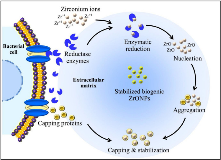
Suriyaraj et al. (2019) reported the one-step biofabrication of ZrO2 nanoparticles using extremophilic Acinetobacter sp. bacterial community. This green source was firstly cultivated in glucose solution, and added by 100 mM ZrOCl2·8H2O for incubation for 12 h. Interestingly, highly crystalline ZrO2 nanomaterial was facilely precipitated under room-temperature condition (37 °C). Moreover, this study found biocompatibility and ineligible cytotoxicity with mouse fibroblast cells. The fluorescein staining was carried out to confirm the major role of extracellular proteins released from Acinetobacter sp. for biosynthesis of ZrO2. Very recently, Ahmed et al. (2021) successfully synthesized zirconia nanoparticles using Enterobacter sp. strain. Notably, the bacterial strain was facilely isolated from the paddy soils, and then, incubated at room temperature. After the bacterial purification, 5 mM ZrOCl2·8H2O was addedto the supernatant under agitating incubation for 24 h. Visible precipitation was finally formed at the bottom of the flask. It was proposed that nitrate reductase enzyme was in charge of reducing Zr4+ ions into zirconia and protein secreted by Enterobacter sp. acts as a capping agent (Fig. 2). As-biosynthesized ZrO2 nanoparticles revealed the promising antifungal activity with 75.5% inhibition against Pestalotiopsis Versicolor disease pathogen. These publications have demonstrated the great potentials and biocompatibility of the bacterially synthesized ZrO2 nanoparticles.
Fig. 2.
Extracellularly biosynthesized mechanism of zirconia nanoparticles using Enterobacter sp. strain. Bacteria extracellularly secrete some specific enzymes such as reductase, which take responsibility for enzymatic bioreduction of Zr4+ ions. The underlying mechanism is attributable to the redox of nicotinamide adenine dinucleotide (NAD+/NADH), which supplies electrons to the reduction of Zr4+ ions during nucleation. Capping proteins can also be released into the extracellular environment and aid the biocapping and biostabilization of zirconia nanoparticles. Reprinted with the permission of Elsevier from reference (Ahmed et al. 2021). Abbreviations: ZrONPs, zirconia nanoparticles; NAD+, an oxidized form of nicotinamide adenine dinucleotide; NADH, a reduced form of nicotinamide adenine dinucleotide
Fungi
Fungi include the microbial organisms that absorb nutrients by decomposing organic matters, without the photosynthesis pathways (Chu et al. 2021). They play the principal role in nutrient cycling and exchanging as well as mitigation of metals-contaminated soils (Riaz et al. 2021). During these processes, fungal communities secrete digestive enzymes into their foods to acquire the nutrients essential for growth (Ferreira et al. 2020). Taking advantage of such features, fungi can be ideal biotemplates for the synthesis of ZrO2 nanoparticles. Basically, the mechanism for green synthesis of ZrO2 nanoparticles using fungal species is closely similar to that using bacterial species. However, the utilization of fungi for ZrO2 production offers many advantageous points (Narayanan and Sakthivel 2010). Firstly, they can endure the harsh synthesis conditions such as flow pressure or agitation in the bioreactor, easily handling the fabrication of ZrO2 (Abinaya et al. 2021). Secondly, fungi have accelerated growth in controllable ways, i.e., through adjusting their nutrient ingredients. Ultimately, almost all fungi are better resistant to genetic or environmental mutations, facilitating the culturing process (Bansal et al. 2011). These features of fungal species are more favorable for biosynthesis of ZrO2 than other biological synthesis methods such as bacteria and plants.
Ghomi et al. (2019) reported that the formation of ZrO2 by extracellular secretion of Penicillium fungal species including P. aculeatum, P. notatum, and P. purpurogenome. Under the optimum conditions at pH 9, and 1.5 mM Zr4+, the solution color changed from pale yellow into deep yellow after seven-day incubation, affirming the generation of colloidal ZrO2. In terms of the ZrO2 biofabrication mechanism, the enzymes presenting in the fungal supernatants may aid to transform Zr4+ ions into ZrO2 nanoparticles. Bansal et al. (2004) cultured another fungal species named Fusarium oxysporum for the successful biosynthesis of ZrO2 nanoparticles from the ZrF62− source at room temperature. To gain insight into the major role of proteins secreted from this fungus for the extracellular hydrolysis of ZrF62−, the resulting filtrates were lyophilized to test for hydrolytic activity. The findings confirmed the cationic nature of proteins to bind easily to ZrF62− anions, which could not be found in other fungal species (e.g., C. lunata, C. gloeosporioides, Phomopsis sp., A. niger). More importantly, ZrF62− anions showed a non-toxicity to Fusarium oxysporum, opening the enormous prospects for large-scale biosynthesis of ZrO2 nanoparticles. To sum up, the highlighted eco-friendly, biocompatible, and energy-conserving advantages of the fungal biosynthesis of ZrO2 nanoparticles are superior to chemical methods, and hence, should not be overlooked.
Algae
Great difference from fungal species, algae are a group of dominantly aquatic, eukaryotic, and autotrophic organisms that acquires their nutrients from the photosynthetic processes. Algae use chlorophyll for their primary photosynthesis; therefore, they are sometimes considered as terrestrial plants. Some algal species such as brown algae, or seaweeds excrete fucoidans from their cell walls (Filote et al. 2021). Fucoidans refer to multifunctional polysaccharides containing a substantial amount of fucose and sulfated esters, exhibiting many bioactivities involving antiviral, antioxidant, and anticoagulant (Ponce and Stortz 2020). These biomolecules can therefore act as ideal bioreducers for the green synthesis of ZrO2. Indeed, Kumaresan et al. (2018) successfully bioprepared tetragonally nanostructured ZrO2 from the aqueous extract of seaweed Sargassum wightii. The biosynthesis procedure was tunable with the grind of ZrO(NO3)2·H2O with Sargassum wightii extract within 20 min. ZrO2 nanoparticles could be formed after the calcination at 400 °C. This nanomaterial showed the spherical morphology with the particle size of 5 nm and was utilized for investigating several potential antibacterial activities against gram-positive and gram negative-bacteria. The researchers demonstrated that large surface area and nanosize (4.8 nm) of ZrO2 obtained from this green strategy contributed considerably to enhancing growth inhibitory effects against antibacterial pathogens. Although many works reported the use of algae for biosynthesizing various nanoparticles, there is still an exception for ZrO2. It is also necessary to elucidate the underlying mechanisms for the formation of ZrO2 nanoparticles using algal species.
Plants
Plants are the most prevalent, locally available, highly cost-effective resource for biosynthesis of ZrO2 nanoparticles (Yadi et al. 2018). Compared with other green materials such as bacteria, fungi, and algae, the use of plants as biotemplates attains many notable benefits (Saravanan et al. 2021). Firstly, there are less risks to health than microbial ZrO2 synthesis strategies. Almost plants are substantially more benign than bacteria or fungi. Some microbial species can even secrete toxins or metabolites during cultivation (Yang and Chiu 2017; Xu et al. 2020). Secondly, the botanical synthesis of ZrO2 nanoparticles is considerably accelerated in comparison with microbial routes. The bioreduction of Zr4+ by biomolecules from plant extract occurs within a few minutes to several hours, while microbial communities mostly prolong these bioprocesses by many days under ambient incubation (Vijayaraghavan and Ashokkumar 2017). This results in the higher kinetic of overall ZrO2 production process. Thirdly, the synthesis of ZrO2 nanomaterial by plant extracts is controllable and easy to manipulate. Diverse parts of plants such as leaves, barks, roots, flowers, etc. can be effortlessly collected for biosynthesis of ZrO2 nanoparticles. The green solvents for extracting phytochemical compounds from plant tissues are mainly water and ethanol. Meanwhile, the microbial culture needs to be carried out under strict conditions to avoid the generation of colonies, unfavorable to ZrO2 fabrication.
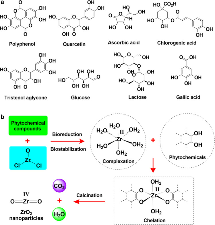
Phytochemicals involving reducing sugars, polyphenols, flavonoids, saponins, and amino acids are inherent in the plant tissues such as flowers, leaves, and roots (Fig. 3a). These compounds are known as strong antioxidants or bioreductants, which can play pivotal roles as biocapping, biochelating, bioreducing, and biostabilizing agents for the synthesis of ZrO2 nanoparticles (Fig. 3b). The phytochemicals participate in the formation and stabilization of octahedral complex of Zr2+–phytochemicals. The presence of phytochemicals also aids to hamper the aggregation of ZrO2 nanoparticles during thermal calcination. This may boost the surface area of ZrO2 nanoparticles, which are conducive to their catalytic and adsorption properties. Consequently, the phytochemicals in plant extract can exhibit a range of important functionalities including replacing hazardous, expensive chemical antioxidants or reductants, and shortening the number of complicated synthesis steps.
Fig. 3.
Phytochemicals in plant extracts (a), and proposed mechanism for the biosynthesis of ZrO2 nanoparticles using these phytochemicals (b). Phytochemicals act as bioreductants to convert Zr4+ into octahedral complex [Zr(H2O)6]2+. This enhances the stability and hinders the clustering of zirconium complexation during chelation with phytochemicals Zr2+–phytochemicals. The calcination of Zr2+–phytochemicals complex produces ZrO2 nanoparticles in tandem with the release of CO2, H2O, N2, and many decomposed products
The general botanical biosynthesis of ZrO2 nanoparticles initiates with the collection and pretreatment of plant tissues to eliminate the irrelevant parts (Table 1). The procedure is followed by extracting phytochemicals using several common kinds of solvents such as water, ethanol, or hexane from cell walls (Saraswathi and Santhakumar 2017; Goyal et al. 2021; Alagarsamy et al. 2022). Water is a prevalently used, and highly polar extractor which recovers the aqueously soluble phytochemical compounds such as gallic acid, polyphenols, glucose, etc. while ethanol exhibits a poorer polarity than water, but higher than hexane. There are two major steps including complexation and calcination to reach the final ZrO2 products. The former is carried out under the incorporation of plant extract into zirconium source, mainly ZrOCl2·8H2O. The proper temperature and time for forming Zr2+–phytochemicals are 55–75 °C, and 1–4 h, respectively. This step can also be assisted by microwave irradiation (900 W) to accelerate the complexation rate up to 15 min (Shinde et al. 2018), or by alkaline addition (Joshi et al. 2021). The Zr2+–phytochemicals complex is totally separated by centrifugation or paper filtration (Gowri et al. 2014). The latter experiences the thermal calcination at high temperature (500–800 °C) for 2–4 h to convert this complex into ZrO2 nanoparticles.
Table 1.
Synthesis of green ZrO2 nanoparticles using plants
| Plant extract and zirconium inputs | Complexation step | Calcination step | Refs. | |||||
|---|---|---|---|---|---|---|---|---|
| Plant species | Plant tissue | Zr source | Zr/extract ratio (v/v) | Temperature | Time | Temperature | Time | |
| Euclea natalensis | Roots | ZrOCl2 | 1:1 | Not reported | 3 h | 550 °C | 3 h | Silva et al. (2019) |
| Aloe vera | Leaves | ZrOCl2 | Not reported | Room | 4 h | 500 °C | Not reported | Gowri et al. (2014) |
| Salvia rosmarinus | Leaves | ZrOCl2 | 1:4 | 70 °C | 1 h | 600 °C | 2 h | Davar et al. (2018) |
| Sapindus mukorossi | Pericarp | ZrOCl2 | 1:1 | 60 °C | 3–4 h | 500–700 °C | Not reported | Alagarsamy et al. (2022) |
| Wrightia tinctoria | Leaves | ZrOCl2 | 1:1 | 75 °C | 3–4 h | 800 °C | Not reported | Al-Zaqri et al. (2021) |
| Ficus benghalensis | Leaves | ZrOCl2 | 1:1 | Microwave | 15 min | 500 °C | 3 h | Shinde et al. (2018) |
| Moringa oleifera | Leaves | ZrOCl2 | 1:1 | 60 °C | 3–4 h | 700–800 °C | Not reported | Annu et al. (2020) |
| Helianthus annuus | Seeds | ZrOCl2 | 1:5 | Not reported | 2–3 h | 600 °C | 4 h | Goyal et al. (2021) |
| Tinospora cordifolia | Leaves | ZrOCl2 | 10:1 | 55 °C | 40 min | Not reported | Not reported | Joshi et al. (2021) |
| Nephelium lappaceum | Fruit | Zr(NO3)4 | 7:1 | 180 °C | 18 h | 500 °C | 4 h | Isacfranklin et al. (2020) |
| Nyctanthes arbor-tristis | Flower | ZrOCl2 | 2:1 | Not reported | 2 h | 300–500 °C | 3 h | Gowri et al. (2015) |
Properties of green ZrO2 nanoparticles
Crystallinity
Bulk ZrO2 nanoparticles exist in three common crystalline phases involving monoclinic, tetragonal, and cubic. Biologically synthesized ZrO2 nanoparticles can also attain such crystalline phases based on X-Ray diffraction analysis (Table 2). Specifically, Davar et al. (2018) reported a cubic phase of ZrO2 nanoparticles bioproduced from Salvia Rosmarinus plant extract with the reflecting planes at (111), (200), (220), (311), (222), and (400). Another study indicated the monoclinic phase of ZrO2 synthesized from Helianthus annuus plant extract (Goyal et al. 2021). However, tetragonal phase is the most prevalent single-crystal phase of green ZrO2 nanoparticles. Green ZrO2 nanoparticles can also have multiphase crystallinity, which includes at least two phases (Sathishkumar et al. 2013; Debnath et al. 2020; Isacfranklin et al. 2020). The presence of crystal phase in green ZrO2 possibly affects their electronic properties, optical band gap, and surface energy, and stability. It is therefore essential for further studies to clarify the role of synthesis conditions, e.g., the ratio between zirconium and plant extract in the formation of ZrO2 phase.
Table 2.
Properties of green ZrO2 nanoparticles
| Green material | Species | Particle size (nm) | Morphology | Crystal phase | Optical band gap (eV) | Surface chemistry | Refs. |
|---|---|---|---|---|---|---|---|
| Alga | Sargassum wightii | 5.0 | Nanosphere | Tetragonal | 4.4 | Hydroxyl, carbonyl, carboxylate | Kumaresan et al. (2018) |
| Bacterium | Enterobacter sp. | 33–75 | Nanosphere | Not reported | Not reported | Hydroxyl, carbonyl, alkene | Ahmed et al. (2021) |
| Bacterium | Acinetobacter sp. | 44 ± 7 | Nanosphere | Monoclinic, tetragonal | 4.9 | Hydroxyl | Suriyaraj et al. (2019) |
| Bacterium | Pseudomonas aeruginosa | 6.14–15 | Nanosphere | Monoclinic, tetragonal | Not reported | Hydroxyl, zirconium hydroxide | Debnath et al. (2020) |
| Fungus | Penicillium | < 100 | Nanosphere | Not reported | Not reported | Hydroxyl, carbonyl, amine | Ghomi et al. (2019) |
| Fungus | Fusarium solani | Not reported | Nanosphere | Tetragonal | Not reported | Hydroxyl, carbonyl | Kavitha et al. (2020) |
| Plant | Euclea natalensis | 5.9–8.54 | Nanosphere | Monoclinic, tetragonal | Not reported | Hydroxyl | Silva et al. (2019) |
| Plant | Aloe vera | < 50 | Nanosphere | Tetragonal | 5.42 | Hydroxyl, carbonyl, zirconium hydroxide | Gowri et al. (2014) |
| Plant | Curcuma longa | 41–45 | Nanochain | Monoclinic, rhombohedral | Not reported | zirconium hydroxide | Sathishkumar et al. (2013) |
| Plant | Aloe vera | 18–42 | Not reported | Face centered cubic | Not reported | Hydroxyl, carbonyl, amine, alkene | Prasad et al. (2014) |
| Plant | Rosmarinus officinalis | 12–17 | Semi-nanosphere | Cubic | Not reported | Hydroxyl, carboxylic, alkyl | Davar et al. (2018) |
| Plant | Sapindus mukorossi | 5–10 | Nanosphere | Tetragonal | 5.535 | Hydroxyl | Alagarsamy et al. (2022) |
| Plant | Wrightia tinctoria | 17 | Nanosphere | Monoclinic, tetragonal | 3.78 | Hydroxyl, alkyl, carboxylic acid | Al-Zaqri et al. (2021) |
| Plant | Lagerstroemia speciosa | 56.8 | Oval | Tetragonal | Not reported | Hydroxyl, carbonyl, amine | Saraswathi and Santhakumar (2017) |
| Plant | Ficus benghalensis | 14.7 | Nanosphere | Monoclinic, tetragonal | 4.9 | Hydroxyl, alkyl, carbonyl | Shinde et al. (2018) |
| Plant | Moringa oleifera | < 10 | Nanosphere | Not reported | Not reported | Hydroxyl | Annu et al. (2020) |
| Plant | Helianthus annuus | 35.45 | Nanosphere | Monoclinic | 3.7 | Carboxyl, alkyl, alkene, amide | Goyal et al. (2021) |
| Plant | Laurus nobilis | 20–100 | Nanosphere | Monoclinic, tetragonal | Not reported | Hydroxyl | Chau et al. (2021) |
| Plant | Tinospora cordifolia | 73 | Nanosphere | Not reported | Not reported | Hydroxyl, carbonyl | Joshi et al. (2021) |
| Plant | Nephelium lappaceum | 50 | Nanorod | Monoclinic, cubic | Not reported | Hydroxyl | Isacfranklin et al. (2020) |
| Plant | Nyctanthes arbor-tristis | < 150 | Nanoflake | Tetragonal | 5.31 | Not reported | Gowri et al. (2015) |
Optical bandgap
The bandgap energy of a semiconductor reflects the energy disparity between the top of valence band and the bottom of conduction band, or the minimum energy to excite an electron from the valence band to the conduction band (Makuła et al. 2018). It can be determined from diffuse reflectance spectra. While the bulk ZrO2 nanoparticles have a wide bandgap energy (~ 5.0 eV), green ZrO2 nanoparticles exhibit values ranging from 3.7 to 5.5 eV (Table 2). Indeed, Goyal et al. (2021) fabricated ZrO2 nanoparticles using methanolic extract of Helianthus annuus seeds with very narrow bandgap (3.7 eV). Al-Zaqri et al. (2021) found the same small value of ZrO2 nanoparticles biosynthesized from Wrightia tinctoria leaf extract. They also gave an assumption about more contribution from external surface defects ZrO2 nanoparticles. Meanwhile, obtaining higher bandgap energies (5.4–5.5 eV) of other green ZrO2 nanoparticles may be ascribed by quantum confinement effects (Gowri et al. 2014; Alagarsamy et al. 2022). The bandgap augments for the smaller size nanoparticles due to the deformity phenomena. Overall, biologically synthesized ZrO2 nanoparticles acquire sufficient gap energies, capable of exhibiting their excellent catalytic and biomedical activities through the formation of reactive oxygen species.
Particle size
Many physicochemical techniques such as X-Ray diffraction, dynamic light scattering, transmission electron microscopy, etc. can be used to measure the average particle size of ZrO2 nanoparticles. From the database of previous studies (Table 2), the particle size of ZrO2 nanoparticles is distributed from 5 to 150 nm, commonly less than 50 nm. Compared with chemical approaches, biological methods generally create ZrO2 nanoparticles with considerably smaller sizes (Suriyaraj et al. 2019; Ahmed et al. 2021). It can be understandable that biomolecules from the secretion of microbial sources, or phytochemicals from plant extracts lessen the assembly of ZrO2 nanoparticles (Kumaresan et al. 2018; Silva et al. 2019; Debnath et al. 2020). Small-size ZrO2 nanoparticles offer many biomedical benefits since they facilitate the penetration into cell walls, performing better antibacterial, antifungal, and anticancer activities (Khatoon et al. 2015).
Morphology
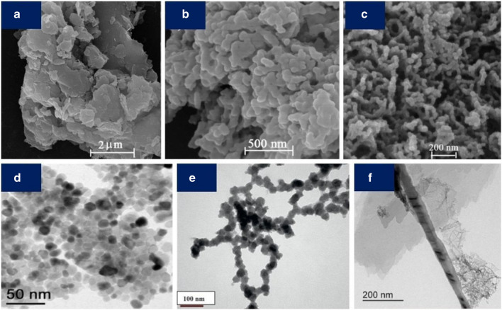
The shape of ZrO2 nanocrystals biosynthesized from biological routes can be explored by scanning/transmission electron microscopy analysis. Accordingly, green ZrO2 nanoparticles can present a wide range of morphological properties such as nanospheres (mainly), nanochains, nanorods, semi-nanospheres, nano-sized ovals, and nanoflakes (Fig. 4). Generally, the ZrO2 morphology is significantly dependent on the synthesis conditions including the ratio between zirconium source and green extract. Spherical ZrO2 can be synthesized by using microbial sources with the long incubation (1–3 days) at room temperature or by several botanical sources such as Euclea natalensis roots, Aloe vera leaves, Ficus benghalensis leaves, Moringa oleifera leaves, Helianthus annuus seeds, Tinospora cordifolia leaves, and Laurus nobilis leaves (Table 2). Meanwhile, other ZrO2 morphologies are relatively rare, mostly synthesized by plant sources. For example, Sathishkumar et al. (2013) obtained ZrO2 nano-chains by the hydrolysis of ZrF62− in the presence of Curcuma longa tuber extract. Gowri et al. (2015) also successfully biosynthesized ZrO2 nanoflakes by the hydrolysis of ZrOCl2·8H2O using aqueously soluble carbohydrates extracted from Nyctanthes arbor-tristis flower. Semi-spherical and oval ZrO2 can be formed from many plant extracts such as Salvia Rosmarinus and Lagerstroemia speciose, respectively (Saraswathi and Santhakumar 2017; Davar et al. 2018). More importantly, diverse morphologies of ZrO2 morphologies can bring many benefits for practical applications. As an example, (Isacfranklin et al. 2020) indicated that ZrO2 nanorods possess sufficient cellular uptake and toxicity, offering the prospect for biomedical application.
Fig. 4.
Field emission scanning electron microscope photographies of ZrO2 nanoparticles produced at 600 °C without Rosmarinus officinalis leaf extract (a), and from Rosmarinus officinalis leaf extract with the ratio between the extract and zirconium
source 3:1 (b), and 6:1 (c). The role of plant extract is to improve the dispersion of ZrO2 nanoparticles during biosynthesis. Reproduced and adapted with the permission of Elsevier from reference (Davar et al. 2018). Transmission electron microscopy photographs of ZrO2 nanospheres (d), nano chains (e), and nanorods (f) biosynthesized from Acinetobacter sp. bacterial community (Suriyaraj et al. 2019), Curcuma longa tuber extract (Sathishkumar et al. 2013), and Nephelium lappaceum fruit extract (Isacfranklin et al. 2020), respectively. Reproduced and adapted with the permission of Elsevier from references (Sathishkumar et al. 2013; Suriyaraj et al. 2019; Isacfranklin et al. 2020)
Surface chemistry
The surface functionalization by chemical groups aids to extend the applications of nanomaterials (Tran et al. 2021). Surface chemistry of ZrO2 nanoparticles can be identified by several physicochemical techniques such as Fourier-transform infrared spectroscopy and X-ray photoelectron spectroscopy or quantified by Boehm titrations (Nguyen et al. 2021b). The surface functional groups on ZrO2 nanoparticles may be originated from the phytochemical compounds. Considering the calcination of the complex of Zr2+–phytochemicals, almost all organic components are decomposed into volatiles or simple products such as CO2, N2, and H2O. A tiny proportion of phytochemicals may still remain due to the chelating with zirconium ions. They can be thermally modified to generate simpler biomolecules with more diverse functional groups such as carbonyl, amine, carboxylate, and alkyl (Table 2). However, hydroxyl groups are detected partly owing to H2O-adsorbed surface of ZrO2 (Kumaresan et al. 2018; Silva et al. 2019; Annu et al. 2020). The functionalization of ZrO2 surface possibly can lead to the improvement of adsorption efficiency towards many pollutants through many key interactions such as electrostatic attraction, hydrogen bond, Yoshida hydrogen bond, and π–π interaction (Suresh et al. 2015; Nguyen et al. 2021c).
Surface area
Theoretically, surface area of nanomaterials links closely to the number of active sites on the edges, resulting in a significant influence on their catalytic activity (Jiang et al. 2011). The higher surface area of ZrO2 nanoparticles can also facilitate the better favorable adsorption process. The bio compounds such as proteins, enzymes, polysaccharides, polyphenols, etc. present in the biological source extract chelating with zirconium ions can prevent the aggregation of ZrO2 nanoparticles during the thermolysis. This enhances the surface area and the number of active sites of ZrO2 nanoparticles. For example, Shinde et al. (2018) reported the surface area (88 m2/g) of ZrO2 nanoparticles biosynthesized from Ficus benghalensis leaf extract using BET (Brunauer, Emmett and Teller) analysis. It was also witnessed an enhancement of the photocatalytic activities of ZrO2 to methylene blue (91%) and methyl orange (69%) for 240 min. These results of green ZrO2 were remarkably higher than those obtained by the chemical methods such as anodization in H2O2/NH4F/ethylene glycol electrolyte (Rozana et al. 2017), decomposition of Zr(OH)4-urea complex (Sudrajat et al. 2016), polymer-assisted sol–gel synthesis (Dhandapani et al. 2016).
Stability
Compared with chemically produced nanomaterials, biologically produced ZrO2 nanoparticles not only exhibit good dispersion but also show better stability. For example, Sathishkumar et al. (2013) found the surprising stability of zirconia nanoparticles up to five months after green fabrication. This phenomenon may be elucidated by the presence of phytochemicals acting as efficient biostabilizers and biocapping agents. The stability of ZrO2 nanoparticles can be assessed by the zeta potential measurements. Basically, the zeta potential values are different with zero, normally minimum ± 30 mV, indicating the high stability of ZrO2 nanoparticles (Jameel et al. 2020). Indeed, Suriyaraj et al. (2019) demonstrated the well-dispersed, anti-clustering, and nanostructured ZrO2 suspension by the measurement of zeta potential (36.5 ± 5.5 mV). Chau et al. (2021) also measured the high zeta potential (− 32.8 mV) of ZrO2 nanoparticles biosynthesized from Laurus nobilis leaf extract by dynamic light scattering analysis, proposing the good anti-sedimentation of the nanomaterials for antimicrobial activities.
Applications of green ZrO2 nanoparticles
Biomedical applications
Biomedical compatibility of green ZrO2 nanoparticles
Bacterial and fungal species attack the immune system of humans and animals and become the major factors causing infections and even deaths (Humbal et al. 2018). Many antibiotics and antifungal pharmaceuticals have been developed over the centuries, but mankind is increasingly encountering the huge problems of antibiotic resistance (Kovalakova et al. 2020). Among the efforts to search for alternatives to antibiotics, bionanotechnology exhibits its high reliability and performance contributing proactively to the abatement of infections (Ong et al. 2018). Many metallic nanomaterials (e.g., silver nanoparticles) possess their inherent antibacterial and antifungal activities; hence, they are widely utilized in biomedical fields (Abbasi et al. 2014). However, the lack of biocompatibility, biostability, and effectivity may restrain the biomedical application of these nanomaterials. Green ZrO2 nanoparticles biosynthesized from environmentally friendly botanical and microbial sources show unique properties, adequate for fabricating biomedical devices. Based on the published literature, this section will discuss some potential activities of green ZrO2 nanoparticles.
Antibacterial activity
The plasma membrane of bacteria is mostly constituted of proteins (e.g., peptidoglycan macromolecules) that charge negatively (Li et al. 2019). ZrO2 nanoparticles charge positively on their surface, leading to electrostatic interactions with bacterial membranes (Xing et al. 2018). This facilitates the biosorption and bioaccumulation of ZrO2 nanoparticles on the cell walls. With the nanoscale size, high surface area, and good biocompatibility of green ZrO2 nanoparticles, they easily pass through cell membrane, inhibit the key metabolic functions, and finally deactivate bacterial cells (Kumaresan et al. 2018). It is suggested that the generation of reactive oxygen species (e.g., ·O2−, and ·OH) is the underlying mechanism (Akintelu and Folorunso 2020). Accordingly, these species damage the genetic material such as deoxyribonucleic and ribonucleic acids, disordering the transcription and translation processes in bacteria (Slavin et al. 2017). Bacterial death occurs as a result of cell division failure.
There are many publications that reported the excellent antibacterial activity of green ZrO2 nanoparticles against both gram-negative and positive bacteria (Table 3). For example, Gowri et al. (2015) investigated the antibacterial activity against E. coli and S. aureus using ZrO2 biosynthesized from Nyctanthes arbor-tristis plant extract. The inhibition zones were used to measure the antibacterial activity of green ZrO2 nanoparticles. The values were significantly improved in the case of the cotton fabric treated with ZrO2 nanoparticles. With very low green ZrO2 concentration (10 μg/mL), Al-Zaqri et al. (2021) revealed good antibacterial activities against gram-negative and positive bacteria. They assumed the major role of biomolecules presented in Wrightia tinctoria leaf extract in improving the properties of green ZrO2 nanoparticles, e.g., small size particles and high surfaces, enhancing their antibacterial activities. In addition to plant extract, ZrO2 can be biosynthesized from other green materials such as fungi and algae for investigating antibacterial activities. Indeed, Kumaresan et al. (2018) reported the use of Sargassum wightii alga for the synthesis of ZrO2, exhibiting the wide zones of inhibition (19–21 mm) against E. coli, S. typhi, and B. subtilis. Meanwhile, Ghomi et al. (2019) utilized the fungus Penicillium to synthesize the ZrO2 nanoparticles. Minimum inhibitory concentrations against P. aeruginosa and E. coli were found, at 0.375, and 0.75 mmol/L, respectively.
Table 3.
Biomedical applications of green ZrO2 nanoparticles
| Green material | Species | Type of activity | Target | Main findings | Ref |
|---|---|---|---|---|---|
| Plant | Nyctanthes arbor-tristis | Antibacterial | Gram negative (E. coli), Gram positive (S. aureus) | Zones of inhibition against E. coli (17 mm), and S. aureus (15 mm) for ZrO2 NPs, and those against E. coli (30 mm), and S. aureus (22 mm) for ZrO2-treated cotton fabric | Gowri et al. (2015) |
| Fungus | Penicillium | Antibacterial | Gram negative (E. coli, P. aeruginosa) and Gram positive (S. aureus) | Minimum inhibitory concentrations for E. coli (0.75 mM), P. aeruginosa (0.375 mM), but not found for S. aureus | Ghomi et al. (2019) |
| Plant | Wrightia tinctoria | Antibacterial | Gram negative (E. coli, P. aeruginosa) and Gram positive (S. aureus, B. subtilis) | Zones of inhibition observed against E. coli (22 ± 0.5 mm), P. aeruginosa (21 ± 0.3 mm), S. aureus (21 ± 0.4 mm), and B. subtilis (20 ± 0.2 mm) at a ZrO2 concentration of 10 μg/mL | Al-Zaqri et al. (2021) |
| Strong antibacterial may be due to the flavonoids, protein, tannins, alkaloids, amino acids and carbohydrates present in the leaf extract | |||||
| Plant | Moringa oleifera | Antibacterial | Gram negative (E. coli, P. aeruginosa) and Gram positive (S. aureus, B. subtilis) | Zone of inhibition against E. coli (4 mm), P. aeruginosa (10 mm), S. aureus (not observed), and B. subtilis (9 mm) at a concentration of 20 mg | Annu et al. (2020) |
| Plant | Helianthus annuus | Antibacterial | Gram negative (E. coli, P. aeruginosa) and Gram positive (S. aureus, K. pneumonie) | Zone of inhibition against E. coli (13 mm), P. aeruginosa (13.5 mm), S. aureus (12.0 mm), and K. pneumonie (12.5 mm) at a ZrO2 concentration of 100 μg/mL | Goyal et al. (2021) |
| Alga | Sargassum wightii | Antibacterial | Gram negative (E. coli, S. typhi) and Gram positive (B. subtilis) | Zone of inhibition (mm) against E. coli (19 mm), S. typhi (19 mm) and B. subtilis (21 mm) at a ZrO2 concentration of 15 μg/mL | Kumaresan et al. (2018) |
| Plant | Enterobacter sp. | Antifungal | Bayberry twig blight pathogen P. versicolor | Wide antifungal inhibition zone (25.18 ± 1.52 mm) against P. versicolor disease at 20 μg mL−1 zirconia nanoparticles | Ahmed et al. (2021) |
| Plant | Laurus nobilis | Antibacterial, antifungal | Gram negative (K. pneumonia, E. coli), Gram positive (B. subtilis, S. aureus), and fungi strain (A. niger) | Zone of inhibition (mm) against B. subtilis (14 mm), S. aureus (13 mm), K. pneumonia (15 mm), E. coli (14 mm), and A. niger (15 mm) | Chau et al. (2021) |
| Plant | Tinospora cordifolia | Antibacterial, antifungal | Gram negative (P. aeruginosa, E. coli, A. fumigatus), Gram positive (B. subtilis, S. mutans), and fungi strain (A. niger) | Zone of inhibition against P. aeruginosa (32 mm), E. coli (34 mm), A. fumigatus (34 mm), B. subtilis (36 mm), S. mutans (28 mm), and A. niger (32 mm) | Joshi et al. (2021) |
| Plant | Nephelium lappaceum | Anticancer | Human breast cancer cell lines (MCF-7) | Half-maximum inhibitory concentration of 55.32 μg mL−1 | Isacfranklin et al. (2020) |
| Plant | Lagerstroemia speciosa | Anticancer | Breast cancer cell lines (MCF-7) | The cells viability with 18% inhibition was observed at 500 μg/mL of zirconia nanoparticles | Saraswathi and Santhakumar (2017) |
| Nearly 30–40% of the cells showed blebbing |
Antifungal activity
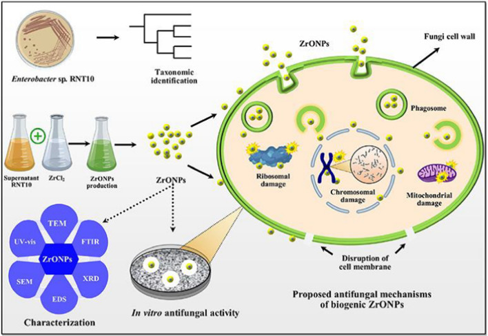
Basically, the ZrO2 nanoparticles may have the inhibition mechanism against fungi as same as that against bacteria (Ahmed et al. 2021). The main mechanism of the disruption of fungal cell division may rely on the generation of reactive oxygen species (e.g., ·O2−, and ·OH) and free radicals (Akintelu and Folorunso 2020). With the high biocompatibility, green ZrO2 nanoparticles can pervade through the fungal cell membranes by endocytosis. In the intracellular environment, ZrO2 nanoparticles account for detrimental functions such as ribosomal, chromosomal, and mitochondrial damages through the formation of reactive oxygen species (Fig. 5). Moreover, ZrO2 nanoparticles possibly break the integrity of fungal cell by changing the physicochemical conditions to cause cell stress and leaking the intracellular components (Reddy et al. 2015).
Fig. 5.
Synthesis, characterization, and proposed antifungal mechanisms of green zirconia nanoparticles. Green zirconia nanoparticles may pervade through the fungal cell membranes, causing many detrimental effects on ribosomal, chromosomal, and mitochondrial parts. They may change the intracellularly physicochemical properties to cause cell stress and break the integrity of fungal cells. Reprinted with the permission of Elsevier from reference (Ahmed et al. 2021). Abbreviations: TEM, transmission electron microscope; FTIR, Fourier-transform infrared spectroscopy; XRD, X-ray diffraction; EDS, energy-dispersive X-ray spectroscopy; SEM, scanning electron microscopy; UV–Vis, ultraviolet–visible spectroscopy; ZrONPs, zirconia nanoparticles; RNT10, an isolated strain of Enterobacter
Although the mechanisms of damaging the fungal cells by ZrO2 nanoparticles have not been fully understood, many attempts were still made to demonstrate their antifungal efficiency. Ahmed et al. (2021) obtained the promising practical outcomes of ZrO2 nanoparticles biosynthesized from Enterobacter sp. against bayberry fungal pathogen. Moreover, the antifungal inhibition zones were very wide 25.18 ± 1.52 mm at the dilute concentration of ZrO2 (only 20 μg/mL). They also observed the clear deconstruction of P. Versicolor fungal cell by extracellular leakage of deoxyribonucleic acid (DNA) and proteins under microscopy imaging techniques. Several works clarified the strength of green ZrO2 for inhibiting both fungal and bacterial species (Chau et al. 2021; Joshi et al. 2021). Regardless of some limitations of the scope of fungal and bacterial species, those may provide more insightful evidence into the antifungal activities of green zirconia nanoparticles. It is also recommended to better elucidate the underlying mechanisms of inhibiting the fungal cells by green ZrO2 nanoparticles for future researches (Tables 4 and 5).
Table 4.
Environmental remediation applications of green ZrO2 nanoparticles
| Green material | Species | Type of treatment | Target pollutants | Main findings | Refs. |
|---|---|---|---|---|---|
| Bacterium | Pseudomonas aeruginosa | Adsorption | Tetracycline | Optimum pH 6.0 | Debnath et al. (2020) |
| High adsorption capacity of 526.32 mg/g, adsorption model fitted best with Langmuir model | |||||
| Reusability up to 5 cycles (81.55% after 5th cycle) | |||||
| Plant | Euclea natalensis | Adsorption | Tetracycline | The best adsorptive capacity of 30.45 mg/g was obtained by response surface methodology | Silva et al. (2019) |
| Plant | Aloe vera | Adsorption | Fluoride | Exothermic and spontaneous adsorption between 20 and 50 °C | Prasad et al. (2014) |
| Nearly 99% F– ions were adsorbed by ZrO2-based adsorbent | |||||
| Chemisorption capacity of 96.58 mg/g | |||||
| Plant | Sapindus mukorossi | Adsorption | Methylene blue | Optimum adsorption conditions at pH 10, adsorbent dosage of 0.3 g, initial methylene blue concentration of 20 mg/L, and average time of 300 min | Alagarsamy et al. (2022) |
| 94% removal efficiency for methylene blue dye and adsorptive capacity of 23.26 mg/g | |||||
| Good recyclability: 0.1 M HCl as an efficient eluent, and three consecutive cycles | |||||
| Plant | Wrightia tinctoria | Catalytic degradation | Reactive yellow 160 dye | 94.58% degradation for Reactive yellow 160 azo dye | Al-Zaqri et al. (2021) |
| 0.9837 min−1 for first order rate constant (k1) | |||||
| Plant | Lagerstroemia speciosa | Catalytic degradation | Methyl orange | Degradation percentage was at 94.58% after irradiating under the sunlight for 290 min | Saraswathi and Santhakumar (2017) |
| Plant | Ficus benghalensis | Catalytic degradation | Methylene blue | Optimum conditions: catalyst loading of 1.5 g/L at pH 7 | Shinde et al. (2018) |
| Removal of 91.22% after 240 min | |||||
| Plant | Ficus benghalensis | Catalytic degradation | Methyl orange | Optimum pH 7 | Shinde et al. (2018) |
| Methyl orange was degraded at 69.23% after 240 min |
Table 5.
Potential applications of green ZrO2-based nanocomposites
| Green material | Species | Composites | Properties | Potential applications | Ref |
|---|---|---|---|---|---|
| Plant | Centaurea cyanus | Ag/Fe3O4/ZrO2 | Particle sizes: 30–90 nm, saturation magnetization: 10 emu/g | Catalytic reduction of 4-nitrophenol and methyl orange, 3 cycles with swift reaction time (7.5–8 min) | Rostami-Vartooni et al. (2019) |
| Plant | Justicia adhatoda | CeO2/ZrO2 | Nano-stick like structure (10–15 nm) | Antibacterial against S. aureus, and E. coli, antioxidant ability, anti-biofilm | Pandiyan et al. (2018) |
| Narrow band gap (3.37 eV) | |||||
| Plant | Hevea brasiliensis | Ni-doped ZrO2 | Energy gap of 2.4–2.75 eV | Nanoelectronics | Yadav et al. (2021) |
| Plant | Leucas aspera | Sm-doped ZrO2 (Sm3+/ZrO2+ = 3–11 mol.%) | Highly symmetric nanocubic (Fm-3 m) | High stable and reusability with six consecutive recycles | Gurushantha et al. (2016) |
| Band gap of 5.3–5.9 eV | High sunlight-driven degradation of Sm/ZrO2 with 11 mol.% dopant against acid green dye | ||||
| Plant | Anacardium occidentale | Ag/ZrO2 | Monoclinic and tetragonal phases of ZrO2 | Not reported | Vivekanandhan et al. (2015) |
| Face-centered cubic crystal phase of Ag NPs (5–20 nm) on ZrO2 surface | |||||
| Plant | Commelina diffusa (-7-hydroxy-4´-methoxy-isoflavon from extract as a reducing and stabilizing agent | Cu/ZrO2 | Reduced agglomeration of Cu/ZrO2 with particle size of 18–25 nm | Catalytic reduction of 2,4- dinitrophenilhydrazine, various organic dyes such as congo red, nigrosin, methyl orange in the presence of NaBH4 at room temperature | Hamad et al. (2019) |
| Cu/ZrO2 exhibited the good stability up to three days after the synthesis process | Ultrafast reaction time for methyl orange (1 s), dinitrophenilhydrazine (40 s), and congo red (150 s) | ||||
| High recyclability at least 5 times | |||||
| Plant | Daphne alpine | V2O5/ZrO2 | Surface area of 214 m2/g, particle size of 41.74 nm, band gap 3.93 eV | The degradation efficiencies of V2O5/ZrO2 against methyl orange (76.9%) and picloram (86%) for 75 min | Rasheed et al. (2020) |
| Thermal stability up to 1000 °C | |||||
| Plant | Ageratum conyzoides | Ag/ZrO2 | Particle size of 50 nm | The reduction of 2,4-dinitrophenylhydrazine, 4-nitrophenol, nigrosin and congo red | Maham et al. (2020) |
| Face centered cubic of Ag on ZrO2 surface | High catalytic performance for reduction of 4-NP into 4-AP (100%) for 6 min, and 2,4-DAPH for 50 s | ||||
| At least 5 cycles for reusability study | |||||
| Plant | Aloe Vera | Mg-doped ZrO2 | Shape: hollow microspheres, and tetragonal phase | ZrO2–Mg (2 mol.%) gave the highest degradation efficiency (93%) | Renuka et al. (2016) |
| Total organic carbon test gave the mineralization rate of 79% after 60 min of reaction | |||||
| Five consecutive cycle runs |
Anticancer
On average, one-third people are diagnosed with cancer in their lifetime, causing one of the world’s leading deaths (Sung et al. 2021). The evolution of cancer disease is the growth of abnormal cells to invade other tissue of the body. Many therapies such as surgery, chemotherapy, stem cell, and radiotherapy have been developed to treat this disease (Mooney et al. 2018). Bionanotechnology has recently played a vital role in designing novel anticancer agents and nanocarriers for drug delivery (Dai et al. 2017). Accordingly, nanoparticles can preferentially approach and accumulate in tumors thanks to the permeable effects, but this accumulation process is mostly influenced by physicochemical factors in the body (Beik et al. 2019). Green ZrO2 nanoparticles have several emerging advantages such as nanoscale size, high surface area, stability, and good biocompatibility to overcome these barriers. On the tumor surface, green ZrO2 nanoparticles bind to the macromolecules such as proteins and then penetrate into the tumor cells (Ranji-Burachaloo et al. 2018). The chief mechanisms of cell damage by green ZrO2 nanoparticles are not elucidated insightfully in the published literatures. According to Table 3, there are a limited number of works that evaluated the cytotoxicity activity of green ZrO2 nanoparticles biosynthesized from Lagerstroemia speciose leaf and Nephelium lappaceum fruit extracts against the breast cancer cell lines (Saraswathi and Santhakumar 2017; Isacfranklin et al. 2020). Therefore, many further investigations of anticancer activity of green ZrO2 nanoparticles are required.
Environmental remediation
Removal of antibiotic drugs
Environmental pollution is currently presenting as one of the most globally critical issues, demanding many urgent actions (Crini and Lichtfouse 2019). In particular, the presence of emerging pollutants such as antibiotics can reduce the quality of water sources, and generate many potential threats for human and aquatic animals (Tahrani et al. 2015; Oberoi et al. 2019; Kovalakova et al. 2020). Several works reported the evolution of antibiotic resistance genes in rivers, hospital effluents, wastewater treatment plants, and sediments (Kairigo et al. 2020; Böger et al. 2021). Therefore, the treatment of antibiotic pharmaceuticals from water media by effective methods is necessarily required. Considering the non-degradable and chemically stable structure of antibiotics, adsorption may be the best remediation method due to its enormous advantages including high efficiency, fast kinetic, and tunable procedure (Tran et al. 2019b, 2020d, b; Dang et al. 2021; Nguyen et al. 2021e). However, the selection and development of novel advanced materials for water treatment are still challenging. Green ZrO2 nanoparticles can meet the requirements for a good adsorbent since they obtain many outstanding properties. Indeed, biologically synthesized ZrO2 nanoparticles have proved their superior functionality of the surface, stability, and eco-friendly synthesis, and hence, exhibiting their capacity for antibiotic adsorption (Table 4).
Silva et al. (2019) optimized the tetracycline removal efficiency of ZrO2 nanoparticles biosynthesized from Euclea natalensis roots by response surface methodology. In this experiment, the conditions of ZrO2 nanofabrication were set up 23 factorial design for extract content, precursor content, and pyrolysis temperature. The calcination temperature at 550 °C was found the most suitable. Under optimized removal conditions at initial concentration of 20 mg/L and adsorbent dose of 0.5 g/L, green ZrO2 nanoparticles obtained the tetragonal phase with extremely small particle size (5.90–8.54 nm) and exhibited the best adsorptive capacity of 30.45 mg/g for tetracycline. However, this capacity value was relatively lower than that obtained by most adsorbents. To improve such situation, Debnath et al. (2020) investigated the synthesis of green ZrO2 nanoparticles by bacterial community, specifically, Pseudomonas aeruginosa species for the treatment of tetracycline in aqueous solutions. Green ZrO2 owned both monoclinic and tetragonal crystal phases with small size (6.41 nm). They optimized the operating tetracycline adsorption parameters such as pH 7 and contact time of 15 min to reach the very promising capacity of 526.32 mg/g. Importantly, green ZrO2 could be easily regenerated with dilute NaOH solution and distilled water, bringing good reusability up to 5 cycles. The authors also proposed the major role of electrostatic interaction for their adsorption mechanisms. With the high performance of stability, reusability, and removal efficiency, green ZrO2 nanoparticles can be ideal nanomaterials for water purification.
Removal of textile dyes
In addition to the contamination of antibiotics in water, many textile dyes are becoming persistent pollutants. It was estimated that a large amount of textile dyes is yearly discharged into water sources, causing many adverse impacts on human health (Zhou et al. 2019). They are also known to dismiss the solar light penetration, resulting in the reduction of photosynthesis efficiency as well as metabolism processes in several aquatic species (Tran et al. 2019a, 2020c). As same as antibiotics, most of these organic dyes are chemically stable, and difficult to degrade by biological methods such as phytoremediation (Nguyen et al. 2021f). However, another feasible solution is to apply the nanomaterials for the remediation of textile dyes through the adsorption and photocatalytic routes (Tran et al. 2020e; Nguyen et al. 2021c, a). Table 4 summarizes the main findings of recent publications on the treatment of dyes using green ZrO2 nanoparticles.
By adsorption pathway, Alagarsamy et al. (2022) demonstrated the high efficiency of green ZrO2 nanoparticles biosynthesized from the pericarp extract of Sapindus mukorossi for the removal of methylene blue. They optimized the adsorption conditions such as pH 10, ZrO2 dose of 0.3 g/L, and 20 mg/L of tetracycline to remove 94% methylene blue for 300 min. Although the adsorption capacity was low (23.25 mg/g), this adsorbent could be reused for three consecutive cycles. In terms of catalytic degradation by green ZrO2 nanoparticles, textile dyes were investigated more widely (Table 4). Take the study by Shinde et al. (2018) for example, both methylene blue and methylene orange dyes could be sufficiently degraded (61–91%) by green ZrO2 nanoparticles biosynthesized from the Ficus benghalensis leaves under ultraviolet light irradiation. Moreover, the authors reported the catalytic optimal conditions at pH 7 and catalyst loading of 1.5 g/L. The plausible catalytic mechanism was suggested by the incorporation of reactive oxygen species such as ·O2−, and ·OH into the degradation process of dyes. However, carrying out the dyes degradation under ultraviolet light irradiation as mentioned by Shinde et al. (2018) may be unfavorable for practical applications. Al-Zaqri et al. (2021) improved this harsh input by using green ZrO2 nanoparticles biosynthesized from Wrightia tinctoria leaves. The outcomes were very promising because this dye was mostly removed with the high first-order rate constant (k1 = 0.9837 min−1) in the presence of sunlight irradiation. The plant extract may be contributed significantly to the formation of green ZrO2 nanoparticles with smaller particle size (17 nm) as well as narrower bandgap energy (3.78 eV), which exhibited better catalytic performance.
Removal of other pollutants
The presence of other emerging pollutants such as fluoride can cause several serve diseases including skeletal fluorosis, teeth mottling, bones deformation, and many neurological issues (Affonso et al. 2020). The process of chemical fertilizing using hydrofluoric acid generates the amount of fluoride ions in water. It is therefore necessary to treat this pollutant before being discharged from the aqueous media. Prasad et al. (2014) assessed the adsorptive efficiency of adopting green ZrO2 nanoparticles from Aloe vera extract. Nearly 99% of F– ions were adsorbed ZrO2-based adsorbent, and fluoride chemisorption capacity was obtained at 96.58 mg/g. The authors also found the exothermic, spontaneous, and recyclability nature of the fluoride adsorption process, suggesting the potential of green ZrO2 in fluoride decontamination. Although the high performance of ZrO2 nanoparticles produced from green extract sources for the treatment of antibiotics, textile dyes, and fluoride has been presented in the previous works, their great potentials for removing other emerging pollutants such as heavy metal ions, chlorinated compounds, persistent organic pollutants, pharmaceuticals, and personal care products have not yet exploited. It is recommended for further studies to explore the strength of green ZrO2 nanoparticles for the treatment of other emerging pollutants in the environmental remediation field.
Other applications
Silicon and many commercialized semiconductors-based solar cells have exhibited their highly efficient performance, but their production process is generally expensive (Roy et al. 2020). Moreover, these processes use chemically synthesized protocols, hindering sustainable development. To reach the purposes of green production, the biological synthesis of low-cost dye-sensitized solar cells should be of great interest (Alami et al. 2019). Green ZrO2 nanoparticles can become one of the prospective electronic nanomaterials for solar energy harvesting. Several studies reported the biosynthesis of green ZrO2 nanoparticles for solar cell applications. For example, Majedi et al. (2016) synthesized the cubic-phase zirconia nanoparticle with small particle size (21 nm) using lemon juice. They reported the electrical conductivity and activation energy of ZrO2 nanoparticles of 0.0034 S/cm, and 31.015 kJ/mol, respectively. These results exhibited a relatively electronic performance of green ZrO2 as potential electrolytes. Vennila et al. (2018) also have investigated the synthesis of ZrO2 using Gloriosa Superba tuber powder and found the potential to create high-efficiency electrodes. However, the applications of green ZrO2 nanoparticles as solar cells are still limited. The reaction mechanism, as well as the ways to improve the energy harvesting efficiency, are sufficiently unexplored. It is recommended that further studies should be investigated.
ZrO2-based nanocomposites
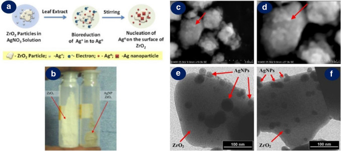
The incorporation of dopants can considerably change the properties of ZrO2 nanoparticles in terms of particle size, uniformity, electronic nature, and morphologies. Green synthesis of ZrO2-based nanocomposites allows improving some disadvantages of bandgap energy, surface area, surface chemistry, and stability of inherent ZrO2 nanoparticles. To reach a green and sustainable approach, the synthesis of ZrO2-based nanocomposites uses microbial or botanical sources, which play a key role in the bioreduction, biocapping, biochelating, and biostabilizing process for the transformation into composites. For example, Vivekanandhan et al. (2015) functionalized the surface of ZrO2 nanocrystallites with silver nanoparticles using Anacardium occidentale leaf extract (Fig. 6). The bio compounds from this extract might participate in the bioreduction of Ag+ into Ag and nucleation of Ag nanospheres (5–20 nm) on ZrO2 surface. The authors also reported the inhibition of the deformation of Ag/ZrO2 nanocomposites in the larger presence of leaf extract. This phenomenon may be attributable to swift bioreduction of Ag+ ions before their dispersion on the surface of ZrO2. Therefore, a sufficient amount of plant extract is important to obtain the structure of ZrO2-based nanocomposites. Table 5 summaries several potential applications of green ZrO2-based nanocomposites.
Fig. 6.
The bioreduction of Ag+ into Ag and nucleation of on ZrO2 surface for the biosynthesis of green Ag/ZrO2 nanocomposites using Anacardium occidentale leaf extract (a); the photograph of as-synthesized green ZrO2 and Ag/ZrO2 nanocomposites (b); scanning electron microscopy (c, d) and transmission electron microscopy (e, f) microphotographies of green Ag/ZrO2 nanocomposites. The presence of leaf extract efficiently leads to the good dispersion and high biostabilization of Ag nanospheres (5–20 nm) on the surface of ZrO2. However, larger addition of leaf extract inhibits the formation of Ag/ZrO2 nanocomposites. Reproduced and adapted with the permission of Elsevier from reference (Vivekanandhan et al. 2015). Abbreviations: AgNP, silver nanoparticles
Because ZrO2 nanoparticles exhibit a wide bandgap (~ 5.0 eV), the absorption of visible light is difficult, hampering their photocatalytic and nanoelectronic applications. The modification of ZrO2 nanoparticles aims to suffer this major drawback. For example, Yadav et al. (2021) inserted an amount of transition metal nickel (Ni) into ZrO2 nanoparticles using rubber latex extract to obtain monoclinic, tetragonal, and cubic Ni-doped ZrO2 polycrystalline. The band gaps were very narrowed between 2.4 and 2.75 eV, making Ni-doped ZrO2 as potential nanoelectronic devices. Rasheed et al. (2020) reported the utilization of Daphne alpine leaf extract to synthesize green V2O5/ZrO2 nanocomposite. They not only observed the increase in surface area (214 m2/g) but also found the improvement of bandgap energy (3.93 eV) and thermal stability (1000 °C). It was suggested that V2O5 efficiently enhanced the electron separation, resulting in good photocatalytic activities against orange (76.9%) and picloram (86%) of green V2O5/ZrO2.
Doping other transition metals (Sm, Cu, Ag) significantly changes the electronic nature and crystalline phases of the nanocomposites, leading to the enhancement of their stability, recyclability, and photocatalytic activities. Indeed, Gurushantha et al. (2016) fabricated the green ZrO2 with samarium dopant (Sm3+/ZrO2+ = 3–11 mol.%) using Leucas Aspera extract. Sm/ZrO2 composites with the doping of 11% dopant obtained highly symmetric nanocubics, and the highest photocatalytic degradation of acid green with six consecutive recycles in the presence of solar energy. This suggested that the Sm/ZrO2 composites are highly stable and reusable. Hamad et al. (2019) demonstrated the presence of 7-hydroxy-4´-methoxy-isoflavon from Commelina diffusa leaves reduced the agglomeration of Cu/ZrO2 nanoparticles (18–25 nm) with good stable ability during the post-synthesis. Moreover, the green Cu/ZrO2 nanoparticles catalyzed the reduction and degradation of 2,4-dinitrophenylhydrazine, and organic dyes including congo red, nigrosin, methyl orange with ultrafast reaction times (1–150 s). In another study, Maham et al. (2020) obtained green Ag/ZrO2 nanocomposites (50 nm) using Ageratum conyzoides plant extract. The catalyst could reduce and degrade a range of substrates such as 2,4-dinitrophenylhydrazine, 4-nitrophenol, nigrosin, and congo red, indicating the greatly catalytic performance of green Ag/ZrO2 nanocomposite.
Although green ZrO2 nanoparticles exhibit their very high catalytic performance, the separation and recovery of these non-magnetically small-size nanomaterials from the aqueous post-reaction is disadvantageous. The intercalation of magnetic components (e.g., Fe3O4) into ZrO2 nanomaterials should be therefore required. Inspired by this idea, Rostami-Vartooni et al. (2019) have successfully the Centaurea cyanus-biofabricated Ag/Fe3O4/ZrO2 nanocomposites with average particle size between 30 and 90 nm. The nanocomposite could be easily separated from the reaction solutions because its saturation magnetization value was obtained at 10 emu/g. The authors also reported some good results of catalytic reduction of 4-nitrophenol and degradation of methyl orange green Ag/Fe3O4/ZrO2 with a swift reaction time between 7.5 and 8 min. Therefore, this advantage may urge the recyclability experiments of magnetic ZrO2 nanocomposites.
ZrO2-based nanocomposites biosynthesized from the green extract sources can offer a high diversity of morphology and open new applications. For example, Pandiyan et al. (2018) employed the green synthesis of CeO2 doped ZrO2 nano sticks with small particle size (10–15 nm) and bandgap (3.37 eV) from Justicia adhatoda leaf extract. This nanocomposite exhibited high antibacterial activities against S. aureus, and E. coli and high antioxidant activity. They also reported the application of CeO2/ZrO2 nano sticks as potential biofilms against many pathogeneses caused by bacteria such as S. marcescens. Renuka et al. (2016) successfully created the hollow structure of Mg-doped ZrO2 microspheres using Aloe Vera leaf extract. They assumed that bio compounds such as polysaccharides from Aloe Vera leaf might chelate with Zr4+ and Mg2+ ions during the formation of Mg/ZrO2 microspheres. The authors assumed that bio compounds such as polysaccharides from Aloe Vera leaf might chelate with Zr4+ and Mg2+ ions during the reaction of complexation. The burning calcination of these complexes allowed to acquire the hollow structure of Mg/ZrO2 microspheres. They found the Mg/ZrO2 composites with the magnesium doping rate of 2 mol% gave the highest photocatalytic activity against Rhodamine B (93%) in the presence of ultraviolet irradiation. Controlled oxygen vacancies on active sites may be attributable to the enhanced catalytic performance of green Mg/ZrO2.
Perspective
Green synthesis of ZrO2 nanoparticles from microbial and botanical sources has gained great attention because they offer many advantages such as eco-friendly and low-cost manufacturing compared with chemical synthesis methods. Green ZrO2 nanoparticles possess many distinctive structure properties including small size, good dispersion, diverse surface chemistry, large surface area, and sufficient bandgap energy, making them extend excellent applications regarding biomedical and environmental fields. In many cases, the green ZrO2 nanoparticles experimentally exhibit better antibacterial, antifungal, catalytic and adsorptive performance than chemically synthesized ZrO2 nanomaterials. These superior prospects of green ZrO2 nanoparticles may pay the way for developing advanced ZrO2-based nanosensors for smart agriculture, high-efficiency solar cells for energy harvesting, and electronic nanodevices for many high-technological manufacturing industries.
Although green ZrO2 nanoparticles hold many key advantages, there are still several limitations that need further investigation. Firstly, regarding the synthesis procedure for ZrO2 nanoparticles microbial sources, the control of biomass growth rate, metabolite generation, and biochemical properties of cultivation environment should be investigated vigorously. For the botanical sources, the determination of bioactive compound ingredients in the plant extract should be conducted to better elucidate the formation mechanism of ZrO2 nanoparticles. Moreover, the formation of green ZrO2 nanoparticles should be optimized to obtain their expectable properties of particle size, charge nature, surface energy, and surface chemistry. Secondly, regarding the treatment of pollutants such as antibiotics, and textile dyes, the treatment of reused ZrO2 nanoparticles has not been addressed. This action may generate secondary pollution; hence, it is necessary to solve the post-treatment problems. Moreover, most studies on the application of green ZrO2 nanoparticles only reported their findings under laboratory modes, resulting in the difference between simulated and practical results. This drawback can be therefore addressed if the tests are conducted under practical conditions. Thirdly, to the best of our knowledge, the potential of green ZrO2 nanoparticles is substantially unexploited in the many environmental fields, e.g., the treatment of emerging persistent organic pollutants, pharmaceuticals and personal care products, nonsteroidal anti-inflammatory drugs, and toxic gas detection. With the excellent properties of green ZrO2 nanoparticles, it is expected that they will contribute significantly to the development of new environmental remediation approaches. Ultimately, the combination of dopants with green ZrO2 nanoparticles can accelerate their inherent properties of the nanocomposites, expanding many promising applications. However, it is generally difficult to control the interactions of green extract with the dopants during the ZrO2 based nanocomposites. The optimization of the biomass proportion should be evaluated to avoid these interactions. Even though the formation mechanism and application of green ZrO2 based nanocomposites have been better understood in some published works, the number of researches is still limited. The knowledge of the overall interactions between ZrO2 nanomaterials and the environment is just burgeoning. The profound evaluations of the ecotoxicity of green ZrO2 nanoparticles should be continuously conducted to avoid possible future challenges.
Conclusion
The present review addressed some aspects of synthesis, properties, and potentials of green ZrO2 nanoparticles and their nanocomposites for biomedical, environmental remediation, and other applications. The bioactive compounds derived from microbial and botanical extracts are attributable to the bioreduction, biocapping, biochelating, and biostabilization during the transformation of zirconium sources into green ZrO2 nanoparticles. Through the structural characterization techniques, green ZrO2 nanoparticles are found with many morphologies such as nanospheres, nanorods, nano chains. They possess diverse surface chemistry, high stability, and wide bandgap energy possibly due to the effect of green extracts. Some applications regarding the antibacterial, antifungal, anticancer, photocatalytic, and adsorptive activities of green ZrO2 nanoparticles have been overviewed and discussed systematically. The advanced properties and applications of green ZrO2-based nanocomposites were also mentioned. To orientate the future researchers, some discussions of knowledge gaps and prospects of green ZrO2 nanoparticles have been clarified. The green ZrO2 nanoparticles and their nanocomposites are highly expected to obtain many good results in various applications.
Acknowledgments
The authors would love to appreciate the effort of researchers all over the world in the fight against the COVID-19 pandemic.
Author contributions
TVT contributed to Conceptualisation; Data curation; Investigation; Methodology; Writing—original draft; Writing—review & editing. DTCN contributed to Conceptualisation; Data curation; Investigation; Methodology; Writing—original draft; Writing—review & editing. PSK contributed to Writing—review & editing; Data curation; Supervision. ATMD contributed to Writing—review & editing; Data curation; Supervision. AAJ contributed to Writing—review & editing; Data curation; Supervision. D-VNV contributed to Writing—review & editing; Data curation; Supervision; Project administration. All authors read and approved the final manuscript.
Funding
There was no external funding for this study.
Availability of data and material
The authors declare that all data and materials support their published claims and comply with field standards.
Code availability
The authors declare that software application or custom code supports their published claims and comply with field standards.
Declarations
Conflict of interest
The authors declare that there are no conflict of interest.
Consent for publication
The manuscript has not been published anywhere nor submitted to another journal. The manuscript is not currently being considered for publication in any another journal. All authors have been personally and actively involved in substantive work leading to the manuscript, and will hold themselves jointly and individually responsible for its content.
Human and animal rights
Research does not involve any Human Participants and/or Animals.
Footnotes
Publisher's Note
Springer Nature remains neutral with regard to jurisdictional claims in published maps and institutional affiliations.
Thuan Van Tran and Duyen Thi Cam Nguyen have contributed equally to this work.
References
- Abbasi E, Milani M, Fekri Aval S, et al. Silver nanoparticles: synthesis methods, bio-applications and properties. Crit Rev Microbiol. 2014;42:1–8. doi: 10.3109/1040841X.2014.912200. [DOI] [PubMed] [Google Scholar]
- Abinaya S, Kavitha HP, Prakash M, Muthukrishnaraj A. Green synthesis of magnesium oxide nanoparticles and its applications: a review. Sustain Chem Pharm. 2021;19:100368. doi: 10.1016/j.scp.2020.100368. [DOI] [Google Scholar]
- Affonso LN, Marques JL, Lima VVC, et al. Removal of fluoride from fertilizer industry effluent using carbon nanotubes stabilized in chitosan sponge. J Hazard Mater. 2020;388:122042. doi: 10.1016/j.jhazmat.2020.122042. [DOI] [PubMed] [Google Scholar]
- Agarwal H, Venkat Kumar S, Rajeshkumar S. A review on green synthesis of zinc oxide nanoparticles – An eco-friendly approach. Resour Technol. 2017;3:406–413. doi: 10.1016/j.reffit.2017.03.002. [DOI] [Google Scholar]
- Ahmed T, Ren H, Noman M, et al. Green synthesis and characterization of zirconium oxide nanoparticles by using a native Enterobacter sp. and its antifungal activity against bayberry twig blight disease pathogen Pestalotiopsis versicolor. NanoImpact. 2021;21:100281. doi: 10.1016/j.impact.2020.100281. [DOI] [PubMed] [Google Scholar]
- Akintelu SA, Folorunso AS. A review on green synthesis of zinc oxide nanoparticles using plant extracts and its biomedical applications. Bionanoscience. 2020;10:848–863. doi: 10.1007/s12668-020-00774-6. [DOI] [Google Scholar]
- Alagarsamy A, Chandrasekaran S, Manikandan A. Green synthesis and characterization studies of biogenic zirconium oxide (ZrO2) nanoparticles for adsorptive removal of methylene blue dye. J Mol Struct. 2022;1247:131275. doi: 10.1016/j.molstruc.2021.131275. [DOI] [Google Scholar]
- Alami AH, Aokal K, Zhang D, et al. Low-cost dye-sensitized solar cells with ball-milled tellurium-doped graphene as counter electrodes and a natural sensitizer dye. Int J Energy Res. 2019;43:5824–5833. doi: 10.1002/er.4684. [DOI] [Google Scholar]
- Al-Zaqri N, Muthuvel A, Jothibas M, et al. Biosynthesis of zirconium oxide nanoparticles using Wrightia tinctoria leaf extract: characterization, photocatalytic degradation and antibacterial activities. Inorg Chem Commun. 2021;127:108507. doi: 10.1016/j.inoche.2021.108507. [DOI] [Google Scholar]
- Annu A, Sivasankari C, Krupasankar U. Synthesis and characerization of Zro2 nanoparticle by leaf extract bioreduction process for its biological studies. Mater Today Proc. 2020;33:5317–5323. doi: 10.1016/j.matpr.2020.02.975. [DOI] [Google Scholar]
- Bandeira M, Giovanela M, Roesch-Ely M, et al. Green synthesis of zinc oxide nanoparticles: a review of the synthesis methodology and mechanism of formation. Sustain Chem Pharm. 2020;15:100223. doi: 10.1016/j.scp.2020.100223. [DOI] [Google Scholar]
- Bansal V, Rautaray D, Ahmad A, Sastry M. Biosynthesis of zirconia nanoparticles using the fungus Fusarium oxysporum. J Mater Chem. 2004;14:3303–3305. doi: 10.1039/B407904C. [DOI] [Google Scholar]
- Bansal V, Ramanathan R, Bhargava SK. Fungus-mediated biological approaches towards ‘green’synthesis of oxide nanomaterials. Aust J Chem. 2011;64:279–293. doi: 10.1071/CH10343. [DOI] [Google Scholar]
- Beik J, Khateri M, Khosravi Z, et al. Gold nanoparticles in combinatorial cancer therapy strategies. Coord Chem Rev. 2019;387:299–324. doi: 10.1016/j.ccr.2019.02.025. [DOI] [Google Scholar]
- Böger B, Surek M, de O Vilhena R, et al. Occurrence of antibiotics and antibiotic resistant bacteria in subtropical urban rivers in Brazil. J Hazard Mater. 2021;402:123448. doi: 10.1016/j.jhazmat.2020.123448. [DOI] [PubMed] [Google Scholar]
- Bolade OP, Williams AB, Benson NU. Green synthesis of iron-based nanomaterials for environmental remediation: a review. Environ Nanotechnol Monit Manag. 2020;13:100279. doi: 10.1016/j.enmm.2019.100279. [DOI] [Google Scholar]
- Cazado ME, Goldberg E, Togneri MA, et al. A new irradiation growth model for Zr-based components of nuclear reactors for the DIONISIO code. Nucl Eng Des. 2021;373:111009. doi: 10.1016/j.nucengdes.2020.111009. [DOI] [Google Scholar]
- Chau TP, Kandasamy S, Chinnathambi A, et al. Synthesis of zirconia nanoparticles using Laurus nobilis for use as an antimicrobial agent. Appl Nanosci. 2021 doi: 10.1007/s13204-021-02041-w. [DOI] [Google Scholar]
- Chen F, Wu Y-R, Wu J-M, et al. Preparation and characterization of ZrO2-Al2O3 bioceramics by stereolithography technology for dental restorations. Addit Manuf. 2021;44:102055. doi: 10.1016/j.addma.2021.102055. [DOI] [Google Scholar]
- Chu R, Li S, Zhu L, et al. A review on co-cultivation of microalgae with filamentous fungi: efficient harvesting, wastewater treatment and biofuel production. Renew Sustain Energy Rev. 2021;139:110689. doi: 10.1016/j.rser.2020.110689. [DOI] [Google Scholar]
- Crini G, Lichtfouse E. Advantages and disadvantages of techniques used for wastewater treatment. Environ Chem Lett. 2019;17:145–155. doi: 10.1007/s10311-018-0785-9. [DOI] [Google Scholar]
- da Silva AFV, Fagundes AP, Macuvele DLP, et al. Green synthesis of zirconia nanoparticles based on Euclea natalensis plant extract: optimization of reaction conditions and evaluation of adsorptive properties. Colloids Surfaces A Physicochem Eng Asp. 2019;583:123915. doi: 10.1016/j.colsurfa.2019.123915. [DOI] [Google Scholar]
- Dai Y, Xu C, Sun X, Chen X. Nanoparticle design strategies for enhanced anticancer therapy by exploiting the tumour microenvironment. Chem Soc Rev. 2017;46:3830–3852. doi: 10.1039/C6CS00592F. [DOI] [PMC free article] [PubMed] [Google Scholar]
- Dang HH, Nguyen DTC, Nguyen TT, et al. Zeolitic-imidazolate framework-derived N-self-doped porous carbons with ultrahigh theoretical adsorption capacities for tetracycline and ciprofloxacin. J Environ Chem Eng. 2021;9:104938. doi: 10.1016/j.jece.2020.104938. [DOI] [Google Scholar]
- Davar F, Majedi A, Mirzaei A. Polyvinyl alcohol thin film reinforced by green synthesized zirconia nanoparticles. Ceram Int. 2018;44:19377–19382. doi: 10.1016/j.ceramint.2018.07.167. [DOI] [Google Scholar]
- Debnath B, Majumdar M, Bhowmik M, et al. The effective adsorption of tetracycline onto zirconia nanoparticles synthesized by novel microbial green technology. J Environ Manage. 2020;261:110235. doi: 10.1016/j.jenvman.2020.110235. [DOI] [PubMed] [Google Scholar]
- Dhandapani C, Narayanasamy R, Karthick SN, et al. Drastic photocatalytic degradation of methylene blue dye by neodymium doped zirconium oxide as photocatalyst under visible light irradiation. Optik (stuttg) 2016;127:10288–10296. doi: 10.1016/j.ijleo.2016.08.048. [DOI] [Google Scholar]
- Fariq A, Khan T, Yasmin A. Microbial synthesis of nanoparticles and their potential applications in biomedicine. J Appl Biomed. 2017;15:241–248. doi: 10.1016/j.jab.2017.03.004. [DOI] [Google Scholar]
- Ferreira JA, Varjani S, Taherzadeh MJ. A critical review on the ubiquitous role of filamentous fungi in pollution mitigation. Curr Pollut Reports. 2020;6:295–309. doi: 10.1007/s40726-020-00156-2. [DOI] [Google Scholar]
- Filote C, Santos SCR, Popa VI, et al. Biorefinery of marine macroalgae into high-tech bioproducts: a review. Environ Chem Lett. 2021;19:969–1000. doi: 10.1007/s10311-020-01124-4. [DOI] [Google Scholar]
- Gahlawat G, Choudhury AR. A review on the biosynthesis of metal and metal salt nanoparticles by microbes. RSC Adv. 2019;9:12944–12967. doi: 10.1039/C8RA10483B. [DOI] [PMC free article] [PubMed] [Google Scholar]
- Ghomi GAR, Mohammadi-Khanaposhti M, Vahidi H, et al. Fungus-mediated extracellular biosynthesis and characterization of zirconium nanoparticles using standard penicillium species and their preliminary bactericidal potential: a novel biological approach to nanoparticle synthesis. Iran J Pharm Res. 2019;18:2101–2110. doi: 10.22037/ijpr.2019.112382.13722. [DOI] [PMC free article] [PubMed] [Google Scholar]
- Gowri S, Gandhi RR, Sundrarajan M. Structural, optical, antibacterial and antifungal properties of zirconia nanoparticles by biobased protocol. J Mater Sci Technol. 2014;30:782–790. doi: 10.1016/j.jmst.2014.03.002. [DOI] [Google Scholar]
- Gowri S, Gandhi RR, Senthil S, Sundrarajan M. Effect of calcination temperature on nyctanthes plant mediated zirconia nanoparticles; optical and antibacterial activity for optimized zirconia. J Bionanoscience. 2015;9:181–189. doi: 10.1166/jbns.2015.1297. [DOI] [Google Scholar]
- Goyal P, Bhardwaj A, Mehta BK, Mehta D. Research article green synthesis of zirconium oxide nanoparticles (ZrO2NPs) using Helianthus annuus seed and their antimicrobial effects. J Indian Chem Soc. 2021;98:100089. doi: 10.1016/j.jics.2021.100089. [DOI] [Google Scholar]
- Gurushantha K, Anantharaju KS, Sharma SC, et al. Bio-mediated Sm doped nano cubic zirconia: photoluminescent, Judd-Ofelt analysis, electrochemical impedance spectroscopy and photocatalytic performance. J Alloys Compd. 2016;685:761–773. doi: 10.1016/j.jallcom.2016.06.105. [DOI] [Google Scholar]
- Hamad SM, Mahmud SA, Sajadi SM, Omar ZA. Biosynthesis of Cu/ZrO2 nanocomposite using 7-hydroxy-4´-methoxy-isoflavon extracted from Commelina diffusa and evaluation of its catalytic activity. Surfaces and Interfaces. 2019;15:125–134. doi: 10.1016/j.surfin.2019.02.008. [DOI] [Google Scholar]
- Hassan NS, Jalil AA. A review on self-modification of zirconium dioxide nanocatalysts with enhanced visible-light-driven photodegradation of organic pollutants. J Hazard Mater. 2022;423:126996. doi: 10.1016/j.jhazmat.2021.126996. [DOI] [PubMed] [Google Scholar]
- Humbal C, Gautam S, Trivedi U. A review on recent progress in observations, and health effects of bioaerosols. Environ Int. 2018;118:189–193. doi: 10.1016/j.envint.2018.05.053. [DOI] [PubMed] [Google Scholar]
- Indiarto R, Indriana LPA, Andoyo R, et al. Bottom–up nanoparticle synthesis: a review of techniques, polyphenol-based core materials, and their properties. Eur Food Res Technol. 2021 doi: 10.1007/s00217-021-03867-y. [DOI] [Google Scholar]
- Isacfranklin M, Dawoud T, Ameen F, et al. Synthesis of highly active biocompatible ZrO2 nanorods using a bioextract. Ceram Int. 2020;46:25915–25920. doi: 10.1016/j.ceramint.2020.07.076. [DOI] [Google Scholar]
- Jadoun S, Arif R, Jangid NK, Meena RK. Green synthesis of nanoparticles using plant extracts: a review. Environ Chem Lett. 2021;19:355–374. doi: 10.1007/s10311-020-01074-x. [DOI] [Google Scholar]
- Jameel MS, Aziz AA, Dheyab MA. Comparative analysis of platinum nanoparticles synthesized using sonochemical-assisted and conventional green methods. Nano-Structures & Nano-Objects. 2020;23:100484. doi: 10.1016/j.nanoso.2020.100484. [DOI] [Google Scholar]
- Jiang H-L, Liu B, Lan Y-Q, et al. From metal-organic framework to nanoporous carbon: toward a very high surface area and hydrogen uptake. J Am Chem Soc. 2011;133:11854–11857. doi: 10.1021/ja203184k. [DOI] [PubMed] [Google Scholar]
- Jiang X, Nie X, Gong Y, et al. A combined experimental and DFT study of H2O effect on In2O3/ZrO2 catalyst for CO2 hydrogenation to methanol. J Catal. 2020;383:283–296. doi: 10.1016/j.jcat.2020.01.014. [DOI] [Google Scholar]
- Joshi NC, Chaudhary N, Rai N. medicinal plant leaves extract based synthesis, characterisations and antimicrobial activities of ZrO2 nanoparticles (ZrO2 NPs) Bionanoscience. 2021;11:497–505. doi: 10.1007/s12668-021-00829-2. [DOI] [Google Scholar]
- Kairigo P, Ngumba E, Sundberg L-R, et al. Occurrence of antibiotics and risk of antibiotic resistance evolution in selected Kenyan wastewaters, surface waters and sediments. Sci Total Environ. 2020;720:137580. doi: 10.1016/j.scitotenv.2020.137580. [DOI] [PubMed] [Google Scholar]
- Kavitha NS, Venkatesh KS, Palani NS, Ilangovan R. Synthesis and characterization of zirconium oxide nanoparticles using Fusarium solani extract. AIP Conf Proc. 2020;2265:30057. doi: 10.1063/5.0017117. [DOI] [Google Scholar]
- Khatoon N, Ahmad R, Sardar M. Robust and fluorescent silver nanoparticles using Artemisia annua: biosynthesis, characterization and antibacterial activity. Biochem Eng J. 2015;102:91–97. doi: 10.1016/j.bej.2015.02.019. [DOI] [Google Scholar]
- Kovalakova P, Cizmas L, McDonald TJ, et al. Occurrence and toxicity of antibiotics in the aquatic environment: a review. Chemosphere. 2020;251:126351. doi: 10.1016/j.chemosphere.2020.126351. [DOI] [PubMed] [Google Scholar]
- Kumaresan M, Vijai Anand K, Govindaraju K, et al. Seaweed Sargassum wightii mediated preparation of zirconia (ZrO2) nanoparticles and their antibacterial activity against gram positive and gram negative bacteria. Microb Pathog. 2018;124:311–315. doi: 10.1016/j.micpath.2018.08.060. [DOI] [PubMed] [Google Scholar]
- Li Z, Ma J, Ruan J, Zhuang X. Using positively charged magnetic nanoparticles to capture bacteria at ultralow concentration. Nanoscale Res Lett. 2019;14:195. doi: 10.1186/s11671-019-3005-z. [DOI] [PMC free article] [PubMed] [Google Scholar]
- Maham M, Nasrollahzadeh M, Mohammad Sajadi S. Facile synthesis of Ag/ZrO2 nanocomposite as a recyclable catalyst for the treatment of environmental pollutants. Compos Part B Eng. 2020;185:107783. doi: 10.1016/j.compositesb.2020.107783. [DOI] [Google Scholar]
- Majedi A, Abbasi A, Davar F. Green synthesis of zirconia nanoparticles using the modified Pechini method and characterization of its optical and electrical properties. J Sol-Gel Sci Technol. 2016;77:542–552. doi: 10.1007/s10971-015-3881-3. [DOI] [Google Scholar]
- Makuła P, Pacia M, Macyk W. How to correctly determine the band gap energy of modified semiconductor photocatalysts based on UV–Vis spectra. J Phys Chem Lett. 2018;9:6814–6817. doi: 10.1021/acs.jpclett.8b02892. [DOI] [PubMed] [Google Scholar]
- Mooney R, Hammad M, Batalla-Covello J, et al. Concise review: neural stem cell-mediated targeted cancer therapies. Stem Cells Transl Med. 2018;7:740–747. doi: 10.1002/sctm.18-0003. [DOI] [PMC free article] [PubMed] [Google Scholar]
- Muhammad S, Long X, Salman M. COVID-19 pandemic and environmental pollution: A blessing in disguise? Sci Total Environ. 2020;728:138820. doi: 10.1016/j.scitotenv.2020.138820. [DOI] [PMC free article] [PubMed] [Google Scholar]
- Narayanan KB, Sakthivel N. Biological synthesis of metal nanoparticles by microbes. Adv Colloid Interface Sci. 2010;156:1–13. doi: 10.1016/j.cis.2010.02.001. [DOI] [PubMed] [Google Scholar]
- Nguyen DTC, Dang HH, Vo D-VN, et al. Biogenic synthesis of MgO nanoparticles from different extracts (flower, bark, leaf) of Tecoma stans (L.) and their utilization in selected organic dyes treatment. J Hazard Mater. 2021;404:124146. doi: 10.1016/j.jhazmat.2020.124146. [DOI] [PubMed] [Google Scholar]
- Nguyen DTC, Le HTN, Nguyen TT, et al. Engineering conversion of Asteraceae plants into biochars for exploring potential applications: a review. Sci Total Environ. 2021;797:149195. doi: 10.1016/j.scitotenv.2021.149195. [DOI] [PubMed] [Google Scholar]
- Nguyen DTC, Le HTN, Nguyen TT, et al. Multifunctional ZnO nanoparticles bio-fabricated from Canna indica L. flowers for seed germination, adsorption, and photocatalytic degradation of organic dyes. J Hazard Mater. 2021;420:126586. doi: 10.1016/j.jhazmat.2021.126586. [DOI] [PubMed] [Google Scholar]
- Nguyen DTC, Nguyen TT, Le HTN, et al. The sunflower plant family for bioenergy, environmental remediation, nanotechnology, medicine, food and agriculture. Rev Environ Chem Lett. 2021;19:3701–3726. doi: 10.1007/s10311-021-01266-z. [DOI] [Google Scholar]
- Nguyen DTC, Vo D-VN, Nguyen CNQ, et al. Box-Behnken design, kinetic, and isotherm models for oxytetracycline adsorption onto Co-based ZIF-67. Appl Nanosci. 2021;11:2347–2359. doi: 10.1007/s13204-021-01954-w. [DOI] [Google Scholar]
- Nguyen LTT, Vo D-VN, Nguyen LTH, et al. Synthesis, characterization, and application of ZnFe2O4@ZnO nanoparticles for photocatalytic degradation of Rhodamine B under visible-light illumination. Environ Technol Innov. 2021 doi: 10.1016/j.eti.2021.102130. [DOI] [Google Scholar]
- Oberoi AS, Jia Y, Zhang H, et al. Insights into the fate and removal of antibiotics in engineered biological treatment systems: a critical review. Environ Sci Technol. 2019;53:7234–7264. doi: 10.1021/acs.est.9b01131. [DOI] [PubMed] [Google Scholar]
- Ong CB, Ng LY, Mohammad AW. A review of ZnO nanoparticles as solar photocatalysts: synthesis, mechanisms and applications. Renew Sustain Energy Rev. 2018;81:536–551. doi: 10.1016/j.rser.2017.08.020. [DOI] [Google Scholar]
- Pandiyan N, Murugesan B, Sonamuthu J, et al. Facile biological synthetic strategy to morphologically aligned CeO2/ZrO2 core nanoparticles using Justicia adhatoda extract and ionic liquid: Enhancement of its bio-medical properties. J Photochem Photobiol B Biol. 2018;178:481–488. doi: 10.1016/j.jphotobiol.2017.11.036. [DOI] [PubMed] [Google Scholar]
- Patil MP, Kim G-D. Marine microorganisms for synthesis of metallic nanoparticles and their biomedical applications. Colloids Surf B Biointerfaces. 2018;172:487–495. doi: 10.1016/j.colsurfb.2018.09.007. [DOI] [PubMed] [Google Scholar]
- Ponce NMA, Stortz CA. A comprehensive and comparative analysis of the fucoidan compositional data across the phaeophyceae. Front Plant Sci. 2020;11:1844. doi: 10.3389/fpls.2020.556312. [DOI] [PMC free article] [PubMed] [Google Scholar]
- Prasad KS, Amin Y, Selvaraj K. Defluoridation using biomimetically synthesized nano zirconium chitosan composite: kinetic and equilibrium studies. J Hazard Mater. 2014;276:232–240. doi: 10.1016/j.jhazmat.2014.05.038. [DOI] [PubMed] [Google Scholar]
- Ram S, Mitra M, Shah F, et al. Bacteria as an alternate biofactory for carotenoid production: a review of its applications, opportunities and challenges. J Funct Foods. 2020;67:103867. doi: 10.1016/j.jff.2020.103867. [DOI] [Google Scholar]
- Rambabu K, Bharath G, Arangadi AF, et al. ZrO2 incorporated polysulfone anion exchange membranes for fuel cell applications. Int J Hydrogen Energy. 2020;45:29668–29680. doi: 10.1016/j.ijhydene.2020.08.175. [DOI] [Google Scholar]
- Rana A, Yadav K, Jagadevan S. A comprehensive review on green synthesis of nature-inspired metal nanoparticles: mechanism, application and toxicity. J Clean Prod. 2020;272:122880. doi: 10.1016/j.jclepro.2020.122880. [DOI] [Google Scholar]
- Ranji-Burachaloo H, Gurr PA, Dunstan DE, Qiao GG. Cancer treatment through nanoparticle-facilitated fenton reaction. ACS Nano. 2018;12:11819–11837. doi: 10.1021/acsnano.8b07635. [DOI] [PubMed] [Google Scholar]
- Rasheed P, Haq S, Waseem M, et al. Green synthesis of vanadium oxide-zirconium oxide nanocomposite for the degradation of methyl orange and picloram. Mater Res Express. 2020;7:25011. doi: 10.1088/2053-1591/ab6fa2. [DOI] [Google Scholar]
- Reddy GB, Madhusudhan A, Ramakrishna D, et al. Green chemistry approach for the synthesis of gold nanoparticles with gum kondagogu: characterization, catalytic and antibacterial activity. J Nanostructure Chem. 2015;5:185–193. doi: 10.1007/s40097-015-0149-y. [DOI] [Google Scholar]
- Renuka L, Anantharaju KS, Sharma SC, et al. Hollow microspheres Mg-doped ZrO2 nanoparticles: green assisted synthesis and applications in photocatalysis and photoluminescence. J Alloys Compd. 2016;672:609–622. doi: 10.1016/j.jallcom.2016.02.124. [DOI] [Google Scholar]
- Riaz M, Kamran M, Fang Y, et al. Arbuscular mycorrhizal fungi-induced mitigation of heavy metal phytotoxicity in metal contaminated soils: a critical review. J Hazard Mater. 2021;402:123919. doi: 10.1016/j.jhazmat.2020.123919. [DOI] [PubMed] [Google Scholar]
- Rostami-Vartooni A, Moradi-Saadatmand A, Bagherzadeh M, Mahdavi M. Green synthesis of Ag/Fe3O4/ZrO2 nanocomposite using aqueous Centaurea cyanus flower extract and its catalytic application for reduction of organic pollutants. Iran J Catal. 2019;9:27–35. [Google Scholar]
- Roy P, Kumar Sinha N, Tiwari S, Khare A. A review on perovskite solar cells: evolution of architecture, fabrication techniques, commercialization issues and status. Sol Energy. 2020;198:665–688. doi: 10.1016/j.solener.2020.01.080. [DOI] [Google Scholar]
- Rozana M, Soaid NI, Kian TW, et al. Photocatalytic performance of freestanding tetragonal zirconia nanotubes formed in H2O2/NH4F/ethylene glycol electrolyte by anodisation of zirconium. Nanotechnology. 2017;28:155604. doi: 10.1088/1361-6528/aa5fac. [DOI] [PubMed] [Google Scholar]
- Saraswathi VS, Santhakumar K. Photocatalytic activity against azo dye and cytotoxicity on MCF-7 cell lines of zirconium oxide nanoparticle mediated using leaves of Lagerstroemia speciosa. J Photochem Photobiol B Biol. 2017;169:47–55. doi: 10.1016/j.jphotobiol.2017.02.023. [DOI] [PubMed] [Google Scholar]
- Saravanan A, Kumar PS, Karishma S, et al. A review on biosynthesis of metal nanoparticles and its environmental applications. Chemosphere. 2021;264:128580. doi: 10.1016/j.chemosphere.2020.128580. [DOI] [PubMed] [Google Scholar]
- Sathishkumar M, Sneha K, Yun Y-S. Green fabrication of zirconia nano-chains using novel Curcuma longa tuber extract. Mater Lett. 2013;98:242–245. doi: 10.1016/j.matlet.2013.02.036. [DOI] [Google Scholar]
- Shafey AME (2020) Green synthesis of metal and metal oxide nanoparticles from plant leaf extracts and their applications: A review. Green Process Synth 9:304–339. 10.1515/gps-2020-0031
- Shinde HM, Bhosale TT, Gavade NL, et al. Biosynthesis of ZrO2 nanoparticles from Ficus benghalensis leaf extract for photocatalytic activity. J Mater Sci Mater Electron. 2018;29:14055–14064. doi: 10.1007/s10854-018-9537-7. [DOI] [Google Scholar]
- Shrimal P, Jadeja G, Patel S. A review on novel methodologies for drug nanoparticle preparation: microfluidic approach. Chem Eng Res Des. 2020;153:728–756. doi: 10.1016/j.cherd.2019.11.031. [DOI] [Google Scholar]
- Slavin YN, Asnis J, Häfeli UO, Bach H. Metal nanoparticles: understanding the mechanisms behind antibacterial activity. J Nanobiotechnology. 2017;15:65. doi: 10.1186/s12951-017-0308-z. [DOI] [PMC free article] [PubMed] [Google Scholar]
- Srivastava M, Srivastava N, Mishra PK, Malhotra BD. Prospects of nanomaterials-enabled biosensors for COVID-19 detection. Sci Total Environ. 2021;754:142363. doi: 10.1016/j.scitotenv.2020.142363. [DOI] [PMC free article] [PubMed] [Google Scholar]
- Sudrajat H, Babel S, Sakai H, Takizawa S. Rapid enhanced photocatalytic degradation of dyes using novel N-doped ZrO2. J Environ Manage. 2016;165:224–234. doi: 10.1016/j.jenvman.2015.09.036. [DOI] [PubMed] [Google Scholar]
- Sung H, Ferlay J, Siegel RL, et al. Global cancer statistics 2020: GLOBOCAN estimates of incidence and mortality worldwide for 36 cancers in 185 countries. CA Cancer J Clin. 2021;71:209–249. doi: 10.3322/caac.21660. [DOI] [PubMed] [Google Scholar]
- Suresh D, Nethravathi PC, Rajanaika H, et al. Green synthesis of multifunctional zinc oxide (ZnO) nanoparticles using Cassia fistula plant extract and their photodegradative, antioxidant and antibacterial activities. Mater Sci Semicond Process. 2015;31:446–454. doi: 10.1016/j.mssp.2014.12.023. [DOI] [Google Scholar]
- Suriyaraj SP, Ramadoss G, Chandraraj K, Selvakumar R. One pot facile green synthesis of crystalline bio-ZrO2 nanoparticles using Acinetobacter sp. KCSI1 under room temperature. Mater Sci Eng C. 2019;105:110021. doi: 10.1016/j.msec.2019.110021. [DOI] [PubMed] [Google Scholar]
- Tahrani L, Van Loco J, Ben Mansour H, Reyns T. Occurrence of antibiotics in pharmaceutical industrial wastewater, wastewater treatment plant and sea waters in Tunisia. J Water Health. 2015;14:208–213. doi: 10.2166/WH.2015.224. [DOI] [PubMed] [Google Scholar]
- Tang Z, Zhang X, Shu Y, et al. Insights from nanotechnology in COVID-19 treatment. Nano Today. 2021;36:101019. doi: 10.1016/j.nantod.2020.101019. [DOI] [PMC free article] [PubMed] [Google Scholar]
- Tran TV, Nguyen DTC, Le HTN, et al. Facile synthesis of manganese oxide-embedded mesoporous carbons and their adsorbability towards methylene blue. Chemosphere. 2019;227:455–461. doi: 10.1016/j.chemosphere.2019.04.079. [DOI] [PubMed] [Google Scholar]
- Tran TV, Nguyen DTC, Le HTN, et al. Response surface methodology-optimized removal of chloramphenicol pharmaceutical from wastewater using Cu3(BTC)2-derived porous carbon as an efficient adsorbent. Comptes Rendus Chim. 2019;22:794–803. doi: 10.1016/j.crci.2019.09.004. [DOI] [Google Scholar]
- Tran TV, Nguyen DTC, Nguyen TT, et al. High performance of Mn2(BDC)2(DMF)2-derived MnO@C nanocomposite as superior remediator for a series of emergent antibiotics. J Mol Liq. 2020;308:113038. doi: 10.1016/j.molliq.2020.113038. [DOI] [Google Scholar]
- Tran TV, Nguyen DTC, Nguyen TT, et al. Metal-organic framework HKUST-1-based Cu/Cu2O/CuO@C porous composite: rapid synthesis and uptake application in antibiotics remediation. J Water Process Eng. 2020;36:101319. doi: 10.1016/j.jwpe.2020.101319. [DOI] [Google Scholar]
- Tran TV, Nguyen H, Le PHA, et al. Microwave-assisted solvothermal fabrication of hybrid zeolitic–imidazolate framework (ZIF-8) for optimizing dyes adsorption efficiency using response surface methodology. J Environ Chem Eng. 2020;8:104189. doi: 10.1016/j.jece.2020.104189. [DOI] [Google Scholar]
- Tran TV, Nguyen VH, Nong LX, et al. Hexagonal Fe-based MIL-88B nanocrystals with NH2 functional groups accelerating oxytetracycline capture via hydrogen bonding. Surf Interfaces. 2020;20:100605. doi: 10.1016/j.surfin.2020.100605. [DOI] [Google Scholar]
- Tran TV, Phan T-QT, Nguyen DTC, et al. Recyclable Fe3O4@ C nanocomposite as potential adsorbent for a wide range of organic dyes and simulated hospital effluents. Environ Technol Innov. 2020;20:101122. doi: 10.1016/j.eti.2020.101122. [DOI] [Google Scholar]
- Tran TV, Nong LX, Nguyen H-TT, et al. Response surface methodology modeling for methylene blue removal by chemically modified porous carbon: adsorption mechanism and role of surface functional groups. Sep Sci Technol. 2021;56:2232–2242. doi: 10.1080/01496395.2020.1820523. [DOI] [Google Scholar]
- Ufnalska S, Lichtfouse E. Unanswered issues related to the COVID-19 pandemic. Environ Chem Lett. 2021;19:3523–3524. doi: 10.1007/s10311-021-01249-0. [DOI] [PMC free article] [PubMed] [Google Scholar]
- Vennila R, Kamaraj P, Arthanareeswari M, et al. Biosynthesis of ZrO nanoparticles and its natural dye sensitized solar cell studies. Mater Today Proc. 2018;5:8691–8698. doi: 10.1016/j.matpr.2017.12.295. [DOI] [Google Scholar]
- Vijayaraghavan K, Ashokkumar T. Plant-mediated biosynthesis of metallic nanoparticles: a review of literature, factors affecting synthesis, characterization techniques and applications. J Environ Chem Eng. 2017;5:4866–4883. doi: 10.1016/j.jece.2017.09.026. [DOI] [Google Scholar]
- Vivekanandhan S, Venkateswarlu M, Rawls HR, et al. Synthesis and characterization of AgNP: ZrO2 functional nanomaterials by leaf extract assisted bioreduction process. Ceram Int. 2015;41:3305–3311. doi: 10.1016/j.ceramint.2014.10.111. [DOI] [Google Scholar]
- Wang M, Chen Z, Liu J, et al. Advanced high-temperature (RT-1100°C) resistant adhesion technique for joining dissimilar ZrO2 ceramic and TC4 superalloys based on an inorganic/organic hybrid adhesive. Ceram Int. 2021 doi: 10.1016/j.ceramint.2021.10.083. [DOI] [Google Scholar]
- Weiss C, Carriere M, Fusco L, et al. Toward nanotechnology-enabled approaches against the COVID-19 pandemic. ACS Nano. 2020;14:6383–6406. doi: 10.1021/acsnano.0c03697. [DOI] [PubMed] [Google Scholar]
- Xing X, Ma W, Zhao X, et al. Interaction between surface charge-modified gold nanoparticles and phospholipid membranes. Langmuir. 2018;34:12583–12589. doi: 10.1021/acs.langmuir.8b01700. [DOI] [PubMed] [Google Scholar]
- Xu C, Huang S, Huang Y, et al. New insights into the harmful algae inhibition by Spartina alterniflora: cellular physiology and metabolism of extracellular secretion. Sci Total Environ. 2020;714:136737. doi: 10.1016/j.scitotenv.2020.136737. [DOI] [PubMed] [Google Scholar]
- Yadav LSR, Ramakrishnappa T, Rayan Pereira J, et al. Rubber latex fuel extracted green biogenic nickel doped ZrO2 nanoparticles and its resistivity. Mater Today Proc. 2021 doi: 10.1016/j.matpr.2021.05.171. [DOI] [Google Scholar]
- Yadi M, Mostafavi E, Saleh B, et al. Current developments in green synthesis of metallic nanoparticles using plant extracts: a review. Artif Cells, Nanomed Biotechnol. 2018;46:S336–S343. doi: 10.1080/21691401.2018.1492931. [DOI] [PubMed] [Google Scholar]
- Yang NJ, Chiu IM. Bacterial signaling to the nervous system through toxins and metabolites. J Mol Biol. 2017;429:587–605. doi: 10.1016/j.jmb.2016.12.023. [DOI] [PMC free article] [PubMed] [Google Scholar]
- Zhang Y, Chen H-X, Duan L, et al. A comparison study of the structural and mechanical properties of cubic, tetragonal, monoclinic, and three orthorhombic phases of ZrO2. J Alloys Compd. 2018;749:283–292. doi: 10.1016/j.jallcom.2018.03.253. [DOI] [Google Scholar]
- Zhou Y, Lu J, Zhou Y, Liu Y. Recent advances for dyes removal using novel adsorbents: a review. Environ Pollut. 2019;252:352–365. doi: 10.1016/j.envpol.2019.05.072. [DOI] [PubMed] [Google Scholar]
Associated Data
This section collects any data citations, data availability statements, or supplementary materials included in this article.
Data Availability Statement
The authors declare that all data and materials support their published claims and comply with field standards.
The authors declare that software application or custom code supports their published claims and comply with field standards.


