Abstract
Graphene: zinc oxide nanocomposite (GN:ZnO NC) platform was tried for the sensitive determination of para-nitrophenol (p-NP) through the electrochemical method. ZnO nanoparticles (NPs) were synthesized by the modified wet-chemical method where in potassium hydroxide and zinc nitrate were used as precursors and starch as a stabilizing agent. A green and facile approach was applied to synthesize GN:ZnO NC in which glucose was employed as a reductant to reduce graphene-oxide to graphene in the presence of ZnO NPs. The synthesized NC was characterized using scanning and high-resolution transmission electron microscopy, energy dispersive x-ray analysis, X-ray diffraction and Raman spectroscopic techniques to examine the crystal phase, crystallinity, morphology, chemical composition and phase structure. GN:ZnO NC layer deposited over the glassy carbon electrode (GCE) was initially probed for its electrochemical performance using the standard 1 mM K3[Fe(CN)6] model complex. GN:ZnO NC modified GCE was monitored based on p-NP concentration. An enhanced current response was observed in 0.1 M phosphate buffer of pH 6.8 for the determination of p-NP in a linear working range of 0.09 × 10⁻⁶ to 21.80 × 10⁻⁶ M with a lower detection limit of 8.8 × 10⁻⁹ M employing square wave adsorptive stripping voltammetric technique at a deposition-potential and deposition-time of − 1.0 V and 300 s, respectively. This electrochemical sensor displayed very high specificity for p-NP with no observed interference from some other possible interfering substances such as 2, 4-di-NP, ortho-NP, and meta-NP. The developed strategy was useful for sensitive detection of p-NP quantity in canals/rivers and ground H2O samples with good recoveries.
Subject terms: Chemistry, Materials science, Nanoscience and technology
Introduction
Para-nitrophenol (p-NP) is an important phenolic compound in the manufacturing industry. It is a starting material in the synthesis of pesicides, dyes, drugs, explosives and is also used in leather darkening1–3. p-NP is also regarded as one of the most priority poisonous toxins because of its toxicity and persistence4. Headache, drowsiness, nausea, and cyanosis are the common symptoms of p-NP inhalation6. It is also a carcinogen and mutagen5. This harmful compound is also present in fresh and manufacturing unused water where it reaches from farming field run-off produced during the organophosphorus compounds/pesticides degradation4,7,8. Therefore, the development of a precise, cheap, and sensitive method for the determination of p-NP in terms of clinical, environmental, and food safety is important. Many analytical methods, like gas and liquid chromatography9–12, spectrophotometry13, fluorescence14, capillary electrophoresis15 are found in the literature for the determination of p-NP. Each process has certain confinements associated with it, such as the heavy instrumentation required, use of costly gases, sample pretreatmen, limt of detection, sensitivity and selectivity.
Voltammetric methods have received considerable attention due to their potential benefits like simplicity, cost-effectiveness, portable, exceptional selectivity and sensitivity, cheap instrumentation, ease of miniaturization, and capability in minimum quantity monitoring16–21. The electrochemical technique is a noticeable quick, economical, easy operatable, extremely sensitive, and most prominently on-site measurement approach to detect wastes like p-NP that needs mild-reaction conditions and has a very low detection limit. These aspects make it a better substitute technique compared to other non- electrochemical approaches. Because of these factors, several electrochemical sensors were reported for the detection of p-NP. Moreover, the aromatic ring of p-NP contains an easily reducible nitro-group which facilitates determining p-NP easily using some efficient electrochemical method.
Different strategies have been established for the qualiaive and quantitative analysis of p-NP employing voltammetric techniques. These include Bi2O3 NPs Decorated Carbon Nanotube22, carbon nanotubes23,24, nano-gold25, crown ether/silver NPs26, ZnO nanorods27, nanostructured CeO228,29 and graphene oxide30 for p-NP detection with satisfactory results.
Recent literature survey reports various types of conductive materials on the surface of which different metal NPs are attached and grown-up. These include carbon nanotubes31–33 and conducting polymers like polyaniline34. Among these, materials graphene has drawn more attention because of its excellent properties such as first-rate electrical conductivity, huge specific surface-area, extraordinary electron-density, high mechanical strength and chemical steadiness. All these things make it an ideal supporting material31,35. Nickel-based nanocomposite and MgFe2O4 NPs have also been found to be worthy electrocatalysts for the reduction of p-nitrophenol36,37. Nanocomposites based on carbon-nanotubes adorned with various metals like platinum, silver, mono-metallic and bi-metallic-gold also exhibit exceptional electrocatalytic activity towards the reduction of p-NP31,38,39, but their small earth richness and high cost inhibit their useful applications. In order to address these problems, an alternative approach was proposed that replaces these precious expensive metals with noble metal zinc having a good stability and cost-effectiveness.
Recent studies have also shown that nanocomposites based on metal NPs decorated graphene where graphene acts as a support to anchor functionalized nanomaterials, find a variety of applications in sensors/biosensors, catalysis, light energy conversion and in fuel cells39–44. This is because of the excellent electron transfer ability, good stability, and wide potential window of graphene18,43. Planar graphene (GN) and graphene oxide-based nano-materials have been extensively used for sensing applications on account of their outstanding charge transfer characteristics, huge specific-surface area, excellent electrocatalytic activity, and low cost45.
Semiconducting zinc oxide (ZnO) NPs have become very popular over the past few years for their applications in optics, optoelectronics, sensors, energy, and biomedical sciences because of their abundant availability, low cost, friendly to the environment, and excellent electro-catalytic activity as compared with other metal oxides46–48. ZnO and graphene both are good electro-catalysts and the activity could be further enhanced by the coupling of two components using graphene as support. ZnO NPs directly synthesized on the graphene surface have been reported as good electrode material for supercapacitors41,49,50.
Here, we separately prepared ZnO NPs by wet chemical method and then loaded on graphene layers by dispersing it with graphene-oxide (GO) for the prepration of GN-ZnO NC employing glucose as a reductant to reduce GO to GN and simultaneously loading ZnO NPs on GN layers. For fabrication of the detection platform, the freshly prepared GN:ZnO NC layer was placed over the glassy carbon electrodes (GCE) through the drop-casting deposition method43,44. The GN:ZnO NC was found to be very effective to electro-chemically catalyze the p-NP concentration. The synergistic effect of GN:ZnO NC improved electrochemical detection of p-NP and enabled its sensitive determination in presence of o-nitrophenol using voltammetric techniques. To the best of our knowledge, no electrochemical method employing GN:ZnO NC was reported for the determination and detection of p-NP in presence of o-NP. In addition, the method is simple and the composite synthesized is clean without any extra stabilizer.
Experimental work
Materials used
Carbon (graphite powder) was supplied from Sigma-Aldrich. Starch, zinc nitrate, potassium hydroxide, ethanol, 4-NP, meta-NP, and 2, 4-di-NP, 2-aminophenol, 4-aminophenol, and chlorophenol, potassium dihydrogen phosphate, dipotassium monohydrogen phosphate, glucose, ethanol, and nitric acid were purchased from Alfa Aesar. Double-distilled water was used throughout for solution preparations.
Synthesis of zinc oxide nanoparticle
The ZnO NPs powder was synthesized by the modified wet-chemical method51. To synthesize ZnO NPs, 2.0 g zinc nitrate was dissolved in 100 cm3 aqueous media under constant mechanical stirring on a hotplate at 60 °C for 30 min. Later on, separately 1.0% starch solution prepared in 10 cm3 dist. water was introduced into the ongoing reaction and the solution was constantly stirred to continue on the hotplate for a further 30 min. 0.74 g of KOH was also dissolved separately in 50 cm3 of double-distilled water and added drop wise touching the walls of the beaker with constant stirring. On complete addition of KOH, the reaction was further carried out for 2 h. After that, the solution was kept overnight to allow the product to get settled down and separate the supernatant. The precipitate formed was separated by centrifugation and washed three times with dist. water to eliminate unreacted reactants. Then it was dried overnight using an electric oven and the temperature was kept at 80 °C51.
Synthesis of GN:ZnO NC
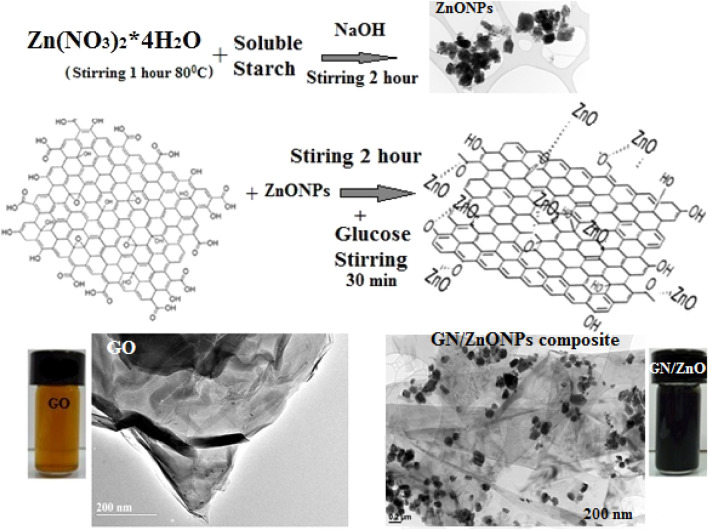
Initially, graphene oxide (GO) was synthesized employing the conventional “Hummer’s method”43,44. For the synthesis of GN:ZnO NC, 50 mg of synthesized GO was dispersed in 100 cm3 of deionized H2O which was exfoliated in an ultrasonic bath using ultrasonication for 15 min. Then to the exfoliated GO dispersion, 50 mg of as-prepared ZnO NPs were added. The 1:1 mixture (by weight) of GO and ZnO NPs was vigorously magnetically stirred for about 2 h for formation of homogeneous suspension. This 100 ml homogenous suspension, 1 g glucose was added followed by stirring for 30 min to reduce GO to GN escaping the use of extremely toxic hydrazine as a reductant. During this process, GO is reduced to GN, and simultaneously the deposition of ZnO NPs onto the GN sheets is achieved. The same procedure was applied for the synthesis of GN but independently and in absence of ZnO NPs to make a comparison. The blackish dispersion obtained was then filtered using a Millipore filtration system and was given washing three times with each of water and ethanol. The obtained GN and the GN:ZnO NC were dried in an oven. Complete representation for the synthesis of GN:ZnO NC is shown in Scheme 1. Finally, 5 mg of the composite as synthesized was dispersed in 2.5 cm3 of ethanol under ultra-sonication for 5 min to make a suspension for electrode fabrication.
Scheme 1.
Schematic representation for synthesis of GN:ZnO NC.
Fabrication of GN:ZnO NC/GCE
A bare GCE was washed in a 1:1 nitric acid: water mixture and then polished by alumina powder to form a clean shiny surface. It was then again washed with the nitric acid–water mixture and ethanol ultrasonically and dried in air. 10 µL of the as-prepared suspension of the composite material was deposited onto the conducting part of GCE surface with the help of a micropipette. The solvent was evaporated using an IR lamp. When the solvent was removed, the testing material was adhered on the electrode surface which was again rinsed thoroughly with distilled water and dried again under an IR lamp. This fabricated electrode was designated as GN:ZnO NC/GCE and was used in successive electrochemical studies.
Characterizations
X-ray diffraction crystallographic studies were carried out using XRD machine (Shimadzu, Japan). S-4800 field emission SEM system was used to take scanning electron microscopic (SEM) images. It is operating at 20.0 kV and equipped with energy-dispersive X-ray (EDX) spectroscopic technique for elemental analysis. Transmission electron microscopic (TEM) and High-resolution-(HR)-TEM (JEOL JEM-2100F microscope at 200 kV with “Orius SC1000” camera) techniques were used to further analyze the micro-structure of the materials. Initially the samples were dispersed in dimethyl formamide and then bath-sonicated for 30 min at room temperature43. The samples were drop-casted on a carbon-grid and allowed to dry. When the solvent got removed by evaporation, the sample got adhered to the grid44. Raman spectra were obtained using (RE-04) Raman spectrometer having a solid state laser diode-pumping at 514 nm.
All the voltammetric and Chronocoulometric experiments were performed using an Electrochemical-workstation (CH-Inst. Model-CHI1100B) with a three-electrode cell. The bare-glassy carbon electrode (GCE, 3 mm diameter) was modified to act as working electrode. Platinum wire and Ag/AgCl were auxiliary and reference electrodes, respectively. Electrochemical-impedance experiments were carried out in 0.1 M KCl soln. along with a mixture of 1.0 mM [Fe(CN)6]−3 and [Fe(CN)6]−4 (1:1) was taken as standard probe employing Electrochemical-workstation (Autolab PGSTAT 30 with Frequency Response Analyzer). The pH readings were taken using ELICO LI-120 digital pH meter.
Results and discussion
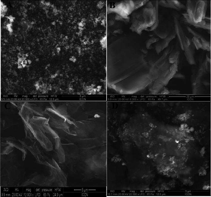
The XRD plots of the synthesized ZnO nanopowder, GO and GN:ZnO NC composite are given in Fig. 1. XRD analysis of ZnO NPs depicts diffraction peaks at 31.80°, 34.4°, 36.3°, 47.5°, 56.6°, 62.8° and 69.0° in the (Fig. 1A), confirming the hexagonal wurtzite structure of ZnO NPs41,52,53 (JPCDS number: 36-1451)52. Also, the presence of quite sharp peaks and the absence of any other peak indicate that the synthesized nanopowder does not contain other impurities. The size of ZnO-NPs was also calculated with the help of “Debye–Scherrer formula, d = 0.89λ/βcosθ”54,55, where, 0.89 is Scherer’s factor, λ is wavelength of x-ray used, ‘θ’ the “Bragg diffraction angle”, and ‘β’ a constant known as the “full width at half-maximum (FWHM)” of a particular diffraction angle. The mean particle size of the ZnO-NPs corresponding to the FWHM of the most intense peak at 36.3° and corresponding to (101) plane using the “Scherrer formula”56,57 was found to be 57.17 nm. The diffraction peak at a 2θ value of 10.81° characteristics of GO (Fig. 1C) disappears in the XRD pattern of as-prepared GN:ZnO NC composite (Fig. 1D). However, a new diffraction peak characteristic of GN at 26.52° with inter-layer spacing (d-spacing) of 0.35 nm was observed in the XRD outline of GN:ZnO NC composite. This confirms that the exfoliated GO has been reduced to GN. The other XRD peaks of the GN:ZnO NC composite can be ascribed to the crystallize ZnO NPs. Further, the XRD peak in case of GN:ZnO NC composite corresponding to GN shifted slightly as compared to individual GN at 26.20° (Fig. 1B) which again confirms GN-ZnO NPs interaction. The ZnO NPs and GN:ZnO NC composites were further analyzed using SEM and EDX techniques. Figure 2A shows the SEM image of as-synthesized ZnO-NPs. As observed, the approximate spherical-shaped ZnO NPs were formed in the range of 40–90 nm size which agrees with the particle size calculated using the “Debye–Scherrer formula” in XRD studies as mentioned above. Figure 2B is the SEM image with distinctive crumpled and wrinkled surface characteristics of GO and Fig. 2C shows the multi-layer structure of graphene sheets stacked together. Figure 2D is the SEM image of GN/ZnO composite which was taken after reduction of GO in presence of ZnO NPs. It is observed that ZnO-NPs are closely spread on the surface of graphene-nanosheets. However, in some regions, few ZnO NPs appear aggregated on the surfaces of GN nanosheets, but most of the ZnONPs are found on the GN sheets and several of which are wrapped by GN sheets.
Figure 1.
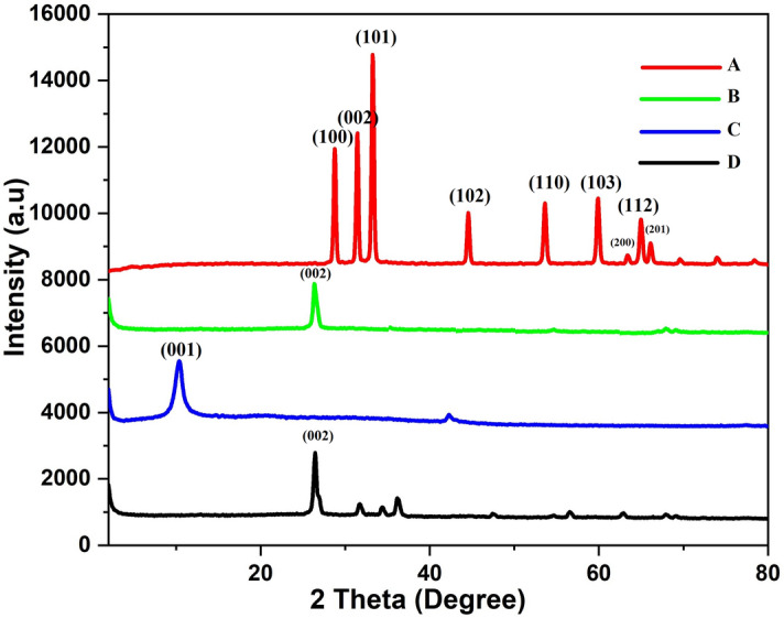
X-ray diffraction pattern depicting peak-indices and 2θ values of (A) ZnO NPs, (B) GN (C) GO and (D) GN:ZnO NC.
Figure 2.
Scanning electron microscopic images of (A) ZnO NPs, (B) GO, (C) GN and (D) GN:ZnO NC.
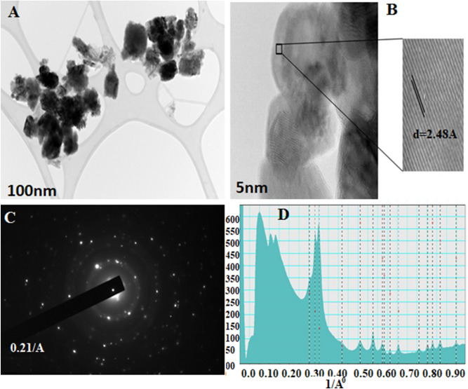
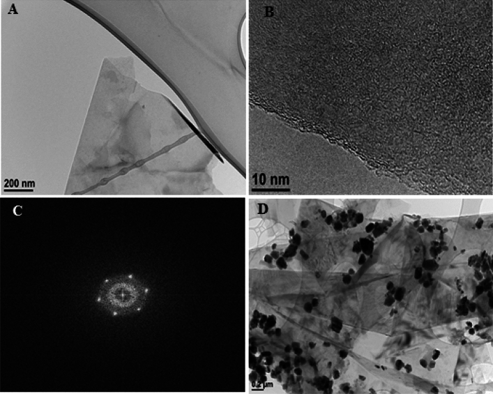
TEM and HRTEM were used to further characterize the microstructure of synthesized ZnO NPs and GN:ZnO NC. Figure 3A shows the TEM image of ZnO NPs which confirms its formation and the particle size varies from 40–100 nm as analyzed by the particle size analyzer which agrees with the results as obtained in SEM images. HR-TEM image of ZnO NPs from a single nanoparticle is shown in Fig. 3B. The selected area electron-diffraction (SAED) outline so obtained is given in Fig. 3C and the rotational average profile of ZnO NPs is given in Fig. 3D. The lattice spacing values obtained from the SAED outline and the standard “JCPDS” values are given in Table S1. The lattice spacing calculated from the selected area is 2.48 Å which matches perfectly with the value obtained from the standard JCPDS card (2.47590 Å) corresponding to 101 planes of hexagonal ZnO. The values given in Table S1 agree very well within the error limit of ± 2.5% confirming the hexagonal phase of the ZnONPs. Figure 3D represents the rotational average profile of ZnO-NPs. Figure 4A represents the TEM image and Fig. 4B shows the HRTEM image of as-synthesized GN and Fig. 4C shows its corresponding Fast Fourier Transform (FFT) pattern. The hexa-gonal FFT outline shows that the as-synthesized GN is highly crystalline in nature with minimum defects. TEM image of GN:ZnO NC is shown in Fig. 4D which shows that the ZnO-NPs are well dispersed in the graphene sheets.
Figure 3.
(A) Transmission electron microscopic and (B) High resolution-TEM images of ZnO NPs, (C) Selected area electron-diffraction outline of ZnO NPs, (D) Rotational average profile of ZnONPs.
Figure 4.
(A) Transmission electron microscopic and (B) HR-TEM images of GNs, (C) Fast Fourier Transform (FFT) pattern of GNs and (D) TEM image of GN:ZnO NC hybrid.
These results show that there is a good interfacial contact of GN and ZnO-NPs. The existence of ZnO-NPs within the GN layers will help to prevent the agglomeration as well as restacking of plane GN layers and simultaneously its available surface-area gets also increased to improve its electrochemical performance. The GN:ZnO NC was also characterized by EDAX technique. Figure 5(1) shows the EDAX spectrum of GN:ZnO NC with peaks characteristic of C, O, and Zn elements. The respective weight percentages using EDAX studies of C, O, and Zn are 66.94, 10.85, and 22.21. This reveals a good loading of ZnO-NPs on graphene nanosheets, which agrees with the SEM and TEM interpretations. In this way, a good dispersion of ZnO NPs with GN is achieved.
Figure 5.
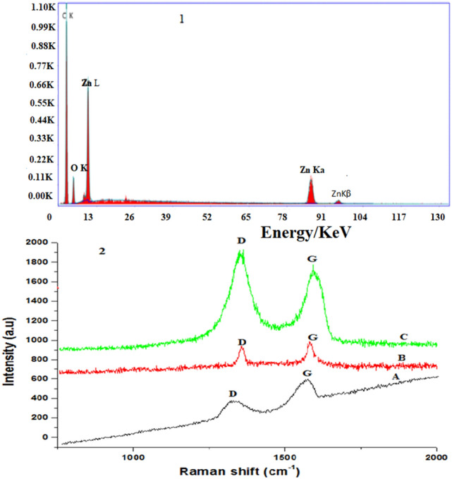
(1) Energy dispersive x-ray spectra of GN:ZnO NC. (2) Raman-spectra of (A) GO, (B) GN and (C) GN:ZnO NC.
Raman spectroscopy is a very sensitive technique for electronic structure and is an essential tool to examine well-ordered and disordered crystalline structures and is widely used to characterize carbonaceous materials like graphene58. Raman spectra of the GO, GN, and GN:ZnO-NC are presented in Fig. 5(2). The two characteristic bands in the Raman spectrum of GN and graphene-based materials are low intensity ‘D-band’ and high intensity ‘G-band’ at 1351 and 1602 cm−1, corresponding to the disordered carbon and in-plane stretching vibration of sp2 C − C bonds, respectively44,59. The ID/IG intensity ratio gives the degree of disorderness and the mean size of the sp2-domains in graphitic materials44,59,60. Figures 5(2A,B) shows that ID/IG intensity ratio is increased for GN as compared to GO which indicates that there is a decrease in the in-plane sp2-domain size on graphene-oxide reduction59. The ID/IG value gets increased from 0.54 in GO to 1.13 in GN:ZnO NC (Fig. 5(2C). This is because on reduction of GO to GN, oxygen functionalities are removed, which decreases the size of the sp2-domains in graphene and interaction between ZnO NPs and graphene nanosheets44,45,61.
During the reduction of GO to GN, most oxygen based functionality are removed and restores the conductivity of graphene, and also increases the stability of GN:ZnO NC62. However, some residual oxygen-containing groups are still left unreduced on GN41. The Zn atoms present in ZnO NPs coordinate with the oxygen atoms of the unreduced functional groups via covalent-interaction which helps in composite formation42. Graphene bearing edges and planes will provide support to spherical ZnO NPs to grow and form GN:ZnO NC.
Electrochemical characterization of GN:ZnO NC/GCE
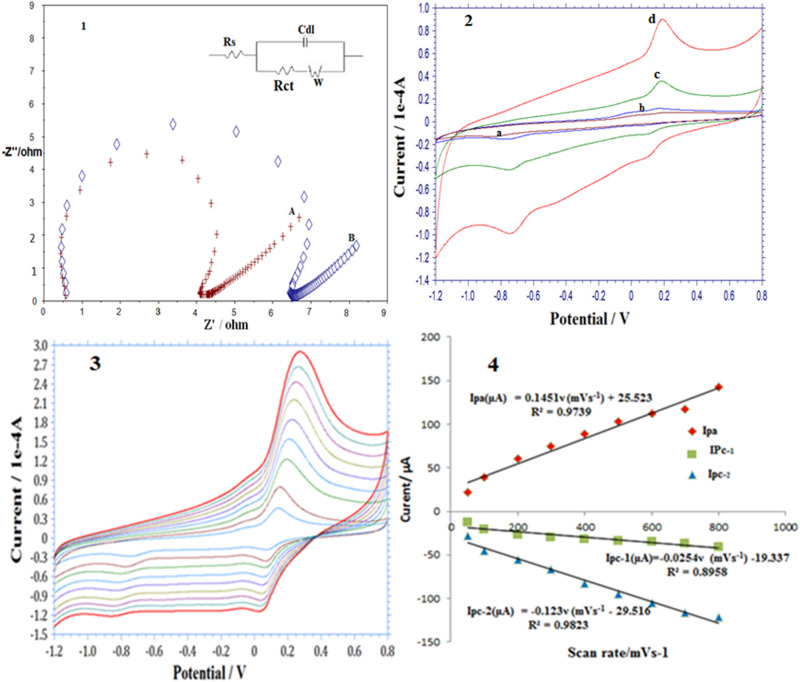
Electrochemical impedance spectroscopic (EIS) and chrono-coulometric (CC) techniques were applied to characterize the GN:ZnO NC modified GCE. EIS is a suitable technique commonly used to characterize a surface modified GCE45. In the Nyquist diagrams, the semicircular-region obtained at high frequency values correspond to the charge transfer limiting phenomena and from the diameter of the semicircular part, charge transfer values (Rct) are calculated63. EIS analysis shows that the Rct value for GN:ZnO NC/GCE (Fig. 6(1A)) is 3.6 Ω which is lower than that of GN/GCE (6.0 Ω, Fig. 6(1B)). The decrease in Rct value confirms that the ZnO-NPs present in the composite improves the charge transfer characteristics of GN:ZnO NC modified GCE42. CC was applied to determine the electrochemically active surface area of the GN and GN:ZnO NC modified electrodes by using Anson charge-time dependence equation Q = nFACD1/2 π−1/2 t1/2, where ‘Q’ is charge in coulombs, ‘n’ number of electrons involved in redox reaction is 1 for K3[Fe(CN)6] system, ’F’ Faraday Constant (96,485 Cmol−1), ‘A’ area of electrode in cm−2, ‘C’ concentration of K3[Fe(CN)6] (1 mM), ‘D’ diffusion coefficient is 7.6 × 10⁻⁶ cm2s−1 for K3[Fe(CN)6] and ’t’ is time in seconds [30]. The slope obtained from the plot of Q against t1/2 using CC and 1 mM K3[Fe(CN)6] standard complex was used to calculate the effective electrochemical surface areas for GN and GN:ZnO NC modified electrodes (Fig. S1A,B, supplementary material). Basd on the linear relationship between Q and t1/2, the slopes obtained for GN:ZnO NC/GCE and GN/GCE were found to be 138.40 Cs−1/2 and 76.44 Cs−1/2, respectively (Fig. S1A,B). The effective electrochemical surface areas for GN:ZnO NC/GCE and GN/GCE were found to be 0.166 cm2 and 0.092 cm2, respectively.
Figure 6.
(1) Nyquist-plots for (A) GN:ZnO NC modified GCE and (B) GN modified GCE in 0.1 M KCl solution in presence of 1.0 mM each of Fe[(CN)6]−3 and Fe[(CN)6]−4 (1:1). Insert: Equivalent circuit for fitting the Nyquist-plot. (2) CV of (a) phosphate-buffer at pH 6.8 (blank) at bare GCE and 1.96 × 10⁻⁶ M p-NP in phosphate buffer at pH 6.8. (b) Bare GCE, (c) GN:GCE and (d) GN:ZnO NC modified GCE, Scan rate100 mVs−1. (A) Effect of scan-rate on peak current of p-NP (1.96 × 10⁻⁶ M) using cyclic voltammetry at 50, 100, 200, 300, 400, 500, 600, 700, 800 mVs−1 using GN:ZnO NC/GCE. (B) Plots of peak current (Ipa, Ipc) vs. potential scan rate (v).
Electrochemical performance of GN:ZnO NC modified GCE for p-NP detrmination
Cyclic voltammetric (CV) study was carried out at a scan-rate of 100 mVs−1 to investigate the electrochemical behavior of 1.96 × 10⁻⁶ M p-NP at bare GCE, GN/GCE, and GN:ZnO NC/GCE. Figure 6 (2a) is the CV of the blank containing phosphate buffer at pH 6.8 at bare GCE. However, using bare-GCE at the same scan-rate and concentration of 1.96 × 10⁻⁶ M, p-NP gives a weak redox couple at 0.20 V ascribed to p-NP (Fig. 2b), while the peak current progressively increased on GN modified GCE (Fig. 2c), and further enhanced with GN:ZnO NC modified GCE (Fig. 2d). Also, the irreversible reduction peak of p-NP at − 0.75 V grows progressively at both GN and GN:ZnO NC modified GCE. This shows that both ZnO NPs and GN had an electrocatalytic effect where ZnO NPs act as spacer to enhance the effective-surface area of GN and improve the electrochemical activity while GN decreases the resistance by providing a conducting path. Therefore, the synergic effect of ZnO NPs and GN as cocatalysts is responsible for the electrocatalytic activity of GN:ZnO NC/GCE and its application in the electro-analysis of para-nitrophenol. This is due to the excellent catalytic capability and large effective-surface area of GN:ZnO-NC/GCE which facilitates p-NP to accommodate at the electrode-surface and enhances the electron-transfer process. The current intensity increased progressively on GCE modified with GN and GN:ZnO NC. Figure 6(2c) shows that one large reduction peak appears at − 0.75 V on the first cathodic sweep for the p-NP and another redox couple with one anodic-peak at 0.19 V and another cathodic-peak at 0.11 V is obtained. It is observed from the cyclic voltammograms (Fig. 6) that this redox couple grows in peak current at the expense of first irreversible reduction-peak at 0.75 V. This indicates that the irreversible reduction product of p-NP stays on the modified-electrode surface and gets oxidized in the anodic sweep. This is also in agreement with the literature employing the conventional electrodes18,21,64,65. The first irreversible reduction-peak obtained is ascribed to the irreversible reduction of the nitroaromatic moiety to yield the hydroxylamine derivative (NHOH) with the four-electron transfer, according to Scheme 2. The anodic redox couple is due to the redox reaction of hydroxylamine and p-NP involving two electrons in the reaction18,30. Figure 6(3) shows the scan rate effect on peak current using CV for p-nitrophenol (1.96 × 10⁻⁶ M) in phosphate-buffer of pH 6.8 at 50, 100, 200, 300, 400, 500, 600, 700, 800 mVs−1 at GN:ZnO NC/GCE. The plot of scan rate against the cathodic and anodic-peak current (Fig. 6(4)) of reduction product of p-NP (hydroxylamine) gives straight lines which indicate that both the electrode processes are adsorption controlled. Also, when reduction peak-current of p-nitrophenol was plotted against scan-rate, a straight line was obtained indicating an adsorption-controlled process.
Scheme 2.
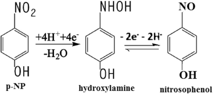
Electrode reaction mechanism of p-nitrophenol at GN:ZnO NC/GCE.
Experimental parameters and their optimization for p-nitrophenol determination
Effect of pH values
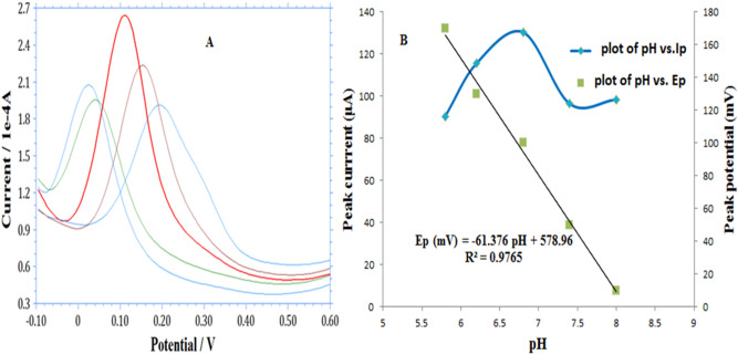
Nature as well as the pH of supporting electrolytes play a vital role in the determination of analytes using any chemically modified electrode. Many supporting electrolytes were analyzed. While choosing a specific supporting electrolyte for p-NP determination, peak current as well as peak shape were taken into consideration. 0.1 M each of borate, acetate, phosphate, and Britton Robinson buffer were tested. In citrate buffers of pH 2.1 and KH2PO4–K2HPO4 buffer solution of pH 6.8, high-intensity anodic peak currents were observed. However, in the citrate buffer of pH 2, the anodic peak was unstable. Therefore, for subsequent studies, the optimum buffer solution chosen was phosphate buffer (pH 6.8). Differential pulse voltammetry, square-wave voltammetry, and cyclic voltammetric techniques were applied to optimize the pH values and it was found that square-wave voltammetry responded the best in respect of peak-current and peak-resolution. Square wave adsorptive stripping voltammograms of p-NP (1.66 × 10⁻⁶ M) at GN:ZnO NC/GCE and various pH values of 0.1 M potassium phosphate buffer are presented in Fig. 7A. It is observed that the peak current goes on increasing with the increase in pH values up to 6.8 but at 7.4 and 8.0 pH values it decreases. It confirms that the redox reaction of p-NP is favored in the acidic medium. Therefore, for further studies phosphate-buffer of pH 6.8 was selected as the supporting-electrolyte. Also, it is observed from Fig. 7A that the peak-potential is shifting towards a more negative side as the pH increase which confirms the proton involvement in the reaction18. A plot of pH against shift in peak potential (Fig. 7B) gives a straight line with a slope equal to 61.3 mV/pH which confirms that protons and electrons are equally taking part in the electrode reaction as shown in Scheme 2.
Figure 7.
(A) Square wave voltammograms of p-NP (1.66 × 10⁻⁶ M) at GN:ZnO NC /GCE at different pH values (5.8, 6.2, 6.8, 7.4, 8 from right to left). Deposition potential: − 1 V. Deposition time 300 s. (B) Plot of pH against peak current (Ip) and peak potential (Ep).
Optimization of deposition potential and deposition time
Deposition potential and deposition time, both effect the degree of adsorption of the electrochemically reduced p-NP at the electrode surface (NH–OH). Therefore, both these paratmeters need to be optimized. The effect of deposition-potential and deposition-time on peak current was investigated using SW-AdSV. It was found that the deposition-potential largely effects the peak current of para-NP at the modified electrode and the peak current enhanced from − 0.1 to − 1.0 V and then diminished. Hence a deposition-potential of − 1.0 V was optimized for subsequent studies. Figure S2 (Supplementary material) presents the effect of deposition-time on the anodic peak current of NH–OH for 10.70 × 10⁻⁷ M p-NP at GN:ZnO NC/GCE under optimized conditions (deposition potential: − 1 V, potassium phosphate-buffer of pH 6.8 as supporting electrolyte) for various deposition times (60, 120, 200, 300, 400 and 500 s). The peak current increases with the increase in deposition-time from 60 to 300 s and then remain almost constant. This is because the modified electrode surface gets saturated due to the accumulation of reduction product (NH–OH). In this way, sensitivity can be improved by increasing the deposition time to determine low concentration levels. A deposition time of 300 s was optimized for subsequent experiments.
Calibration curve
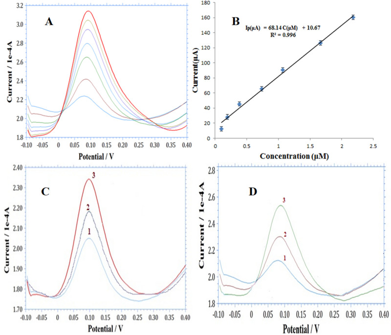
Figure 8A shows the calibration curve for the fabricated sensor using the optimized experimental parameters. The calibration curve was used for the quantitative analysis of p-NP solutions using GN:ZnO NC/GCE employing SW-AdSV using the as-optimized experimental parameters. The stripping-peak current gave a linear relation by changing the concentrations of p-NP from 0.09 × 10⁻⁶ M to 21.80 × 10⁻⁶. Using the linear plot of the peak current (ipa) versus concentration of p-NP (Fig. 8B) the regression equation obtained is; ipa (µA) = 68.14 C(µM) + 10.67 (r2 = 0.996). This equation was used to calculate the limit of detection (LOD) and sensitivity of the developed sensor according to IUPAC recommendations65,66. LOD = 3 s/k where s is the standard deviation of intercept for replicate determination values under the similar conditions (n = 5); k is the sensitivity, namely the slope of the calibration graph. The limit of detection (LOD) and sensitivity of the developed sensor were estimated to be 8.8 × 10⁻⁹ M (S/N = 3) and 68.14 μA μM−1, respectively. This very low detection limit of the sensor shows that the developed method can potentially be applied for the sensitive determination of p-NP. A comparative analysis for the performance of the developed sensor with different electrochemical sensors based on different nanomaterials for the sensitive determination of p-nitrophenol is summarized in Table 1. It is obvious from Table 1 that the sensor based on the present nanocomposite material is superior in respect of its wide linear working range and more important its limit of detection is least compared to those reported by the earlier workers. It can be concluded that the GN:ZnO NC-based electrode is comparatively an first-rate platform for the sensitive determination of p-NP. This improved performance of the electrochemical sensor is attributed to the synergistic effect of GN and ZnONPs based composite material due to their high adsorption ability and excellent electrocatalytic activity.
Figure 8.
(A) Square wave adsorptive stripping voltammograms at GN:ZnO NC/GCE at different concentrations of p-NP using phosphate buffer (pH 6.8). Deposition-potential: − 1.0 V, deposition-time: 300 s. (B) Plot of concentration of p-NP aganist peak current with error bars. (C) SW-AdS voltagrams for p-NP determination using standard adition method spiked with (1) 35.61 × 10⁻⁸ M ,(2) 71.22 × 10⁻⁸ M and 106.83 × 10⁻⁸ M p-NP in river water sample. (D) SW-AdS voltagrams for p-NP determination using standard adition method spiked with (1) 35.61 × 10⁻⁸ M , (2) 71.22 × 10⁻⁸ M and 106.83 × 10⁻⁸ M p-NP in ground water sample.
Table 1.
Comparison of the performances of different electrochemical sensors for p-nitrophenol.
| Electrode material | Linear range (μM) | Correlation coefficient | Detection limit (μM) | References |
|---|---|---|---|---|
| aAuNP/RGO | 0.05–2 | – | 0.01 | 18 |
| Bi2O3NPs/CNT | 1–10 | – | 0.1 | 22 |
| bMWNT-Nafion/GCE | 0.1–10 | 0.998 | 0.04 | 23 |
| alpha-MnO2/MWCNTs | 30–475 | – | 0.186 | 24 |
| Nano-gold/GCE | 10–1000 | – | 8 | 25 |
| Nanostructured CeO2 doped Ag | 7.8–1000 | – | – | 28 |
| GO/GCE | 0.1–120 | 0.997 | 0.02 | 30 |
| cPd NFs/C60-NH2 | 5–1000 | – | 0.09 | 33 |
| Nanoporous gold | 3.59–71.8 | 0.994 | – | 64 |
| GN:ZnO NC/GCE | 0.099–21.8 | 0.996 | 0.0088 | This work |
aGold nanoparticle/reduced graphene oxide, bMulti-wall carbon nanotubes, cPalladium nano-flowers supported on fullerene.
Effect of potentially interfering substances
Selectivity of the prepared GN:ZnO NC-based sensor for the analysis of p-nitrophenol was evaluated by studying the influence of some important interfering substances of p-NP. The interference study was examined in 0.1 M phosphate-buffer of pH 6.8 in presence of 1.07 × 10⁻⁶ M p-NP. Most of the phenols like pyrocatechol, o-aminophenol, hydroquinone, hydroxyphenyl, , p-aminophenol, and chlorophenol did not affect the signals of p-NP with deviations less than 4.0% up to the 100-fold excess. The common nitrophenols that contain the same nitro group as that of p-nitrophenol are o-nitrophenol, m-nitrophenol, and 2, 4, di-nitrophenol. The interference of these nitrophenols was tested on the GN:ZnO NC modified GCE. It was found that o-NP produced a separate reduction peak at − 0.10 V at the same concentration and suppressed the signal of p-NP at higher concentrations as shown in Fig. S3 (Suplementary material). Therefore, it can be concluded that both p-nitrophenol and o-nitrophenol can be detected simultaneously at the said modified GCE at a lower concentration. However, the presence of m-NP and 2, 4, DNP did not affect the determination of p-nitrophenol. The results obtained from interference tests indicate that GN:ZnO NC-based sensor will be appropriate for the analytical determination of p-NP in presence of the above-mentioned phenolic compounds and meta and ortho-nitrophenol.
The fabricated electrode was also evaluated for its reproducibility and stability. The reproducibility for the detection of 1.66 × 10⁻⁶ M p-NP using 0.1 M phosphate-buffer of pH 6.8 employing five electrodes was found to have a relative-standard-deviation (RSD) value of 4.70%, revealing a good reproducibility of the sensor. The stability of the sensor was also examined in 0.1 M phosphate-buffer of pH 6.8 for 1.66 × 10⁻⁶ M p-nitrophenol and the current response obtained was periodically monitored. An initial peak current response of about 93.40% of the sensor was retained for 15 days which proved good stability of the sensor. The repeatability of the as developed sensor was also carried out for 10 successive measurements with an RSD value of 4.0% indicating good repeatability.
Accuracy and precision of the method
The accuracy of the method as developed was determined by spiking an exactly weighed amount (pre-analyzed amount) of p-NP. The accuracy is represented as a mean relative error (Table 2A). The mean percentage recovery was found to be 98.93% and 99.34% for intra-day and inter-day assays, respectively. The recovery values are useful to provide good confidence in the accuracy of the developed method.
Table 2.
(A) Precision and accuracy analysis for p-nitrophenol in pre-analyzed samples by the developed method (SW-AdSV), (B) Percentage recovery assay for p-nitrophenol in real water samples by the developed procedure (SW-AdSV).
| (A) | ||||
|---|---|---|---|---|
| Added (10−8 M) | Found (10−8 M) | (%R) | Precision (% R.S.D, n = 5) | Accuracy (% Bias) |
| Intraday | ||||
| 19.60 | 19.34 | 98.63 | 1.21 | − 1.32 |
| 38.4 | 38.35 | 99.80 | 1.78 | − 0.13 |
| 74.00 | 75.20 | 98.37 | 1.90 | 1.62 |
| Interday | ||||
| 19.6 | 19.80 | 98.97 | 1.89 | 1.02 |
| 38.4 | 38.76 | 100.39 | 2.07 | 0.93 |
| 74.0 | 73.01 | 98.66 | 1.88 | − 1.33 |
| B: | ||||
|---|---|---|---|---|
| Sample | Added(10−8 M) | Founda (10–8 M) (n = 5) | % Recovery | |
| Panchganga river water | 35.61 | 34.78 | 97.66 | |
| 71.22 | 70.01 | 98.30 | ||
| 106.83 | 103.99 | 97.34 | ||
| Ground water | 35.61 | 34.59 | 97.13 | |
| 71.22 | 69.20 | 97.16 | ||
| 106.83 | 104.31 | 97.64 | ||
aAverage of five measurements.
The precision of the developed method was also determined. The concentration of p-nitrophenol was analyzed in pre-analyzed sample solutions five times in intra-day assay and successively for five days in an inter-day assay using the SW-AdSV techniques. The precision and the percentage recovery values of the developed method obtained as the mean of five separate measurements are given in Table 2A. The average values of variation-coefficients for intra-day and inter-day assays based on five measurements were found to be 1.63% and 1.94%, respectively. Thus, the results obtained confirm a good reproducibility of the fabricated electrode and a high precision of the developed method.
Practical application of the developed senor in the determination of para-NP in real samples
The GN:ZnONPs composite based sensor was used to analyze p-NP in various real water samples. Groundwater sample was collected from Andheri (East) and river water sample from Panchganga River, Mumbai. 0.1 M potassium phosphate-buffer (supporting electrolyte) was directly prepared in 100 cm3 of each sample which was used as an analyte. 25 cm−3 of sample volume was taken in an electrochemical cell for detection of p-NP using the standard addition method. The developed method was applied under the optimized experimental parameters (deposition potential: − 1.0 V and deposition time : 300 s) and under the established concentration range. The measurements were performed three times. It was found that neither p-NP nor o-NP was detected in both these water samples. Therefore, the desired amount of standard p-nitrophenol was spiked into the collected water samples for performing the recovery tests. Figure 8C shows the SW-AdSV curves for standard p-nitrophenol spiked in river water sample and Fig. 8D shows the SW-AdSV curves for standard p-nitrophenol spiked in ground water sample under the optimized conditions. The percentage recovery of both the samples is given in Table 2B. A recovery of 97.660% and 97.130% was obtained in river and groundwater samples respectively. These observations validate the suitability of the developed method for the determination of p-nitrophenol in the natural water samples.
Conclusion
A novel electrochemical method employing GN:ZnONC was established for the sensitive determination of p-nitrophenol. The good dispersibility of ZnO NPs with GN demonstrated an improved electrocatalytic performance for the determination of p-nitrophenol in contrast with GN-based GCE. This can be attributed to the outstanding electrical conductivity, large adsorptive capacity, and very high effective surface area of GN and ZnONPs. In addition, it was found that simultaneous detection of ortho and p-NPs can be achived at the said electrode without the interference of other possible interferents at low concentrations. The developed procedure was applied to determine p-nitrophenol in the river and ground water samples using SW-AdSV with acceptable recoveries of 97.660% and 97.130% respectively. It is evident from these results that the GN:ZnO NC composite is a good candidate for advanced electrode materials for the sensitive determination of p-NP.
Supplementary Information
Acknowledgements
Authors are thankful to the D/O Chemistry, University of Mumbai for providing facilities to carry out all electrochemical studies and the author (RAD) is also grateful to the UGC, New Delhi, India for awarding financial assistance under its Dr. D. S. Kothari Post-doctoral fellowship scheme. The authors also acknowledge the US ARMY Research Laboratory for SEM and TEM Characterizations and University of Mumbai, Mumbai, India for providing a research grant under its minor research project (MRP) grant to RAD under project number 673.
Author contributions
R.A.D: Conceptualization, experimental design, sample preparation, measurement, data analysis, methodology and writing, review and supervision, G.A.N: Review, proofreading, editing, similarity index check out, A.K.S: Data interpretation, data analysis using different techniques, review, validation, supervision, I.U.H: Review, proofreading, S.P.K: measurement, data analysis, methodology and writing, review and supervision, L.G: SEM/TEM characterizations and data analysis, A.M.H.S: Review and editing, M.R: Review, validation, editing and proofreading, W.A: Review, proofreading.
Competing interests
The authors declare no competing interests.
Footnotes
Publisher's note
Springer Nature remains neutral with regard to jurisdictional claims in published maps and institutional affiliations.
This article has been retracted. Please see the retraction notice for more detail: https://doi.org/10.1038/s41598-024-73187-0"
Change history
3/14/2024
Editor’s Note: Editor’s Note: Readers are alerted that this Article is the subject of concerns which are currently under consideration by the Editors. Appropriate editorial action will be taken once this matter is resolved.
Change history
9/24/2024
This article has been retracted. Please see the Retraction Notice for more detail: 10.1038/s41598-024-73187-0
Contributor Information
Riyaz Ahmad Dar, Email: riyazabid.2008@rediffmail.com.
Gowhar Ahmad Naikoo, Email: gahmed@du.edu.om.
Supplementary Information
The online version contains supplementary material available at 10.1038/s41598-021-03495-2.
References
- 1.Veerakumar, P. et al. Nickel nanoparticle-decorated porous carbons for highly active catalytic reduction of organic dyes and sensitive detection of Hg(II) ions. ACS Appl. Mater. Interfaces7, 24810–24821 (2015). [DOI] [PubMed] [Google Scholar]
- 2.Rajkumar, C., Veerakumar, P., Chen, S., Thirumalraj, B. & Lin, K. C. Ultrathin sulphur-doped graphitic carbon nitride nano sheets as metal-free catalyst for electrochemical sensing and catalytic removal of 4-nitrophenol. ACS Sustain. Chem. Eng.6, 16021–16031 (2018). [Google Scholar]
- 3.Zhang, Z., Zhua, Y., Wena, R., Dong, G. C. & Li, H. W. Selfassembled palladium nanoflowers supported on fullerene electrochemical catalytic performance for the reduction of 4-nitrophenol. Electrochem. Commun.104, 106484–106491 (2019). [Google Scholar]
- 4.Wu, S., Fan, S., Tan, S., Wang, J. & Li, C. A new strategy for the sensitive electrochemical determination of nitrophenol isomers using β-cyclodextrin derivative-functionalized siliconcarbide. RSC Adv.8, 775–784 (2018). [DOI] [PMC free article] [PubMed] [Google Scholar]
- 5.National Center for Biotechnology Information. PubChem Compound Summary for CID 980 (2021). https://pubchem.ncbi.nlm.nih.gov/compound/4-Nitrophenol.
- 6.Kalaimurugan, D. et al. Novel strategy for biodegradation of 4-nitrophenol by the immobilized cells of Pseudomonas sp. YPS3 with Acacia gum. Saudi. J. Biol. Sci.28, 833–839 (2021). [DOI] [PMC free article] [PubMed] [Google Scholar]
- 7.Kumar, A., Kumar, S. & Gupta, D. V. Adsorption of phenol and 4-nitrophenol on granular activated carbon in basal salt medium: Equilibrium and kinetics. J. Hazard. Mater.147, 155–166 (2007). [DOI] [PubMed] [Google Scholar]
- 8.Prashant, T. D., Dilip, H. L. & Ramakant, S. I. Removal of 4-nitrophenol from aqueous solution by adsorption onto activated carbon prepared from Acacia glauca sawdust. Water Sci. Technol.73(4), 955–966 (2016). [DOI] [PubMed] [Google Scholar]
- 9.Kovacs, A., Mortl, M. & Kende, A. Development and optimization of a method for the analysis of phenols and chlorophenols from aqueous samples by gas chromatography–mass spectrometry, after solid-phase extraction and trimethylsilylation. Microchem. J.99, 125–131 (2011). [Google Scholar]
- 10.Zhang, M. et al. Gas chromatographic determination of three chlorophenols in toilet paper by ultrasonic assisted extraction and synchronous derivative dispersive liquid–liquid microextraction. Anal. Methods6, 207–214 (2014). [Google Scholar]
- 11.Hofmann, D., Hartmann, F. & Herrmann, H. Analysis of nitrophenols in cloud water with a miniaturized light-phase rotary perforator and HPLC–MS. Anal. Bioanal. Chem.391, 161–169 (2008). [DOI] [PubMed] [Google Scholar]
- 12.Yamauchi, Y., Ido, M., Ohta, M. & Maeda, H. High performance liquid chromatography with an electrochemical detector in the cathodic mode as a tool for the determination of p-nitrophenol and assay of acid phosphatase in urine samples. Chem. Pharm. Bull.52, 552–555 (2004). [DOI] [PubMed] [Google Scholar]
- 13.Niazi, A. & Yazdanipour, A. Spectrophotometric simultaneous determination of nitrophenol isomers by orthogonal signal correction and partial least squares. J. Hazard. Mater.146, 421–427 (2007). [DOI] [PubMed] [Google Scholar]
- 14.Zhang, W. & Wilson, C. R. Indirect fluorescent determination of selected nitro-aromatic and pharmaceutical compounds via UV-photolysis of 2-phenylbenzimidazole-5-sulfonate. Talanta74, 1400–1407 (2008). [DOI] [PubMed] [Google Scholar]
- 15.Guo, X., Wang, Z. & Zhou, S. The separation and determination of nitrophenol isomers by high-performance capillary zone electrophoresis. Talanta64, 135–139 (2004). [DOI] [PubMed] [Google Scholar]
- 16.Lupu, S., Lete, C., Marin, M., Totir, N. & Balaure, P. C. Electrochemical sensors based on platinum electrodes modified with hybrid inorganic-organic coatings for determination of 4-nitrophenol and dopamine. Electrochim. Acta.54, 1932–1938 (2009). [Google Scholar]
- 17.Devasenathipathy, R., Mani, V., Chen, S. M., Manibalan, K. & Huang, S. T. Determination of 4-nitrophenol at iron phthalocyanine decorated graphene nanosheets film modified electrode. Int. J. Electrochem. Sci.10, 1384–1392 (2015). [Google Scholar]
- 18.Yanhong, T., Run, H., Chengbin, L., Shanli, Y. & Shenglian, L. Electrochemical detection of 4-nitrophenol based on a glassy carbon electrode modified with a reduced graphene oxide/Au nanoparticle composite. Anal. Methods5, 5508–5514 (2013). [Google Scholar]
- 19.Abaker, M. et al. CuO nanocubes based highly-sensitive 4-nitrophenol chemical sensor. Sci. Adv. Mater.4, 1–8 (2012). [Google Scholar]
- 20.Deylova, D., Yosypchuk, B., Vyskocil, V. & Barek, J. Voltammetric determination of 4 nitrophenol and 5 nitrobenzimidazole using different types of silver solid amalgam electrodes: A comparative study. Electroanalysis23, 1548–1555 (2011). [Google Scholar]
- 21.Hu, S., Xu, C., Wang, G. & Cui, D. Voltammetric determination of 4-nitrophenol at a sodium montmorillonite-anthraquinone chemically modified glassy carbon electrode. Talanta54, 115–123 (2001). [DOI] [PubMed] [Google Scholar]
- 22.Raviraj, P. D., Ajay, V. M., Balaji, B. M. & Bhaskar, R. S. Bi2O3 nanoparticles decorated carbon nanotube: An effective nanoelectrode for enhanced electrocatalytic 4-nitrophenol reduction. Front. Chem.8, 325 (2020). [DOI] [PMC free article] [PubMed] [Google Scholar]
- 23.Huang, W., Yang, C. & Zhang, S. Simultaneous determination of 2-nitrophenol and 4 nitrophenol based on the multi-wall carbon nanotubes Nafion-modified electrode. Anal. Bioanal. Chem.375, 703–707 (2003). [DOI] [PubMed] [Google Scholar]
- 24.Anbumannan, V., Dinesh, M., Rajendrakumar, R. T. & Suresh, K. Hierarchical alpha-MnO2 wrapped MWCNTs sensor for low level detection of p-nitrophenol in water. Ceram. Int.45, 23097–23103 (2019). [Google Scholar]
- 25.Chu, L., Han, L. & Zhang, X. Electrochemical simultaneous determination of nitrophenol isomers at nano-gold modified glassy carbon electrode. J. Appl. Electrochem.41, 687–694 (2011). [Google Scholar]
- 26.RounaghI, G., Kakhki, R. M. & Azizi-toupkanloo, H. Voltammetric determination of 4-nitrophenol using a modified carbon paste electrode based on a new synthetic crown ether/silver nanoparticles. Mater. Sci. Eng. C.32, 172–177 (2012). [Google Scholar]
- 27.Abaker, M. et al. Ce-doped ZnO nanorods for the detection of hazardous chemical. Senor. Actuators B173, 72–78 (2012). [Google Scholar]
- 28.Ansari, A. A. & Alam, M. Electrochemical sensitive detection of hydrazine through cobalt-doped cerium oxide nanostructured platform. J Mater. Sci.10, 13897–13905. 10.1007/s10854-021-05965-9 (2021). [Google Scholar]
- 29.Anees, A. A., Manawwer, A. & Md, A. A. Nanostructured CeO2: Ag platform for electrochemically sensitive detection of nitrophenol. Colloids Surf. A613, 126116. 10.1016/j.colsurfa.2020.126116 (2021). [Google Scholar]
- 30.Li, J. et al. A graphene oxide-based electrochemical sensor for sensitive determination of 4-nitrophenol. J. Hazar. Mater.02, 250–259 (2012). [DOI] [PubMed] [Google Scholar]
- 31.Umar, A., Kim, S., Kumar, R., Algarni, H. & Al-Assiri, M. S. Platinum nanoparticles decorated carbon nanotubes for highly sensitive 2-nitrophenol chemical sensor. Ceram. Int.42, 9257–9263 (2016). [Google Scholar]
- 32.Chen, X. et al. Synthesis of “Clean” and well-dispersive Pd nanoparticles with excellent electrocatalytic property on graphene oxide. J. Am. Chem. Soc.133, 3693–3695 (2011). [DOI] [PubMed] [Google Scholar]
- 33.Li, Z. et al. Selfassembled palladium nanoflowers supported on fullerene electrochemical catalytic performance for the reduction of 4-nitrophenol. Electrochem. Commun.104, 106484 (2019). [Google Scholar]
- 34.Wang, G., Morrin, A., Li, M., Liu, N. & Luo, X. Nanomaterial-doped conducting polymers for electrochemical sensors and biosensors. J. Mater. Chem. B6, 4173–4190 (2018). [DOI] [PubMed] [Google Scholar]
- 35.Liu, C. H. et al. Small and uniform Pd monometallic/bimetallic nanoparticles decorated on multiwalled carbon nanotubes for efficient reduction of 4-nitrophenol. Carbon94, 295–300 (2015). [Google Scholar]
- 36.Baby, J., Sriram, B., Wang, S. & George, M. Effect of various deep eutectic solvents on the sustainable synthesis of MgFe2O4 nanoparticles for simultaneous electrochemical determination of nitrofurantoin and 4-nitrophenol. ACS Sustain. Chem. Eng.8, 1479–1486 (2019). [Google Scholar]
- 37.Mejri, A., Mars, A., Elfil, H. & Hamzaoui, A. H. Reduced graphene oxide nanosheets modified with nickel disulphide and curcumin nanoparticles for non-enzymatic electrochemical sensing of methyl parathion and 4-nitrophenol. Microchim. Acta186, 704 (2019). [DOI] [PubMed] [Google Scholar]
- 38.Ding, Q. K., Zewen, C., Liping, L., Lin, H. M. & Yang, D. Conversion of waste eggshell into difunctional Au/CaCO3 nanocomposite for 4-nitrophenol electrochemical detection and catalytic reduction. Appl. Surf. Sci.510, 145526 (2019). [Google Scholar]
- 39.Zhao, J., Chen, G., Zhu, L. & Li, G. Graphene quantum dots-based platform for the fabrication of electrochemical biosensors. Electrochem. Commun.13, 31–33 (2011). [Google Scholar]
- 40.Wang, J. et al. Green synthesis of graphene nanosheets/ZnO composites and electrochemical properties. J. Solid State Chem.184, 1421–1427 (2011). [Google Scholar]
- 41.Chen, Z., Zhang, N. & Yi-Jun, X. Synthesis of graphene–ZnO nanorod nanocomposites with improved photoactivity and anti-photocorrosion. CrystEngComm15, 3022–3030 (2013). [Google Scholar]
- 42.Dar, R. A., Giri, L., Karna, S. P. & Srivastava, A. K. Performance of palladium nanoparticle–graphene composite as an efficient electrode material for electrochemical double layer capacitors. Electrochim. Acta196, 547–557 (2016). [Google Scholar]
- 43.Dar, R. A., Khare, N. G., Srivastava, A. K., Karna, S. P. & Cole, D. P. Green synthesis of silver nanoparticles–graphene oxide nanocomposite and its application for anodic stripping analysis of As (III). RSC, Adv.4, 14432–14440 (2014). [Google Scholar]
- 44.Khan, M. et al. Graphene based metal and metal oxide nanocomposites: Synthesis, properties and their applications. Mater. Chem. A.3, 18753–18808 (2015). [Google Scholar]
- 45.Chen, W., Zhang, H., Hsing, I. M. & Yanga, S. A new photoanode architecture of dye sensitized solar cell based on ZnO nanotetrapods with no need for calcination. Electrochem. Commun.11, 1050–1060 (2009). [Google Scholar]
- 46.Liu, J. et al. Carbon-decorated ZnO nanowire array: A novel platform for direct electrochemistry of enzymes and biosensing applications. Electrochem. Commun.11, 202–205 (2009). [Google Scholar]
- 47.Yumak, T. et al. Preparation and characterization of zinc oxide nanoparticles and their sensor applications for electrochemical monitoring of nucleic acid hybridization. Colloid Surf. B86, 397–403 (2011). [DOI] [PubMed] [Google Scholar]
- 48.Li, Z. et al. High-performance solid-state supercapacitors based on graphene-ZnO hybrid nanocomposites. Nano. Res. Lett.8, 1–9 (2013). [DOI] [PMC free article] [PubMed] [Google Scholar]
- 49.Haldorai, Y., Voit, W. & Jae-Jin, S. ZnO@reduced graphene oxide composite for high performance supercapacitor: Green synthesis in supercritical fluid. Electrochem. Acta120, 65–72 (2014). [Google Scholar]
- 50.Nuraqeelah, M. S., Boon, S. W., Suk, F. C. & Kuan, Y. K. Synthesis and characterization of zinc oxide nanoparticles with small particle size distribution. Acta Chim. Slov.65, 578–585 (2008). [PubMed] [Google Scholar]
- 51.Zhou, F., Zhao, Y., Wang, Y. & Yang, L. Size-controlled synthesis of ZnO nanoparticles and their photoluminescence properties. J. Luminescence.123, 195–197 (2007). [Google Scholar]
- 52.Khoshhesab, Z. M., Sarfaraz, M. & Asadabad, M. A. Preparation of ZnO nanostructures by chemical precipitation method. Synthesis and Reactivity in Inorg. Metal-Org. Nano-Metal Chem.41, 814–819 (2011). [Google Scholar]
- 53.JCPDS. Powder Diffraction File, Alphabetical Index Inorganic Compounds (International Centre for Diffraction Data, 1977). [Google Scholar]
- 54.Cullity, B. D. Elements of X-Ray Diffraction 3rd edn. (Addison-Wesley, 1967). [Google Scholar]
- 55.Cullity, B. D. Elements of X-ray Diffraction (Addison-Wesley Company, 1956). [Google Scholar]
- 56.Bindu, P. & Thomas, S. Estimation of lattice strain in ZnO nanoparticles: X-ray peak profile analysis. J. Theor. Appl. Phys.8, 123–134 (2014). [Google Scholar]
- 57.Vadukumpully, S., Paul, J. & Valiyaveettil, S. Cationic surfactant mediated exfoliation of graphite into graphene flakes. Carbon47, 3288–3294 (2009). [Google Scholar]
- 58.ONeill, A., Khan, U., Nirmalraj, P. N., Boland, J. & Coleman, J. N. Graphene dispersion and exfoliation in low boiling point solvents. J. Phys. Chem. C115, 5422–5428 (2011). [Google Scholar]
- 59.Goomez-Navarro, C. et al. Electronic transport properties of individual chemically reduced graphene oxide sheets. Nano Lett.7, 3499–3503 (2007). [DOI] [PubMed] [Google Scholar]
- 60.Xiaojuan, B. et al. Photocatalytic degradation of deoxynivalenol using graphene/ZnO hybrids in aqueous suspension. Appl. Catal. B204, 11–20 (2017). [Google Scholar]
- 61.Zan, G. et al. Graphene nanosheet/Ni /Al layered double-hydroxide composite as a novel electrode for a supercapacitor. Chem. Mater.23, 3509–3516 (2011). [Google Scholar]
- 62.Chang, B. Y. & Park, S. M. Electrochemical impedance spectroscopy. Annu. Rev. Anal. Chem.3, 207–209 (2010). [DOI] [PubMed] [Google Scholar]
- 63.Liu, Z. et al. Electrochemical sensor for the detection of p-NP based on Nanoporous gold. Electrochem. Commun.11, 1365–1368 (2009). [Google Scholar]
- 64.Mhammedi, M., Achak, M., Bakasse, M. & Chtaini, A. Electrochemical determination of para-nitrophenol at apatite-modified carbon paste electrode: Application in river water samples. J. Hazard. Mater.163, 323–328 (2009). [DOI] [PubMed] [Google Scholar]
- 65.Miller, J. C. & Miller, J. N. Statistics for Analytical Chemistry 3rd edn, 115 (Ellis Horwood- Prentice Hall, 1993). [Google Scholar]
- 66.Riyaz Ahmad, D., Pradeep Kumar, B., Sweety, T. & Krishna Sadashiv, P. Electrochemical determination of atropine at multi-wall carbon nanotube electrode based on the enhancement effect of sodium dodecyl benzene sulfonate. Collds. Surf. B91, 10–17 (2012). [DOI] [PubMed] [Google Scholar]
Associated Data
This section collects any data citations, data availability statements, or supplementary materials included in this article.


