Abstract
Background
Widespread misuse of antibiotics caused bacterial resistance increasingly become a serious threat. Bacteriophage therapy promises alternative treatment strategies for combatting drug-resistant bacterial infections. In this study, we isolated and characterized a novel, potent lytic bacteriophage against multi-drug resistant (MDR) Acinetobacter baumannii and described the lytic capability and endolysin activity of the phage to evaluate the potential in phage therapy.
Methods
A novel phage, pIsf-AB02, was isolated from hospital sewage. The morphological analysis, its host range, growth characteristics, stability under various conditions, genomic restriction pattern were systematically investigated. The protein pattern of the phage was analyzed, and the endolysin activity of the phage was determined under the non-denaturing condition on SDS-PAGE. The optimal lytic titer of phage was assessed by co-culture of the phage with clinical MDR A. baumannii isolates. Finally, HeLa cells were used to examine the safety of the phage.
Results
The morphological analysis revealed that the pIsf-AB02 phage displays morphology resembling the Myoviridae family. It can quickly destroy 56.3% (27/48) of clinical MDR A. baumannii isolates. This virulent phage could decrease the bacterial host cells (from 108 CFU/ml to 103 CFU/ml) in 30 min. The optimum stability of the phage was observed at 37 °C. pH 7 is the most suitable condition to maintain phage stability. The 15 kDa protein encoded by pIsf-AB02 was detected to have endolysin activity. pIsf-AB02 did not show cytotoxicity to HeLa cells, and it can save HeLa cells from A. baumannii infection.
Conclusion
In this study, we isolated a novel lytic MDR A. baumannii bacteriophage, pIsf-AB02. This phage showed suitable stability at different temperatures and pHs, and demonstrated potent in vitro endolysin activity. pIsf-AB02 may be a good candidate as a therapeutic agent to control nosocomial infections caused by MDR A. baumannii.
Supplementary Information
The online version contains supplementary material available at 10.1186/s12941-022-00492-9.
Keywords: Multi-drug resistant Acinetobacter baumannii, Bacteriophage, Phage therapy, Endolysin activity
Background
Acinetobacter baumannii (A. baumannii) is responsible for many health care infections, particularly burn and wound infections [1]. This non-fermentative, non-motile, and aerobic gram-negative bacterium is listed as one of the six most dangerous pathogens, namely ESKAPE (Enterococcus faecium, Staphylococcus aureus, Klebsiella pneumonia, Acinetobacter baumannii, Pseudomonas aeruginosa, and Enterobacter spp.) [2]. ESKAPE pathogens are resistant to antibiotics and are responsible for the majority of nosocomial infections [2, 3]. Recently, some strains of A. baumannii were found to be resistant to nearly all known antibiotics [4, 5]. Multidrug-resistant (MDR) A. baumannii refers to A. baumannii strains resistant to at least three of the five types of antimicrobial agents, including β-lactamase inhibitors, carbapenems, cephalosporins, fluoroquinolones, and aminoglycosides [6]. Therefore, alternative treatments for these infections are urgently needed.
Bacteriophage therapy is a promising alternative treatment for MDR bacterial infections. A bacteriophage (phage) is a virus which infects and lyses the bacterial host. Phage therapy is a century-old therapeutic method applied for the treatment of bacterial infections [7, 8]. With the increasing emergence of antimicrobial resistance, the focus on phage therapy has been renewed [9]. The phages employed for therapy display many advantages, including host specificity (do not affect normal flora and eukaryotic cells), rapid replication inside the bacteria, and killing the host cells [10–12]. In addition to the application of lytic phages in the treatment of bacterial infections, phage-derived antimicrobial substances, such as endolysin, are identified as potent antimicrobial agents and have been utilized as a successful treatment for the bacterial infections in vitro and in animal models [13]. Thus, the isolation and characterization of lytic phages is a potential strategy for fighting Multi-Drug Resistance (MDR) A. baumannii [11, 14]. Prior to clinical application, potential therapeutic phages must be thoroughly examined for safety and effectiveness [15, 16].
Materials and methods
Bacterial isolation and identification
This study included 48 clinical isolates of A. baumannii. All clinical samples were taken from patients admitted to Intensive Care units (ICUs) at the Medical University hospitals of Isfahan, Iran, during 2016–2018. All specimens were cultured initially on blood and MacConkey agar (Merck) and incubated for 24 h at 37 °C. Clinical isolates were identified based on conventional microbiological methods [17] and confirmed by PCR. The genomic DNA of the bacterial isolates were extracted by boiling method, as described by Dashti et al. [18]. PCR was performed based on the amplification of the blaOXA-51 gene for the molecular identification of A. baumannii isolates. The PCR condition and the Primers used in this study were defined previously [19].
Antibiotic susceptibility testing
Agar disk diffusion method was performed to determine the susceptibility of the isolates to various antibiotics, including amikacin (30 μg), cefepime (30 μg), ceftazidime (30 μg), ciprofloxacin (5 μg), and rifampin (5 μg), (Rosco, Denmark). The inhibition of bacterial growth was measured and compared to the reference tables provided by the Clinical and Laboratory Standards Institute (CLSI 2018) [20].
Isolation, purification, and titration of lytic phages
For isolating phages, sewage samples were collected from various water sources in Alzahra General Hospital (Isfahan, Iran). A clinical MDR A. baumannii (MDR-AB02) was used as an indicator for bacteriophage screening of the sewage samples. The phages were isolated and enriched using the enrichment method [21]. Briefly, 50 ml of centrifuged sewage supernatant was filtered through a 0.45 µm pore size membrane and mixed with an equal volume of 2 × nutrient broth containing 1 ml exponential phase of MDR A. baumannii (OD600 = 0.6) to enrich the phages at 35 °C overnight with shaking at 160 rpm. The culture was centrifuged for 10 min at 13,000×g rpm, and then the supernatant was filtered through a 0.45 µm pore-size membrane filter to remove the residual bacteria. Subsequently, 200 µl of the filtrate was mixed with 100 µl of the MDR A. baumannii (OD600 = 0.6) and 2.5 ml of soft nutrient agar (0.7% agar). Then, the mixture was overlayed onto a solidified nutrient agar (1.5% agar) and incubated for 24 h at 37 °C. The clear plaques were picked, and a double-layer agar method was performed to obtain purified phage. Each individual phage was purified by several rounds of plaque picking, and the purification process was repeated until single-plaque morphology was observed [22]. The phage titer was determined by the double-layer agar method, and the titer was reported as a plaque-forming unit (PFU/ml) [23].
Phage concentration and storage
Each single purified plaque was added into 5 ml of nutrient broth containing the MDR-AB02 (OD600 = 0.6) and cultured at 37 °C for 24 h. Then, the suspension was transferred into 500 ml of nutrient broth and shaken overnight at 35 °C. Chloroform was added to a final concentration of 0.1%, mixed gently, and allowed to stand at room temperature for 15 min to kill the bacteria. Solid NaCl was added to the culture to a final concentration of 1 M, mixed and dissolved, and the culture was incubated in an ice bath for one hour. In order to remove cell debris, centrifugation at 10,000×g for 10 min was done, and solid PEG6000 was added to the supernatant to a final concentration of 10% (w/v) while mixed and dissolved slowly at room temperature. The solution was incubated for 1 h on ice to precipitate the phage particles. After centrifugation (10,000×g) for 10 min at 4 °C, the pellet was suspended in 5 ml of SM buffer (50 mM Tris–Cl, 100 mM NaCl, 8 mM MgSO4, pH 7.5) [45]. An equal volume of chloroform was then added to separate the phage particles from PEG6000. After centrifugation at 3000×g for 10 min, the supernatant was passed through a 0.22 μm pore-size membrane filter and stored at 4 °C [24].
Examination of the phage morphology by transmission electron microscopy (TEM)
A drop of phage solution was placed onto a copper mesh grid surface and negatively stained with 2% phosphotungstic acid (PTA). The grid was examined by transmission electron microscopy (Zeiss–EM10c, Germany) at an operating voltage of 100 kV.
pH, thermal, and chloroform stability
For the pH stability test, 1010 PFU/ml of the phage aliquots were treated with various pH buffers (3, 5, 7, 9, and 11) at 37 °C in SM buffer for 1 h. The phage titer was determined by the double-layer agar method, as described above. As for the thermal stability, the phage preparations were incubated at pH 7 in SM buffer at different temperatures (37 °C, 50 °C, and 70 °C) for one hour, and the titer of the virus was assessed. To determine chloroform stability, 1 ml (1 × 10¹⁰ PFU) of the phage was mixed with 0.4 ml chloroform, and the phage was collected and titered after one hour incubation at room temperature [25].
Determination of optimal phage titer
To decrease the bacterial concentration, the optimal titer of the phage was determined. An overnight culture of MDR A. baumannii was transferred to 30 ml of nutrient broth medium grown at 35℃ until the OD600 of the culture reached 1.0. A serial dilution of the isolated phage (106–109 PFU/ml), equal to Multiplicity of infection (MOI) of 0.01, 0.1, 1, and 10, was prepared and inoculated to the fresh MDR A. baumannii culture, separately. The mixtures were incubated at 35℃. One milliliter of the culture sample was removed at interval time and centrifuged at 12,000 for 5 min to separate the pellet from the supernatant. Then the bacterium pellet was washed with phosphate buffer saline (PBS) and resuspended in 1 ml PBS. The bacterial suspension was serial diluted and spread on the nutrient agar (1.5%). The titer was assessed by counting the visible bacteria on the plate and represented as a colony-forming unit (CFU/ml) [26].
One-step growth curve
For the one-step growth curve experiment, one milliliter of the MDR A. baumannii suspension at Nutrient Broth (OD600 = 0.1) in the exponential phase was mixed with the phage with a final concentration of 106 PFU/ml at an MOI 0.01 and let to adsorb for 10 min. The unabsorbed phages were removed by brief centrifugation (6000g, 10 min), and 50 µl of the pellet was transferred to 50 ml of Nutrient Broth medium and placed at 37° C on a shaker (160 rpm). Samples were collected every 10 min over a time period of 120 min, and the number of phages was immediately assessed by the double-layer agar method [26]. This experiment was done in triplicate.
Phage genome analysis with restriction enzymes
The phage DNA was extracted using the Viral Nucleic Extraction Kit II (Geneaid, Taipei, Taiwan). The phage DNA was digested with the HindIII, HincII, EcoRI, and NheI restriction enzymes (Sigma Aldrich) according to the manufacturer's protocol. Restriction digestions were repeated three times. The digested DNA was analyzed by 0.8% agarose gel electrophoresis with 0.5% TBE (Tris–Borate EDTA) running buffer [26].
Phage protein analysis under denaturing conditions
For protein analysis, precipitated purified phage particles were denatured in loading buffer (50 mM Tris–HCl, 1% β-Mercaptoethanol, 2% sodium dodecyl sulfate (SDS), 10% glycerol, and 0.1% bromophenol blue). Samples were heated in a boiling water bath for 3 min and subjected to SDS-PAGE. The separated protein bands were visualized by the coomassie Blue G-250 staining method [27].
Phage protein analysis under non-denaturing conditions
In order to study the lysis protein of the phage, we used SDS-PAGE under non-denaturing conditions [28]. Phage lysates were centrifuged at 13,000×g for 30 min at 4 °C. Then, the supernatant was filtered through a 0.22 μm filter and concentrated by ultracentrifugal filtration (Amiqon, Millipore Sigma-Aldrich, USA) according to the manufacturer’s protocol. The concentrated protein sample was mixed with protein loading buffer without β-mercaptoethanol. The samples were then loaded on an SDS-PAGE without boiling. The resolved gel was placed onto an agar-coated plate, in which soft agar mixed with the MDR AB was previously poured onto the gel and incubated at 35℃ overnight. Clear zones on the overlay indicate endolytic activity.
Bacteriophage host range
The phage host range was evaluated by the spot method. In brief, 43 MDR A. baumannii clinical isolates, P. aeruginosa (ATCC 27853), E. coli (ATCC 25922), K. pneumonia (ATCC 10031) were included for the determination of the lytic spectrum of the isolated phage. Briefly, 200 µl of 108 CFU/ml of each overnight culture of bacteria was mixed separately with 3 ml of 0.6% melted agar (50 °C) and poured onto a solidified nutrient agar coated plate (1.5% agar). After agar was solidified, 10 µl of the filtered phage was spotted on each plate, with A. baumannii clinical isolates. The appearance of lysis plaques was investigated after 12 h [29].
Bacterial reduction assay
We used the method previously described by Ghajavand et al. [30]. Briefly, 1 ml of fresh culture of MDR-AB02 (OD600 = 0.1) was inoculated to two separated flasks containing 100 ml nutrient broth. One flask was inoculated with the isolated phage, and the other one was taken without phage as a negative control. The cultures were incubated at 35 °C at 160 rpm. The optical density (OD 600) of samples was measured at 20 min intervals for 4 h.
Cells survival assay
We investigated the toxicity of the isolated phage to Hela cells. HeLa cell line (ATCC CCL-2) was obtained from the National Cell Bank of Iran, Pasteur Institute of Iran (Tehran, I.R. Iran). The HeLa cells (0.5 × 10⁴ cells /well) were seeded in a 96-well cell culture plate in the presence of 100 µl Dulbecco's modified eagle's medium (DMEM, Gibco, USA) supplemented with 5% fetal calf serum (FCS; Gibco, USA), and incubated for 12 h (37 °C in 5% CO2) [15]. Then, 106 CFU/ml of A. baumannii (AB02) was added to each well, followed by adding the phage at different MOI (0.01, 0.1, 1, 10). As a control, Hela cells were treated with 108 PFU/ml of the phage without the addition of A. baumannii. In a separate experiment, the cells were first infected with 106 CFU of AB02. After 2 h, the phage was added to the infected wells. After incubation for 24 h, the cells were washed twice with PBS, incubated with trypsin solution (0.05% trypsin, 0.5 mM EDTA), and the number of living HeLa cells was counted using Neobar cell count and microscopic observation [26].
Results
Bacterial isolates identification and antibiotic susceptibility
All 48 clinical samples, which identified A. baumannii phenotypically, harbored the blaOXA-51 gene (Fig. 1). Based on agar disk diffusion assay, 82% of the isolates showed resistance to amikacin, 97% to cefepime, 96% to ceftazidime, 99% to ciprofloxacin, and 82% to rifampin. Few samples had the intermediate resistance pattern, while susceptibility was not found among the MDR A. baumannii isolates (Fig. 2).
Fig. 1.
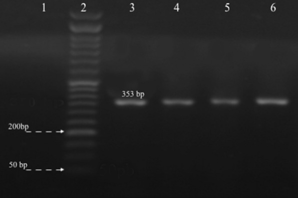
PCR amplification of blaOXA-51 gene. Lane 1: Negative control, Lane 2: size marker 50 bp, Lane 3: Positive control A. baumannii ATCC 19,606, and Lanes 4–6: clinical isolates show bands of amplified DNA at 353 bp
Fig. 2.
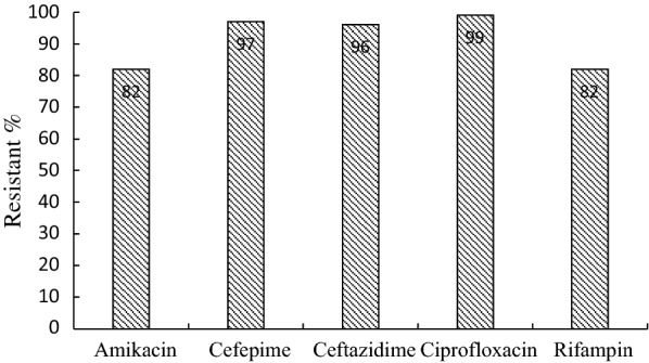
Antimicrobial resistance pattern of Acinetobacter baumannii clinical isolates
Isolation, purification and titration of lytic phages
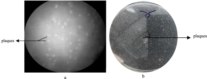
MDR A. baumannii strain, MDR-AB02, isolated from a patient's catheter with pneumonia at Alzahra hospital, was resistant to more than three groups of antibiotics (Additional file 1: Table S1). This (MDR-AB02) was used as an indicator to screen bacteriophages in sewage samples of the same hospital. The isolated phage was labeled as pIsf-AB02. The pIsf-AB02 forms clear, round, 2–3 mm plaques in the double-layer agar, indicating the lytic property of the phage (Fig. 3). Most MDR A. baumannii isolates in this study were sensitive to pIsf-AB02 (27/48); therefore, it was chosen for further study.
Fig. 3.
a Plaque morphology of phage pIsf-AB02 plaques under light microscope, b the clear zone, which demonstrated phage plaque in double-layer agar
Examination phage morphology by TEM
The morphology of pIsf-AB02 was examined by negative staining of the phage and observation under electron microscopy. The phage had an icosahedral head of 70 ± 10 nm and a tail of about 60 nm (Fig. 4). The phage belongs to the order Caudovirales and family Myoviridae following the current guidelines of the ICTV (International Committee on Taxonomy of Viruses, http://ictv.global/taxonomyRelease.s.asp).
Fig. 4.
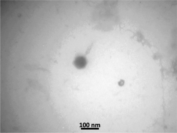
Transmission Electron Microscopy of pIsf-AB02 phage. The bar represents a length of 100 nm
pH, thermal, and chloroform stability
The stability of the pIsf-AB02 to different pH, chloroform, and temperature was tested. The phage pIsf-AB02 lost its infectivity at pH 3 and 11, while pH 7 is the most suitable condition to maintain the phage. The phage was stable at different temperatures ranging from − 20 to 25 °C. However, the phage titer was slightly dropped at 50 °C and reduced dramatically at 70 °C. The activity of the virus was not affected by chloroform treatment (Additional file 1: Figs. S1, S2).
Determination of optimal MOI
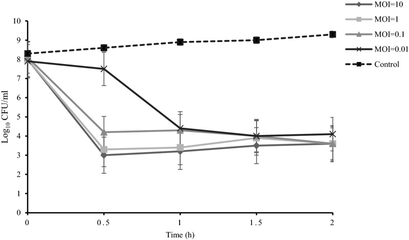
The lytic activity of pIsf-AB02 was assessed by inoculating the phage to MDR-AB02. Different MOIs of the phage were inoculated into AB02 (108 CFU/ml). As shown in Fig. 5, The pIsf-AB02 with MOI of 1 reduces the MDR-AB02 from 108 CFU/ml to 103 CFU/ml in 30 min. Lower MOIs (0.1 and 0.01) decreased the virus titer to the same point in 1.5–2 h. The results indicate that although higher MOI reduced A. baumannii concentration more quickly, but is not necessary for lysis.
Fig. 5.
determination of optimal phage titer. The pIsf-AB02 was used at different titers to infect MDR-AB02 to determine the optimal titer of the host during 2 h
One-step growth curve
One-step growth experiment showed that the latent period of pIsf-AB02 was about 30 min and was followed by the lysis phase, which lasted for 70 min. The burst size was 120 PFU per infected cell (Fig. 6).
Fig. 6.
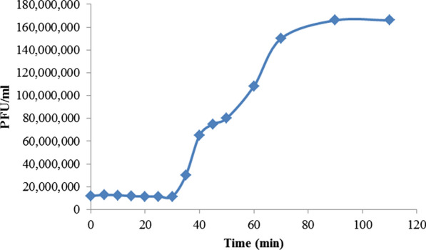
One-step growth curve of pIsf-AB02 phage
Phage genome analysis
The genome analysis indicated that phage pIsf-AB02 has a double-stranded DNA genome (approximately 12.6 kb). The genome of pIsf-AB02 could be digested by HindIII endonuclease (Fig. 7). It was found that HindIII has three cutting sites. Although, endonucleases, HincII, EcoR1and NdeI have no cutting site.
Fig. 7.
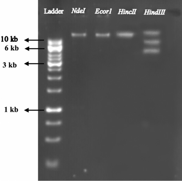
The pIsf-AB02 phage genomic DNA restriction patterns and size determination
Phage protein analysis
The results of pIsf-AB02 phage protein analysis in denaturing conditions showed nine structural protein bands in 12% SDS-PAGE, with a molecular weight ranging from 14.5 to 150 kDa. The most abundant proteins band in the gel were 100 kDa and 15 kDa. The major band was assumed to be the phage putative coat protein. The latter was predicted to be endolysin, which was confirmed with a non-denaturing condition (Fig. 8).
Fig. 8.
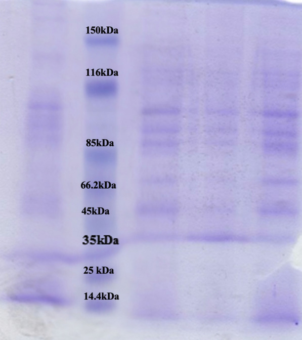
The pIsf-AB02 phage proteins separated by 12% SDS PAGE. Lane 1,4: the phage proteins without boiling, Lane 3,5: the phage proteins with boiling, Lane 2: Ladder
Phage protein analysis under non-denaturing conditions (endolysin activity)
At the time of release, the phages erupt the bacteria, causing the endolysin to flow out into the medium. Proteins of supernatant were concentrated and separated by SDS- PAGE under the non-denaturing condition as described before. The MDR-AB02 overlay on SDS-PAGE showed a clear band at 15 kDa (Fig. 9).
Fig. 9.
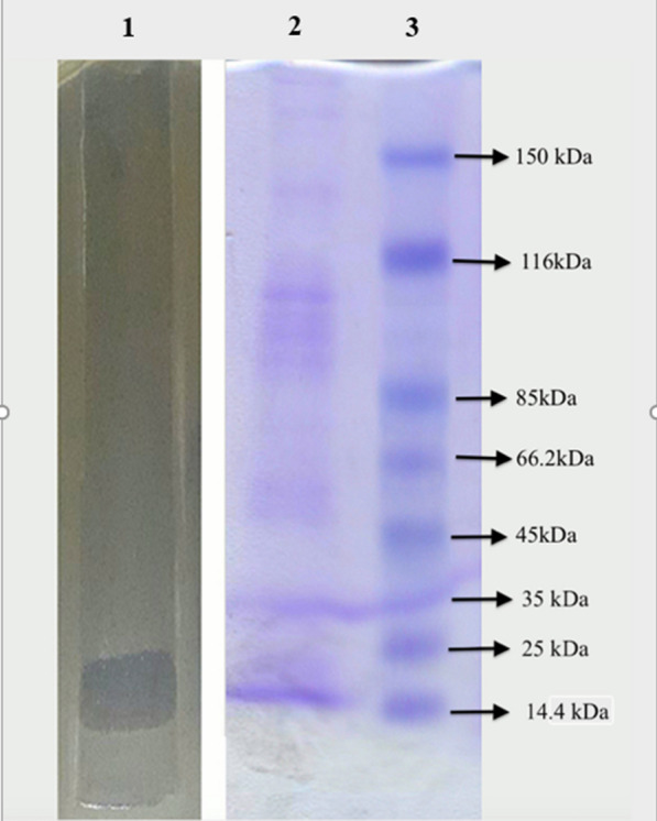
Phage endolysin activity in SDS-PAGE with the non-denaturing condition. The protein from the phage lysate was mixed with sample buffer without β-mercaptoethanol and subjected to SDS-PAGE without boiling. The SDS-PAGE gel was placed onto an agar-containing plate, and soft agar mixed with MDR-AB02 was overlaid. The protein with endolysin activity produced a clear region on the overlay. Lane 1: PAGE overlay on a plate covered by Acinetobacter baumannii. Lane 2: phage proteins without boiling in PAGE. Lane 3: protein Ladder
Bacteriophage host range
Host range spectrum surveyed on forty-eight A. baumannii clinical isolates and showed that the pIsf-AB02 phage could infect and lyse 56.3% of the A. baumannii isolates (S1). The results demonstrated that phage was specific for the A. baumannii and did not affect Klebsiella, Pseudomonas, E. coli.
Bacterial reduction assay
Infections of A. baumannii with a high titer of the lysate (1010 PFU/ml) were monitored for 7.5 h. Phage infection significantly decreased the A. baumannii culture turbidity in comparison to control. However, an increase in turbidity (OD600) was observed after about 4 h of culture incubation. This increase in turbidity was most probably due to the growth of phage-resistant bacteria (Fig. 10).
Fig. 10.
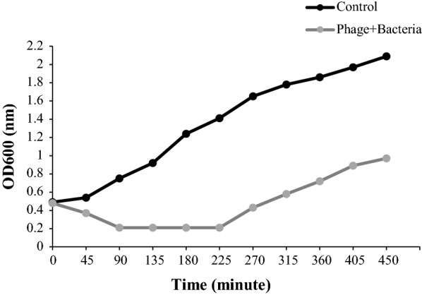
Bacterial reduction assay. Effect of pIsfAB02 on the growth of MDR-AB02 compared with the control
Cell survival assay under bacterial infection
For the safety of phage therapy, HeLa cells were used to examine the cytotoxicity of the isolated phage in the presence of MDR-AB02. In 96-wells plate, different dilutions MOI = 10, 1, 0.1, 0.01 of pIsf-AB02 was added in the presence of 106 CFU/ml MDR-AB02. The phage showed the highest protection against A. baumannii AB02 infection of cells (104 cells/well) (Fig. 11). Phage at an MOI of 10, 1, 0.1 enabled cells inoculated with A. baumannii AB02 (106 CFU) to survive as well as uninoculated controls. Although, the lowest cell viability was found at an MOI of 0.01. In bacterial control (cells inoculated with MDR-AB02 without phage treatment), all cells were completely killed by A. baumannii. Cells treated with the phage at an MOI of 10 (107 PFU), but not inoculated with bacteria, survived as well as the control cells (p > 0.05), indicating that high dose of pIsf-AB02 did not affect HeLa survival. The results showed that pIsf-AB02 eliminated bacteria and protected HeLa cells from immediate killing by A. baumannii AB02 bacteria.
Fig. 11.
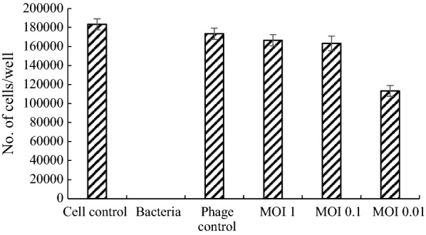
The cell survival assay of pIsf-AB02 on HeLa cells. The cells were infected with 106 CFU of MDR-AB02. Phage at different MOI (10, 1, 0.1, 0.01) was added to the wells. The cell control; Hela cell, Phage control: phage with MOI 10 without bacteria. MOI 1, 0.1, and 0.01 of phage with exposure of A. baumannii
In another experiment, the cell viability was assessed by adding the phage 2 h post-infection of the cells with MDR-AB02. As shown in Fig. 12, the pIsf-AB02 could not protect the cells at different MOIs (p < 0.001).
Fig. 12.
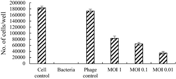
Protection efficacy test of pISF-AB02 on Hela Cell 2 h post infection: The cells (0.05 × 10⁴ cells/well) were first infected with 106 CFU of MDR-AB02, and the Hela cells were counted after 12 h
Discussion
The objective of our study was to demonstrate a new approach for using the bacteriophage to treat life-threatening MDR A. baumannii infections. The antibiotic sensitivity of 48 A. baumannii isolates revealed that the prevalence of MDR and pan-drug A. baumannii is increasing in the region, which is in agreement with other studies in different countries [31, 32].
We isolated and characterized pIsf-AB02, a lytic phage from a clinical MDR A. baumannii isolate. It was able to form clear pinpointed plaques on MDR-AB02 lawns, indicative of strong lytic activity, and a wider host range among MDR A. baumannii clinical isolates (27/48, 56.2%) than our previously screened bacteriophages [33]. The major limitations of phage therapy are their narrow host range and the emergence of bacteriophage-insensitive mutants (BIMs); therefore, a large cocktail of phages is needed to improve therapy by extending the host range and reducing resistance and it is very important to isolate novel phages to enrich the phage supply.
Studying the isolated phage by TEM indicated that phage pIsf-AB02 should be assigned to the family of Myoviridae based on its morphological characteristics. Regeimbal et al. in the United States also isolated a particular A. baumannii phage from sewage and stated that the phage had a shaft-shaped head along with a contractile tail and belonged to the Myoviridae family [33]. Our isolated phage was a 6-cupsid, fractured, and constricted phage in which the morphological results overlapped with the other investigations [23, 34].
In a study by Kusradze et al., an A. baumannii phage from sewage was isolated and introduced through microscopic examination to the Myoviridae family. They also examined the phage stability at different temperatures, pH, and chloroform and showed that after a 24-h incubation of the phage at 37 °C, the potency of the phage remained unchanged and was stable in exposure to chloroform and normal pH [23]. Broadly speaking, high pH stability and high thermal resistance made the phage remarkably pledged for practical usage in the deracination of A. baumannii contaminations and or the treatment of A. baumannii infections. Phage pIsf-AB02 revealed impressive characteristics compared to the other phages. After a 24-h inoculation, the phage exhibited a steady state for chloroform. These results are consistent with the results of Kusradze [23]. Thermal resistant phages were usually isolated from extreme thermal habits [35, 36]; however, they could also be found in other environments. Recently, the thermal resistant phages have been isolated and characterized from various dairy products [37, 38].
One-step growth curve analysis revealed a 30-min latent period, a 70-min lysis period, and a burst size of 120 phage particles per infected host cell. Compared with the isolated phage by Yang et al., pIsf-AB02 had a smaller burst size but a wider host range among the local A. baumannii isolates and a boarder range of temperature and pH stability [39], making pIsf-AB02 a suitable nominee for further application of phage therapy.
In 30 min, pIsf-AB02 with MOI of 1 reduced MDR-AB02 from 108 CFU/ml to 103 CFU/ml and maintained this concentration for about 2 h. However, the lower MOIs (0.1 and 0.01) decreased the virus titer to the same point in 1.5–2 h. The optimal titer of the phage in phage therapy is crucial. Despite the thought that the higher MOI of phage inoculation would get higher efficiency on waning the bacterium, the interaction between hosts and phages should be optimized. It might be possible that the high titer phage would occupy the receptor for phage and the bacterial lysis rate would not rise with an increase in MOI of phage [40]. Another possibility is that sometimes the high titer phage would induce the host immune system and limit phage therapy [41].
Herein, a high concentration of bacteria was applied to infect the HeLa cells to examine the protective efficacy of phages on the cells. The data showed that in the presence of the bacteria, pIsf-AB02 phage in higher MOIs didn’t affect on the survival rate significantly after a 24-h incubation in comparison to the control. On the other hand, the phage pIsf-AB02 did not have an adverse effect on the growth of HeLa cells, indicating that pIsf-AB02 is a suitable candidate for phage therapy. Further animal model experiments should be performed to confirm that phage pIsf-AB02 has a good protection on the animals infected by MDR A. baumannii.
Most double-stranded DNA phages accomplish host cell lysis through the holin-endolysin system. The similarity of bacteriophage endolysin genes is essential for structural analysis, which contributes to the potential of utilizing endolysin as an antimicrobial agent [42]. The endolysins antibacterial activity is generally attributed to their enzymatic function, which ruptures the covalent bonds in peptidoglycan. However, some endolysins, especially those from phages of Gram-negative bacteria, can affect the bacterial cells employing a mechanism completely independent of their enzymatic activity [43–45]. In the current work, the protein causing lysis was estimated to be about 15 kDa through SDS PAGE, which corroborated the previous results [46]. The lysin was utterly stable and constant over a wide range of pHs.
Phage candidates for therapeutic application purposes should not harbor foreign genes, such as virulence or antibiotic resistance genes, integrases, site-specific recombinases, and repressors of the lytic cycle [47]. Phages could serve as a vector for horizontal transfer virulence gene to bacteria, making them more pathogenic or resistant to antibiotics [48].Therefore, the genomic characterization of pIsf-AB02 is very important and should be taken into consideration to guarantee the safety of phage therapy. Phages may show synergistic effects when combined with antibiotics [49, 50]. A reduction in the formation of bacterial biofilms has also been reported when antibiotic treatment is applied in combination with phages [51, 52]. These conclusions should be verified through future studies to further overcome the limitations of phage therapy.
In this study, we isolated and characterized a novel lytic A. baumannii bacteriophage and evaluated the lytic activity of the phage against the isolated MDR A. baumannii. Furthermore, we assessed the efficacy of phage endolysin on MDR A. baumannii clinical isolates. Our findings support the potential application of the phage with the potent endolysin activity against MDR A. baumannii and suggest that this phage could be developed for the treatment of MDR A. baumannii infections.
Conclusion
In this study, we isolated a novel lytic MDR A. baumannii bacteriophage, pIsf-AB02. This phage showed suitable stability at different temperatures and pHs, and demonstrated potent in vitro endolysin activity. pIsf-AB02 may be a good candidate as a therapeutic agent to control nosocomial infections caused by MDR A. baumannii.
Supplementary Information
Additional file 1: Table S1. The antibiotic sensitivity results and the spot test of pISF-AB2 phage, Figure S1. pH stability test, Figure S2: Thermal stability of pISF-AB2 phage.
Acknowledgements
This research benefited from a grant from Isfahan University od medical sciences.
Abbreviations
- MDR
Multidrug resistance
- ICU
Intensive Care Units
- ESKAPE
(Enterococcus faecium, Staphylococcus aureus, Klebsiella pneumonia, Acinetobacter baumannii, Pseudomonas aeruginosa And Enterobacter spp)
- MOI
Multiplicity of infection
- OD
Optimal density
- PFU
Plaque-forming unit
- CFU
Colony-forming unit
- pH
The potential of hydrogen
- TEM
Transmission electron microscopy
- NB
Nutrient broth
- TBE
Tris-boric acid-EDTA
- ATCC
American type culture collection
Authors' contributions
BS was a significant contributor to doing and writing the manuscript. AM, MS, NH and VK collaborated in doing the thesis that results in the paper.SM Designed and supervised the manuscript. All authors read and approved the final manuscript.
Funding
This study was financially supported by Isfahan University of Medical Sciences, Isfahan, I.R. Iran through Grant No. 396188.
Availability of data and materials
The datasets used and/or analyzed during the current study are available from the corresponding author on reasonable request.
Declarations
Ethics approval and consent to participate
Not applicable.
Consent for publication
Not applicable.
Competing interests
The authors declare that they have no competing interests.
Footnotes
Publisher's Note
Springer Nature remains neutral with regard to jurisdictional claims in published maps and institutional affiliations.
References
- 1.Bassetti M, Righi E, Esposito S, Petrosillo N, Nicolini L: Drug treatment for multidrug-resistant Acinetobacter baumannii infections. 2008. [DOI] [PubMed]
- 2.Rice LB. Progress and challenges in implementing the research on ESKAPE pathogens. Infect Control Hosp Epidemiol. 2010;31(S1):S7–S10. doi: 10.1086/655995. [DOI] [PubMed] [Google Scholar]
- 3.Navidinia M: The clinical importance of emerging ESKAPE pathogens in nosocomial infections. 2016.
- 4.Gong Y, Shen X, Huang G, Zhang C, Luo X, Yin S, Wang J, Hu F, Peng Y, Li M. Epidemiology and resistance features of Acinetobacter baumannii isolates from the ward environment and patients in the burn ICU of a Chinese hospital. J Microbiol. 2016;54(8):551–558. doi: 10.1007/s12275-016-6146-0. [DOI] [PubMed] [Google Scholar]
- 5.Huang G, Yin S, Gong Y, Zhao X, Zou L, Jiang B, Dong Z, Chen Y, Chen J, Jin S. Multilocus sequence typing analysis of carbapenem-resistant Acinetobacter baumannii in a Chinese burns institute. Front Microbiol. 2016;7:1717. doi: 10.3389/fmicb.2016.01717. [DOI] [PMC free article] [PubMed] [Google Scholar]
- 6.Falagas ME, Karageorgopoulos DE. Pandrug resistance (PDR), extensive drug resistance (XDR), and multidrug resistance (MDR) among Gram-negative bacilli: need for international harmonization in terminology. Clin Infect Dis. 2008;46(7):1121–1122. doi: 10.1086/528867. [DOI] [PubMed] [Google Scholar]
- 7.Haq IU, Chaudhry WN, Akhtar MN, Andleeb S, Qadri I. Bacteriophages and their implications on future biotechnology: a review. Virol J. 2012;9(1):9. doi: 10.1186/1743-422X-9-9. [DOI] [PMC free article] [PubMed] [Google Scholar]
- 8.Mulani MS, Kamble EE, Kumkar SN, Tawre MS, Pardesi KR. Emerging strategies to combat ESKAPE pathogens in the era of antimicrobial resistance: a review. Front Microbiol. 2019;10:539. doi: 10.3389/fmicb.2019.00539. [DOI] [PMC free article] [PubMed] [Google Scholar]
- 9.Nakai T, Park SC. Bacteriophage therapy of infectious diseases in aquaculture. Res Microbiol. 2002;153(1):13–18. doi: 10.1016/s0923-2508(01)01280-3. [DOI] [PubMed] [Google Scholar]
- 10.Laanto E, Sundberg L-R, Bamford JK. Phage specificity of the freshwater fish pathogen Flavobacterium columnare. Appl Environ Microbiol. 2011;77(21):7868–7872. doi: 10.1128/AEM.05574-11. [DOI] [PMC free article] [PubMed] [Google Scholar]
- 11.Parisien A, Allain B, Zhang J, Mandeville R, Lan C. Novel alternatives to antibiotics: bacteriophages, bacterial cell wall hydrolases, and antimicrobial peptides. J Appl Microbiol. 2008;104(1):1–13. doi: 10.1111/j.1365-2672.2007.03498.x. [DOI] [PubMed] [Google Scholar]
- 12.Matsuzaki S, Rashel M, Uchiyama J, Sakurai S, Ujihara T, Kuroda M, Ikeuchi M, Tani T, Fujieda M, Wakiguchi H. Bacteriophage therapy: a revitalized therapy against bacterial infectious diseases. J Infect Chemother. 2005;11(5):211–219. doi: 10.1007/s10156-005-0408-9. [DOI] [PubMed] [Google Scholar]
- 13.Lai M-J, Soo P-C, Lin N-T, Hu A, Chen Y-J, Chen L-K, Chang K-C. Identification and characterisation of the putative phage-related endolysins through full genome sequence analysis in Acinetobacter baumannii ATCC 17978. Int J Antimicrob Agents. 2013;42(2):141–148. doi: 10.1016/j.ijantimicag.2013.04.022. [DOI] [PubMed] [Google Scholar]
- 14.Fan J, Zeng Z, Mai K, Yang Y, Feng J, Bai Y, Sun B, Xie Q, Tong Y, Ma J. Preliminary treatment of bovine mastitis caused by Staphylococcus aureus, with trx-SA1, recombinant endolysin of S aureus bacteriophage IME-SA1. Veter Microbiol. 2016;191:65–71. doi: 10.1016/j.vetmic.2016.06.001. [DOI] [PubMed] [Google Scholar]
- 15.Lin DM, Koskella B, Lin HC. Phage therapy: An alternative to antibiotics in the age of multi-drug resistance. World J Gastrointest Pharmacol Ther. 2017;8(3):162. doi: 10.4292/wjgpt.v8.i3.162. [DOI] [PMC free article] [PubMed] [Google Scholar]
- 16.Philipson CW, Voegtly LJ, Lueder MR, Long KA, Rice GK, Frey KG, Biswas B, Cer RZ, Hamilton T, Bishop-Lilly KA. Characterizing phage genomes for therapeutic applications. Viruses. 2018;10(4):188. doi: 10.3390/v10040188. [DOI] [PMC free article] [PubMed] [Google Scholar]
- 17.Adams-Haduch JM, Paterson DL, Sidjabat HE, Pasculle AW, Potoski BA, Muto CA, Harrison LH, Doi Y. Genetic basis of multidrug resistance in Acinetobacter baumannii clinical isolates at a tertiary medical center in Pennsylvania. Antimicrob Agents Chemother. 2008;52(11):3837–3843. doi: 10.1128/AAC.00570-08. [DOI] [PMC free article] [PubMed] [Google Scholar]
- 18.Dashti AA, Jadaon MM, Abdulsamad AM, Dashti HM. Heat treatment of bacteria: a simple method of DNA extraction for molecular techniques. Kuwait Med J. 2009;41(2):117–122. [Google Scholar]
- 19.Safari M, Saidijam M, BAHADOR A, Jafari R, Alikhani MY: High prevalence of multidrug resistance and metallo-beta-lactamase (MbetaL) producing Acinetobacter baumannii isolated from patients in ICU wards, Hamadan, Iran. 2013. [PubMed]
- 20.Wayne P: Clinical and Laboratory Standards Institute: Performance standards for antimicrobial susceptibility testing: 28th informational supplement. CLSI document M100-S20 2018.
- 21.Standardization IOf: Water Quality: Detection and Enumeration of Bacteriophages: ISO; 2000.
- 22.Kropinski AM, Mazzocco A, Waddell TE, Lingohr E, Johnson RP: Enumeration of bacteriophages by double agar overlay plaque assay. In Bacteriophages. Springer; 2009: 69–76. [DOI] [PubMed]
- 23.Kusradze I, Karumidze N, Rigvava S, Dvalidze T, Katsitadze M, Amiranashvili I, Goderdzishvili M. Characterization and testing the efficiency of Acinetobacter baumannii phage vB-GEC_Ab-M-G7 as an antibacterial agent. Front Microbiol. 2016;7:1590. doi: 10.3389/fmicb.2016.01590. [DOI] [PMC free article] [PubMed] [Google Scholar]
- 24.Lockington RA, Attwood GT, Brooker JD. Isolation and characterization of a temperate bacteriophage from the ruminal anaerobe Selenomonas ruminantium. Appl Environ Microbiol. 1988;54(6):1575–1580. doi: 10.1128/aem.54.6.1575-1580.1988. [DOI] [PMC free article] [PubMed] [Google Scholar]
- 25.Capra M, Quiberoni A, Reinheimer J. Phages of Lactobacillus casei/paracasei: response to environmental factors and interaction with collection and commercial strains. J Appl Microbiol. 2006;100(2):334–342. doi: 10.1111/j.1365-2672.2005.02767.x. [DOI] [PubMed] [Google Scholar]
- 26.Shen G-H, Wang J-L, Wen F-S, Chang K-M, Kuo C-F, Lin C-H, Luo H-R, Hung C-H. Isolation and characterization of φkm18p, a novel lytic phage with therapeutic potential against extensively drug resistant Acinetobacter baumannii. PLoS ONE. 2012;7:10. doi: 10.1371/journal.pone.0046537. [DOI] [PMC free article] [PubMed] [Google Scholar]
- 27.Laemmli UK, Favre M. Maturation of the head of bacteriophage T4: I. DNA packaging events. J Mol Biol. 1973;80(4):575–599. doi: 10.1016/0022-2836(73)90198-8. [DOI] [PubMed] [Google Scholar]
- 28.Jyothisri K, Deepak V, Rajeswari MR. Purification and characterization of a major 40 kDa outer membrane protein of Acinetobacter baumannii. FEBS Lett. 1999;443(1):57–60. doi: 10.1016/s0014-5793(98)01679-2. [DOI] [PubMed] [Google Scholar]
- 29.Lee C-N, Tseng T-T, Lin J-W, Fu Y-C, Weng S-F, Tseng Y-H. Lytic myophage Abp53 encodes several proteins similar to those encoded by host Acinetobacter baumannii and phage phiKO2. Appl Environ Microbiol. 2011;77(19):6755–6762. doi: 10.1128/AEM.05116-11. [DOI] [PMC free article] [PubMed] [Google Scholar]
- 30.Ghajavand H, Esfahani BN, Havaei A, Fazeli H, Jafari R, Moghim S. Isolation of bacteriophages against multidrug resistant Acinetobacter baumannii. Res Pharm Sci. 2017;12(5):373. doi: 10.4103/1735-5362.213982. [DOI] [PMC free article] [PubMed] [Google Scholar]
- 31.Mahmoudi Monfared A, Rezaei A, Poursina F, Faghri J. Detection of genes involved in biofilm formation in MDR and XDR Acinetobacter baumannii isolated from human clinical specimens in Isfahan. Iran. Arch Clin Infect Dis. 2019;14:2. [Google Scholar]
- 32.Mirzaei B, Bazgir ZN, Goli HR, Iranpour F, Mohammadi F, Babaei R. Prevalence of multi-drug resistant (MDR) and extensively drug-resistant (XDR) phenotypes of Pseudomonas aeruginosa and Acinetobacter baumannii isolated in clinical samples from Northeast of Iran. BMC Res Notes. 2020;13(1):1–6. doi: 10.1186/s13104-020-05224-w. [DOI] [PMC free article] [PubMed] [Google Scholar]
- 33.Regeimbal JM, Jacobs AC, Corey BW, Henry MS, Thompson MG, Pavlicek RL, Quinones J, Hannah RM, Ghebremedhin M, Crane NJ. Personalized therapeutic cocktail of wild environmental phages rescues mice from Acinetobacter baumannii wound infections. Antimicrob Agents Chemother. 2016;60(10):5806–5816. doi: 10.1128/AAC.02877-15. [DOI] [PMC free article] [PubMed] [Google Scholar]
- 34.Merabishvili M, Vandenheuvel D, Kropinski AM, Mast J, De Vos D, Verbeken G, Noben J-P, Lavigne R, Vaneechoutte M, Pirnay J-P. Characterization of newly isolated lytic bacteriophages active against Acinetobacter baumannii. PLoS ONE. 2014;9(8):e104853. doi: 10.1371/journal.pone.0104853. [DOI] [PMC free article] [PubMed] [Google Scholar]
- 35.Rice G, Stedman K, Snyder J, Wiedenheft B, Willits D, Brumfield S, McDermott T, Young MJ. Viruses from extreme thermal environments. Proc Natl Acad Sci. 2001;98(23):13341–13345. doi: 10.1073/pnas.231170198. [DOI] [PMC free article] [PubMed] [Google Scholar]
- 36.Arnold HP, Zillig W, Ziese U, Holz I, Crosby M, Utterback T, Weidmann JF, Kristjanson JK, Klenk HP, Nelson KE. A novel lipothrixvirus, SIFV, of the extremely thermophilic crenarchaeon Sulfolobus. Virology. 2000;267(2):252–266. doi: 10.1006/viro.1999.0105. [DOI] [PubMed] [Google Scholar]
- 37.Capra ML, Binetti AG, Mercanti DJ, Quiberoni A, Reinheimer JA. Diversity among Lactobacillus paracasei phages isolated from a probiotic dairy product plant. J Appl Microbiol. 2009;107(4):1350–1357. doi: 10.1111/j.1365-2672.2009.04313.x. [DOI] [PubMed] [Google Scholar]
- 38.Atamer Z, Dietrich J, Müller-Merbach M, Neve H, Heller KJ, Hinrichs J. Screening for and characterization of Lactococcus lactis bacteriophages with high thermal resistance. Int Dairy J. 2009;19(4):228–235. [Google Scholar]
- 39.Yang Z, Yin S, Li G, Wang J, Huang G, Jiang B, You B, Gong Y, Zhang C, Luo X. Global transcriptomic analysis of the interactions between phage φAbp1 and extensively drug-resistant acinetobacter baumannii. MSystems. 2019;4(2):e00068–e119. doi: 10.1128/mSystems.00068-19. [DOI] [PMC free article] [PubMed] [Google Scholar]
- 40.Vieira A, Silva Y, Cunha A, Gomes N, Ackermann H-W, Almeida A. Phage therapy to control multidrug-resistant Pseudomonas aeruginosa skin infections: in vitro and ex vivo experiments. Eur J Clin Microbiol Infect Dis. 2012;31(11):3241–3249. doi: 10.1007/s10096-012-1691-x. [DOI] [PubMed] [Google Scholar]
- 41.Kocharunchitt C, Ross T, McNeil D. Use of bacteriophages as biocontrol agents to control Salmonella associated with seed sprouts. Int J Food Microbiol. 2009;128(3):453–459. doi: 10.1016/j.ijfoodmicro.2008.10.014. [DOI] [PubMed] [Google Scholar]
- 42.Kitti T, Thummeepak R, Thanwisai A, Boonyodying K, Kunthalert D, Ritvirool P, Sitthisak S. Characterization and detection of endolysin gene from three Acinetobacter baumannii bacteriophages isolated from sewage water. Indian J Microbiol. 2014;54(4):383–388. doi: 10.1007/s12088-014-0472-x. [DOI] [PMC free article] [PubMed] [Google Scholar]
- 43.Düring K, Porsch P, Mahn A, Brinkmann O, Gieffers W. The non-enzymatic microbicidal activity of lysozymes. FEBS Lett. 1999;449(2–3):93–100. doi: 10.1016/s0014-5793(99)00405-6. [DOI] [PubMed] [Google Scholar]
- 44.Orito Y, Morita M, Hori K, Unno H, Tanji Y. Bacillus amyloliquefaciens phage endolysin can enhance permeability of Pseudomonas aeruginosa outer membrane and induce cell lysis. Appl Microbiol Biotechnol. 2004;65(1):105–109. doi: 10.1007/s00253-003-1522-1. [DOI] [PubMed] [Google Scholar]
- 45.Lai M-J, Lin N-T, Hu A, Soo P-C, Chen L-K, Chen L-H, Chang K-C. Antibacterial activity of Acinetobacter baumannii phage ϕAB2 endolysin (LysAB2) against both gram-positive and gram-negative bacteria. Appl Microbiol Biotechnol. 2011;90(2):529–539. doi: 10.1007/s00253-011-3104-y. [DOI] [PubMed] [Google Scholar]
- 46.Zhang H, Buttaro BA, Fouts DE, Sanjari S, Evans BS, Stevens RH. Bacteriophage φEf11 ORF28 endolysin, a multifunctional lytic enzyme with properties distinct from all other identified Enterococcus faecalis phage endolysins. Appl Environ Microbiol. 2019;85(13):e00555–e1519. doi: 10.1128/AEM.00555-19. [DOI] [PMC free article] [PubMed] [Google Scholar]
- 47.Brown N, Cox C. Bacteriophage Use in Molecular Biology and Biotechnology. Bacteriophage Biol Technol Ther. 2021;90:465–506. [Google Scholar]
- 48.Chen J, Novick RP. Phage-mediated intergeneric transfer of toxin genes. Science. 2009;323(5910):139–141. doi: 10.1126/science.1164783. [DOI] [PubMed] [Google Scholar]
- 49.Jo A, Kim J, Ding T, Ahn J. Role of phage-antibiotic combination in reducing antibiotic resistance in Staphylococcus aureus. Food science and biotechnology. 2016;25(4):1211–1215. doi: 10.1007/s10068-016-0192-6. [DOI] [PMC free article] [PubMed] [Google Scholar]
- 50.Chaudhry WN, Concepcion-Acevedo J, Park T, Andleeb S, Bull JJ, Levin BR. Synergy and order effects of antibiotics and phages in killing Pseudomonas aeruginosa biofilms. PLoS ONE. 2017;12(1):e0168615. doi: 10.1371/journal.pone.0168615. [DOI] [PMC free article] [PubMed] [Google Scholar]
- 51.Schooley RT, Biswas B, Gill JJ, Hernandez-Morales A, Lancaster J, Lessor L, Barr JJ, Reed SL, Rohwer F, Benler S. Development and use of personalized bacteriophage-based therapeutic cocktails to treat a patient with a disseminated resistant Acinetobacter baumannii infection. Antimicrob Agents Chemother. 2017;61(10):e00954–e1917. doi: 10.1128/AAC.00954-17. [DOI] [PMC free article] [PubMed] [Google Scholar]
- 52.Nouraldin AAM, Baddour MM, Harfoush RAH, Essa SAM. Bacteriophage-antibiotic synergism to control planktonic and biofilm producing clinical isolates of Pseudomonas aeruginosa. Alexandria J Med. 2016;52(2):99–105. [Google Scholar]
Associated Data
This section collects any data citations, data availability statements, or supplementary materials included in this article.
Supplementary Materials
Additional file 1: Table S1. The antibiotic sensitivity results and the spot test of pISF-AB2 phage, Figure S1. pH stability test, Figure S2: Thermal stability of pISF-AB2 phage.
Data Availability Statement
The datasets used and/or analyzed during the current study are available from the corresponding author on reasonable request.


