Abstract
Posterior fossa aneurysms presenting with isolated subdural haemorrhage (SDH) have scarcely been described with no cases attributed to a vertebrobasilar (VB) location. Non-saccular VB aneurysms are a distinct sub-group and in this report we also discuss the pathophysiology and treatment options for these difficult-to-manage lesions. We present a case of a 49 year-old man who presented with a 7-day history of severe headaches who was found to have an isolated acute clival SDH. Vascular imaging revealed a VB dolicoectatic segment with superimposed fusiform dilatations that contacted the dura adjacent to the SDH. A staged treatment was performed with initial parental vessel occlusion of the ruptured vertebral artery segment and subsequent insertion of a braided stent (LEO) with flow diverting properties into the progressively dilating basilar artery. A third procedure was performed to occlude a recurrent pouch at the lower basilar dilatation. Complete angiographic occlusion was achieved and the patient is under continued surveillance. To our knowledge, this is the first case of a ruptured non-saccular VB aneurysm presenting with radiologically isolated clival SDH. Clinical history will often inform the need for vascular imaging in such atypical presentations. Managing these lesions remains an endovascular challenge and requires a specialist multi-disciplinary approach.
Keywords: Subdural haemorrhage, Dolicoectasia, Vertebrobasilar, Fusiform, Aneurysm
INTRODUCTION
Acute subdural haematoma may require emergent neurosurgical management and is commonly encountered in the setting of trauma. In patients presenting with subdural haemorrhage (SDH) or multi-compartmental intracranial haemorrhage without a clear traumatic history, a high suspicion of other aetiologies is required. Ruptured intracranial aneurysms classically present with acute subarachnoid haemorrhage (SAH) but concomitant SDH is reported in 2-22% of cases [5].
Acute SDH from aneurysm rupture without radiological evidence of SAH is a rare entity occurring in 0.1-2.9% of all presentations [14]. Aneurysmal SDH (AnSDH) has been linked to poor outcomes, in part attributable to delayed or missed diagnosis. Supratentorial AnSDH has become a well-recognised presentation, however posterior fossa SDH from a ruptured Vertebrobasilar (VB) aneurysm has not been hitherto described.
We present an unusual case of a radiologically isolated clival SDH from direct rupture of a non-saccular VB aneurysm (NSVB). These lesions form a rare heterogenous sub-group with a high morbidity and mortality if left untreated. In this report we also review AnSDH in the posterior fossa, the pathophysiology of non-saccular VB aneurysms and discuss treatment options for these difficult-to-manage lesions.
CASE DESCRIPTION
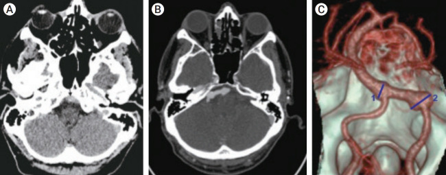
A 49 years-old gentleman with a past medical history of anxiety and labile hypertension presented to the emergency department (ED) with a seven days history of severe right sided headache. At initial assessment, a Glasgow Coma Scale (GCS) score of 15, blood pressure of 211/142 mmHg and no history of trauma were recorded. Computed tomography (CT) Head demonstrated a hyperdense clival subdural haematoma (Fig. 1A). Lumbar puncture revealed xanthochromia prompting a referral to the neurosurgical team.
Fig. 1.
(A) Non-contrast CT demonstrates hyperdense subdural haemorrhage posterior to the clivus (black arrow). (B and C) CTA and 3D volume rendering demonstrating a dolicoectatic V4 segment of the right VA and BA with superimposed fusiform dilatations of the VA segment beyond the PICA measuring up to 10 mm (2) and lower BA measuring up to 8 mm (1). VA, vertebral artery; BA, basilar artery; PICA, posterior inferior cerebellar artery.
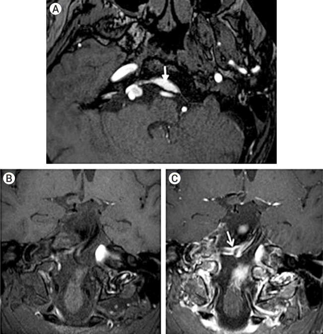
CT intracranial angiogram was initially performed, according to our routine intuitional practice (Fig. 1B and C), revealing a dolicoectatic V4 segment of the right vertebral artery (VA) and basilar artery (BA) with superimposed fusiform dilatations of the VA segment beyond the posterior inferior cerebellar artery (PICA) measuring up to 10 mm and lower BA measuring up to 8 mm. The dilated VA segment made a broad-based contact with the dura raising suspicion of a likely rupture point into the subdural space (Fig. 2A). Subsequently, contrast enhanced magnetic resonance imaging (MRI) with vessel-wall imaging protocol was performed demonstrating vessel-wall enhancement of both superimposed dilated segments (Fig. 2B), further supporting the initial impression of aneurysm rupture.
Fig. 2.

(A) Time-of-flight MRA demonstrated the dilated VA segment (* making a broad based contact with the dura (white arrow indicates SDH). (B) MR T1-weighted fat-saturated vessel wall images pre and post contrast (C). (C) The white arrow denotes vessel wall enhancement in the diseased right vertebrobasilar segment. MRA, magnetic resonance angiogram; VA, vertebral artery; SDH, subdural haemorrhage.
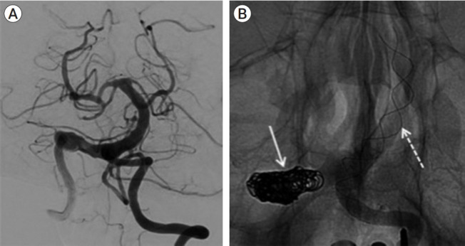
Treatment options (endovascular vs neurosurgical) were discussed at the multi-disciplinary team meeting and the consensus view was to perform endovascular trapping of the diseased VA segment. The patient was consented for right VA sacrifice with a quoted 10-20% estimated risk of disabling stroke. Complete occlusion of the VA diseased segment was achieved with preserved filling of right PICA and good left VA-BA flow (Fig. 3A). The 8 mm lower BA fusiform dilatation was not treated. The patient made a good post-operative recovery with resolution of headaches and no delayed complications at the three and sixmonth magnetic resonance angiogram (MRA) follow-up.
Fig. 3.

(A) Pre-treatment DSA image demonstrates the diseased right vertebrobasilar segment. (B) Image after deployment of the LEO stent showing the coiled right V4 segment after the 1st treatment (solid white arrow) and the LEO stent placement (dashed white arrow) during the 2nd treatment for a progressively dilating diseased lower basilar segment. DSA, digital subtraction angiogram.
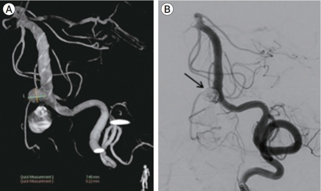
MRA at 30-months follow-up showed interval growth in the lower basilar dilatation. Endovascular treatment options were considered with view to flow diversion (FD). However, following measurement on the 3D rotational angiogram, the BA distal landing-zone was >5.5 mm while the distal left VA diameter was 3.5 mm. These dimensions were deemed unfavourable for conventional FD, and a braided LEO stent was placed (Fig. 3B). Concerns related to lower basilar perforator supply and risk of proximal stent displacement, related to further manipulation, discouraged simultaneous aneurysm coiling and early MRA follow-up was arranged. The initial MRA at 3 months showed partial thrombosis of the lower basilar pouch but at 6 months the aneurysmal segment had enlarged and the patient was successfully re-treated with coil embolisation achieving complete angiographic occlusion of the aneurysm (Fig. 4). The patient made an unremarkable post-operative recovery and long term imaging follow up is currently awaited.
Fig. 4.

(A) The 3D angiogram at 6-month follow-up showed residual enlarging segment. (B) 3rd treatment was performed: Coil embolization of the enlarging proximal basilar segment (solid black arrow).
DISCUSSION
Aneurysmal SDH
Pathogenesis of AnSDH has been a subject of debate with multiple potential mechanisms offered. One explanation is that adhesions can form between symptomatic aneurysm sacs and arachnoid membrane with subsequent direct rupture into the subdural space [9]. This theory is supported by the fact that a sentinel headache before the index event has been demonstrated as an independent risk factor for developing AnSDH [3].
An important role has also been attributed to the perianeurysmal environment (PAE) in determining why some aneurysms are more likely to rupture beyond the subarachnoid space [11].
Aneurysms that demonstrate more unbalanced contact with fixed external structures like bone, dura mater, or other parenchyma may be more prone to rupture and may in part explain why AnSDH is commonly seen at the posterior communicating (PCOM) artery location [3].
The overall lower incidence posterior fossa aneurysms likely accounts for the relative scarcity of reported infratentorial AnSDH however some authors have attributed this in part to the presence of the thicker Lilliquist arachnoid membranes that surround the BA [1].
Clival subdural haematoma is in itself a rare entity and is encountered in cases of severe intracranial trauma but has also been reported with coagulopathy and pituitary apoplexy.
Non-saccular VB aneurysms
Non-saccular VB (NSVB) aneurysms represent an uncommon heterogeneous group of lesions that have varied clinical presentations including infarction, mass effect, with intracranial haemorrhage (ICH) representing around 7% of all VB aneurysms [12]. Flemming et al. used radiological categories to predict their natural history by separating them into three categories which are reproducible in daily practice [6]:
1. Fusiform–aneurysmal dilatation 1.5 times of the normal diameter without a definable neck with any degree of tortuosity.
2. Dolicoechtasia–uniform aneurysmal dilatation of an artery greater than 1.5 times of the normal size with any degree of tortuosity.
3. Transition–uniform aneurysmal dilatation as above with a superimposed dilatation of a portion of the involved arterial segment.
Although overlap can exist, by the above criteria, the aneurysm we present is best described as a transitional type. A recent meta-analysis suggested that fusiform and transitional sub-groups have a significantly higher growth and rupture rate [10].
Treatment options
Ruptured non-saccular VB aneurysms pose a significant treatment challenge. The poor natural history of these aneurysms favours treatment, despite a wide profile of serious procedure-related complications. Factors that add technical complexity include: vascular tortuosity, aneurysm morphology, luminal thrombosis and location of side branches/ perforator vessels.
Open surgical treatments including ligation, bypass and trapping have been used with varied success. Advancements in endovascular technology and techniques have however shown promise in the modern era [2]. These can be categorised into deconstructive, consisting of parent vessel occlusion (PVO), and reconstructive techniques that include single or telescoped laser cut or woven stents (with or without coiling) and flow diverting stents.
Comparing PVO with reconstructive treatments for VB aneurysms has shown a trend towards higher rates of good clinical outcome and lower peri-procedural morbidity-mortality (4% vs 12%) in the reconstructive group, however similar outcomes were found when only patients with ruptured aneurysms were compared [13]. Another important role of PVO in NSVB aneurysms being treated with a stent across the VB junction is occlusion of contralateral distal V4 segment occlusion to prevent endoleak.
A recent meta-analysis reviewing flow diversion in NSVB aneurysms demonstrated long-term occlusion rates of 52% and good neurological outcome in only 50% of patients, with 25% suffering ischaemic complications [8]. Subgroup analysis in this study revealed better outcomes in smaller aneurysms (<10 mm) and in cases of isolated VA involvement (83%). These studies highlight that FD treatments certainly have a role to play in posterior fossa aneurysms, however case selection is paramount in justifying treatment and optimising outcomes.
As the majority of current available evidence for reconstructive techniques is primarily for patients with unruptured NSVB aneurysms, it is difficult to extrapolate reliable conclusions for ruptured aneurysms where dual antiplatelet therapy is often necessitated for stenting procedures, despite ICH. The encouraging results seen with FD in ruptured anterior circulation aneurysms have not translated to the posterior circulation with up to a 50% reported complication rate [4].
Large woven stents with or without supportive coiling have been successfully used for reconstruction of symptomatic VB dolicoectasia [7]. Due to their pore sizes ranging between laser-cut and FD stents, a degree of flow diversion can be achieved when the stent is compacted or placed in an overlapping fashion. In cases of superimposed fusiform dilatation, the risk of growth and perforator infarction remain the main concern as the vessel lumen outside the unopposed stent can continue to fill or thrombose overtime.
Stent assisted coiling improves long-term aneurysm occlusion rates, particularly in unruptured broad-necked saccular aneurysms. However, the use of this technique in ruptured NSVB aneurysms is complicated by antiplatelet requirement and the risks of aneurysm rupture, branch or perforator infarction, coil compartmentalisation, compaction, migration into thrombus and poor wall opposition of the stent. This highlights the potential benefit of staged treatment in ruptured NSVB aneurysms, which has been advocated to balance the risk of re-bleed and treatment related perforator infarcts. He et al reported no brainstem infarcts in their recent series of 19 patients treated with either stenting alone or with adjunct loose coiling [7]. The authors postulated that this method likely allowed for remodelling of dolichoectasia whilst allowing filling of important perforators.
It is important to appreciate the variable natural history of different NSVB aneurysm types, to recognise the complications associated with complex treatments in this anatomy and the variable long-term outcomes achieved by treatment. These considerations influenced the staged approach to treatment in our case where the patient made a choice at each operative stage that favoured the lowest risk profile of each of the treatment options discussed, with the understanding that the treatment outcome may not be definitive before each of the procedures.
While the cumulative risk of 3 procedures is probably equivalent to that of a single treatment, staged treatment avoided the risks associated with dual antiplatelet therapy in the acute setting and enabled evaluation of the evolution of the basilar segment over time. Delayed definitive treatment was associated with an unclear risk of further bleeding from the aneurysm sac during the period of staged treatment.
CONCLUSIONS
To our knowledge, this is the first reported case of a ruptured VB aneurysm presenting with radiologically isolated SDH. As for any patient presenting with unexplained SDH we would advise a low threshold for performing vascular imaging.
Endovascular treatments for symptomatic NSVB aneurysms are challenging for a number of reasons but have emerged as a comparatively safe and effective option for selected cases, with relatively good occlusion rates and long-term neurological outcomes. Deconstructive and reconstructive treatments both have important roles that can be used in conjunction and in a staged manner. A multidisciplinary approach to patient selection and treatment remains paramount for procedural planning.
Footnotes
Disclosure
The authors report no conflicts of interest concerning the materials or methods used in this study or the findings specified in this article.
REFERENCES
- 1.Anik I, Ceylan S, Koc K, Tugasaygi M, Sirin G, Gazioglu N, Sam B. Microsurgical and endoscopic anatomy of Liliequist’s membrane and the prepontine membranes: cadaveric study and clinical implications. Acta Neurochir (Wien) 2011 Aug;153(8):1701–11. doi: 10.1007/s00701-011-0978-5. [DOI] [PubMed] [Google Scholar]
- 2.Awad AJ, Mascitelli JR, Haroun RR, De Leacy RA, Fifi JT, Mocco J. Endovascular management of fusiform aneurysms in the posterior circulation: the era of flow diversion. Neurosurg Focus. 2017 Jun;42(6):E14. doi: 10.3171/2017.3.FOCUS1748. [DOI] [PubMed] [Google Scholar]
- 3.Biesbroek JM, Rinkel GJE, Algra A, van der Sprenkel JWB. Risk factors for acute subdural hematoma from intracranial aneurysm rupture. Neurosurgery. 2012 Aug;71(2):264–8. doi: 10.1227/NEU.0b013e318256c27d. discussion 268. [DOI] [PubMed] [Google Scholar]
- 4.Cagnazzo F, di Carlo DT, Cappucci M, Lefevre PH, Costalat V, Perrini P. Acutely ruptured intracranial aneurysms treated with flow-diverter stents: a systematic review and meta-analysis. Am J Neuroradiol. 2018 Sep;39(9):1669–75. doi: 10.3174/ajnr.A5730. [DOI] [PMC free article] [PubMed] [Google Scholar]
- 5.Caton MT, Wiggins WF, Nuñez D. Non-traumatic subdural hemorrhage: beware of ruptured intracranial aneurysm. Emergency Radiology. 2019 Oct;26(5):567–71. doi: 10.1007/s10140-019-01691-2. [DOI] [PubMed] [Google Scholar]
- 6.Flemming KD, Wiebers DO, Brown RD, Jr, Link MJ, Huston J, 3rd, McClelland RL, et al. The natural history of radiographically defined vertebrobasilar nonsaccular intracranial aneurysms. Cerebrovasc Dis. 2005;20(4):270–9. doi: 10.1159/000087710. [DOI] [PubMed] [Google Scholar]
- 7.He X, Duan C, Zhang J, Li X, Zhang X, Li Z. The safety and efficacy of using large woven stents to treat vertebrobasilar dolichoectasia. J Neurointerv Surg. 2019 Nov;11(11):1162–6. doi: 10.1136/neurintsurg-2019-014933. [DOI] [PubMed] [Google Scholar]
- 8.Kiyofuji S, Graffeo CS, Perry A, Murad MH, Flemming KD, Lanzino G, et al. Meta-analysis of treatment outcomes of posterior circulation non-saccular aneurysms by flow diverters. J Neurointerv Surg. 2018 May;10(5):493–9. doi: 10.1136/neurintsurg-2017-013312. [DOI] [PubMed] [Google Scholar]
- 9.Kocak A, Ates O, Durak A, Alkan A, Cayli S, Sarac K. Acute subdural hematomas caused by ruptured aneurysms: experience from a single Turkish center. Turk Neurosurg. 2009 Oct;19(4):333–7. [PubMed] [Google Scholar]
- 10.Nasr DM, Flemming KD, Lanzino G, Cloft HJ, Kallmes DF, Murad MH, et al. Natural history of vertebrobasilar dolichoectatic and fusiform aneurysms: a systematic review and meta-analysis. Cerebrovascular Diseases. 2018;45(1-2):68–77. doi: 10.1159/000486866. [DOI] [PubMed] [Google Scholar]
- 11.San Millan Ruiz D, Yilmaz H, Dehdashti AR, Alimenti A, de Tribolet N, Rufenacht DA. The perianeurysmal environment: influence on saccular aneurysm shape and rupture. Am J Neuroradiol. 2006;27(3):504–12. [PMC free article] [PubMed] [Google Scholar]
- 12.Shapiro M, Becske T, Riina HA, Raz E, Zumofen D, Nelson PK. Non-saccular vertebrobasilar aneurysms and dolichoectasia: a systematic literature review. J Neurointerv Surg. 2014 Jun;6(5):389–93. doi: 10.1136/neurintsurg-2013-010793. [DOI] [PubMed] [Google Scholar]
- 13.Sonmez O, Brinjikji W, Murad MH, Lanzino G. Deconstructive and reconstructive techniques in treatment of vertebrobasilar dissecting aneurysms: a systematic review and meta-analysis. Am J Neuroradiol. 2015 Jul;36(7):1293–8. doi: 10.3174/ajnr.A4360. [DOI] [PMC free article] [PubMed] [Google Scholar]
- 14.Verhey LH, Wang W, Adel JG. True cortical saccular aneurysm presenting as an acute subdural hematoma. World Neurosurgery. 2018 May;113:58–61. doi: 10.1016/j.wneu.2018.01.187. [DOI] [PubMed] [Google Scholar]


