Abstract
Recently, vaccination against COVID-19 has gained wide diffusion, especially among vulnerable individuals, such as cancer patients. At the same time, patients have been undergoing PET/CT examinations after vaccination in an increasing number, and cases of false-positive axillary nodal uptake have been described, mostly at 18F-FDG PET. Here, we describe the case of both 68Ga-DOTATOC and 18F-FDG axillary nodal uptake in a young woman affected by a metastatic retroperitoneal paraganglioma.
Key Words: nodal uptake, vaccine, DOTA PET/CT, FDG PET/CT
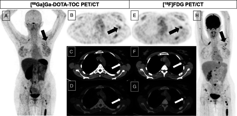
FIGURE 1.

We describe the case of a 29-year-old woman with stage IV retroperitoneal paraganglioma and disseminated bone metastases, who underwent both 68Ga-DOTATOC and 18F-FDG PET/CT during her follow-up. After the injection of 101 MBq of 68Ga-DOTATOC, the PET/CT examination revealed a stable disease in the already known numerous bone and pulmonary metastases; furthermore, a mild uptake was demonstrated in some small left axillary nodes (white and black arrows; A, MIP; B, axial PET image; C, CT image; D, fused PET/CT image). The day after, she underwent 18F-FDG PET/CT confirming bone and lung lesions and a mild uptake (SUVmax, 3.80) in the left axillary region, corresponding to the small nodes with fatty hilus previously observed (white and black arrows; E, axial PET image; F, CT image; G, fused PET/CT image; H, MIP). A fade uptake was also detected within the subcutaneous soft tissues superficial to the left deltoid muscle. The patient presented a history of receiving the Pfizer-BioNTech COVID-19 vaccine in the left deltoid muscle 6 and 7 days before the 68Ga-DOTATOC and the 18F-FDG PET/CT, respectively. Tracer injection was via the right (contralateral) antecubital fossa in both examinations, hence not a potential cause. Clinical correlation and its morphological aspect supported the nodal uptake to be reactive, ascribed to the recent vaccination. Similar findings were described after COVID-19 vaccinations (both Pfizer-BioNTech and AZ) on 18F-FDG PET/CT1–7 and, recently, also on 18F-choline PET/CT examination.8 To the best of our knowledge, this is the first case of a COVID-19 vaccine–related nodal uptake on 68Ga-DOTATOC PET/CT, as very recently another case involving 68Ga-DOTATATE PET/CT had been reported.9 This case highlights another potential pitfall associated with the current COVID-19 pandemic vaccination program, which may result in incorrect image interpretation and inadvertent disease upstaging.
ACKNOWLEDGMENTS
The authors would like to thank the technicians, the nurses, and all the members of the nuclear medicine unit.
Footnotes
Conflicts of interest and sources of funding: none declared.
Authors contribution: P.G., S.M., and S.B. contributed to the data analysis, manuscript writing, and manuscript reviewing. A.S., F.P., and M.G. contributed to the manuscript reviewing.
Contributor Information
Simona Muccioli, Email: simona.muccioli@iov.veneto.it.
Sara Berti, Email: sara.berti@iov.veneto.it.
Alida Sartorello, Email: alida.sartorello@iov.veneto.it.
Fiammetta Pesella, Email: fiammetta.pesella@iov.veneto.it.
Michele Gregianin, Email: michele.gregianin@iov.veneto.it.
REFERENCES
- 1.Nawwar AA Searle J Hagan I, et al. COVID-19 vaccination induced axillary nodal uptake on 18F-FDG PET/CT. Eur J Nucl Med Mol Imaging. 2021;48:2655–2656. [DOI] [PMC free article] [PubMed] [Google Scholar]
- 2.Avner M Orevi M Caplan N, et al. COVID-19 vaccine as a cause for unilateral lymphadenopathy detected by 18F-FDG PET/CT in a patient affected by melanoma. Eur J Nucl Med Mol Imaging. 2021;48:2659–2660. [DOI] [PMC free article] [PubMed] [Google Scholar]
- 3.Doss M Nakhoda SK Li Y, et al. COVID-19 vaccine–related local FDG uptake. Clin Nucl Med. 2021;46:439–441. [DOI] [PubMed] [Google Scholar]
- 4.Ahmed N Muzaffar S Binns C, et al. COVID-19 vaccination manifesting as incidental lymph nodal uptake on 18F-FDG PET/CT. Clin Nucl Med. 2021;46:435–436. [DOI] [PubMed] [Google Scholar]
- 5.McIntosh LJ Bankier AA Vijayaraghavan GR, et al. COVID-19 vaccination–related uptake on FDG PET/CT: an emerging dilemma and suggestions for management. AJR Am J Roentgenol. 2021;1–9. [DOI] [PubMed] [Google Scholar]
- 6.Eifer M, Eshet Y. Imaging of COVID-19 vaccination at FDG PET/CT. Radiology. 2021;299:E248. [DOI] [PMC free article] [PubMed] [Google Scholar]
- 7.Hanneman K, Iwanochko RM, Thavendiranathan P. Evolution of lymphadenopathy at PET/MRI after COVID-19 vaccination [published correction appears in Radiology. 2021 Aug;300(2):E338]. Radiology. 2021;299:E282. [DOI] [PMC free article] [PubMed] [Google Scholar]
- 8.Nawwar AA Searle J Singh R, et al. Oxford-AstraZeneca COVID-19 vaccination induced lymphadenopathy on [18F]choline PET/CT—not only an FDG finding. Eur J Nucl Med Mol Imaging. 2021;48:2657–2658. [DOI] [PMC free article] [PubMed] [Google Scholar]
- 9.Lu Y. DOTATATE-avid bilateral axilla and subpectoral lymphadenopathy induced from COVID-19 mRNA vaccination visualized on PET/CT. Clin Nucl Med. 2021. [Online ahead of print]. doi: 10.1097/RLU.0000000000003697. [DOI] [PMC free article] [PubMed] [Google Scholar]


