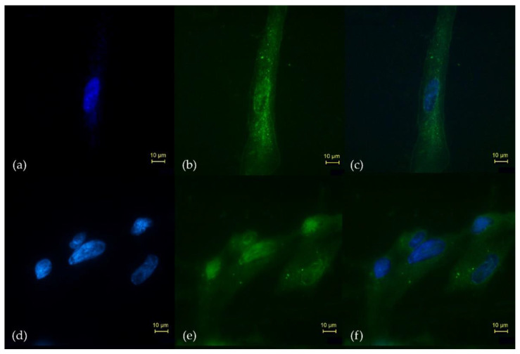
Figure 5.
Microscopic fields (40×) of the in vitro uptake and internalization in the N30 cells (FAP-positive) of (b) Lu2O3-iFAP and (e) Lu2O3 nanoparticles, detailing (a,d) the Hoechst-stained nucleus, and the merged images of nuclear and N30 cells treated with (c) Lu2O3-iFAP or (f) Lu2O3 (control). Observe the qualitative difference regarding the number of lutetium oxide nanoparticles (bright spots) between the Lu2O3-iFAP (b) and nanoparticles without iFAP (e) in the membrane and cytoplasm of N30 cells.

