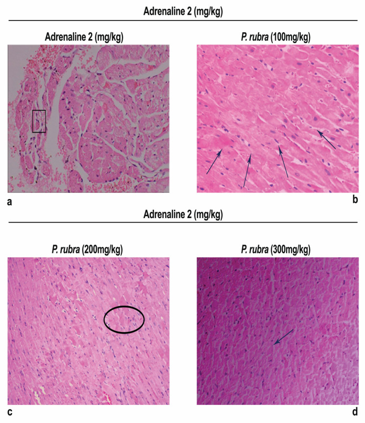
Figure 6.
Photomicrograph showing histopathological variations of ventricle section of rabbit heart: (a) ADR-intoxicated group; (b) P.rubra 100 mg/kg + ADR; (c) P. rubra 200 mg/kg; (d) P. rubra 300 mg/kg + ADR. In comparison to the ADR-intoxicated group, less inflammatory cells, cardiomyocyte deterioration, infiltration and fibrosis were observed in a dose-dependent manner. Rectangular shape indicates enlarged cardiomyocytes, upward arrows indicate cellular infiltration, oval shape indicates cardiac fibrosis and downward arrows indicate dense normal cluster of cardiomyocytes. Magnification is ×100.

