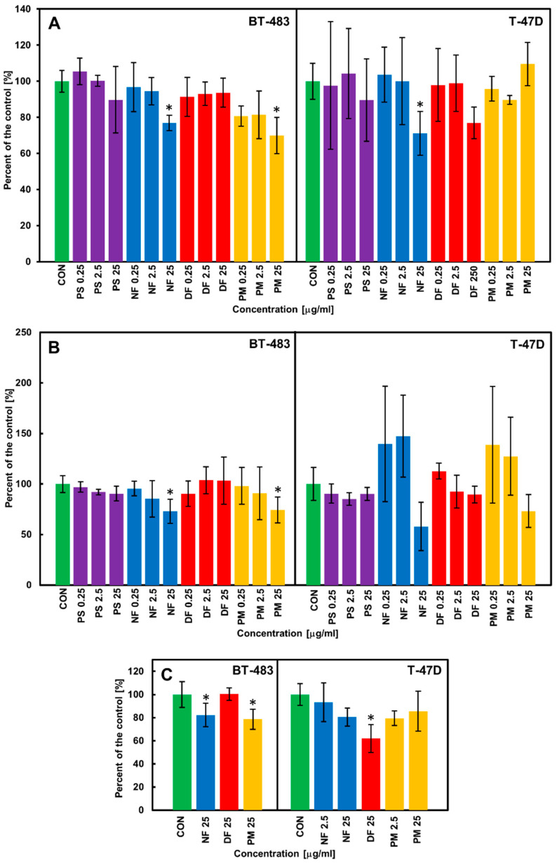
Figure 2.
The DS variants affect the viability (A), DNA biosynthesis (B), and cell counts (C) of luminal breast cancer cells (BT-483 and T-47D) in a manner that is dependent on their structure and cell type. (A) The breast cancer cells were exposed to the structural variants of DS that had been applied at the indicated concentrations for 24 h. Then, the cell viability was evaluated using the test based on measurements of the activity of the mitochondrial dehydrogenases. (B) The cancer cells were grown for 48 h in the presence of the tested variants of DS that had been used at the indicated concentrations. The cell proliferation was evaluated by measuring the incorporation to DNA of the bromodeoxyuridine that had been added to the cultures after the first 24 h of incubation. (C) The cancer cells were grown for 48 h in the exposure to the selected DS variants that had been applied at the indicated concentrations. Then, the cell number in the cultures was estimated using the crystal violet test. The results are expressed as the percentage of effect that was visible in the control cultures and are presented as the mean ±SD of at least three independent experiments in which n = 3 for each DS concentration. *—difference statistically significant (p ≤ 0.05) versus the control. CON—control (cultures untreated), PS—DS from porcine skin, NF—DS from normal human fascia, DF—DS from fibrosis affected fascia, PM—DS from porcine intestinal mucosa.

