. 2022 Jan 6;11:e67301. doi: 10.7554/eLife.67301

Figure 7. Localization of [111In]In-4497-IgG1 to a subcutaneous implant pre-colonized with biofilm.

Two days after implantation, mice were injected with 7.5 MBq [111In]In-4497-IgG1 (n = 7) and imaged at 24 hr, 72 hr, and 120 hr after injection. (A) Maximum intensity projection (corrected for decay) of a mouse subcutaneously bearing pre-colonized (C; left flank) and sterile (S; right flank) catheter. Additional scans can be seen in the supplementary information (Figure 7—figure supplement 2). (B) The activity detected in regions of interests was expressed as a percentage of total body activity. Each data point represents one mouse. A two-tailed paired t-test was performed to test for differences in activity in sterile versus colonized implants displayed as *p≤0.05, **p≤0.01, ***p≤0.001, or ****p≤0.0001. Exact p-values are displayed in Supplementary file 2.
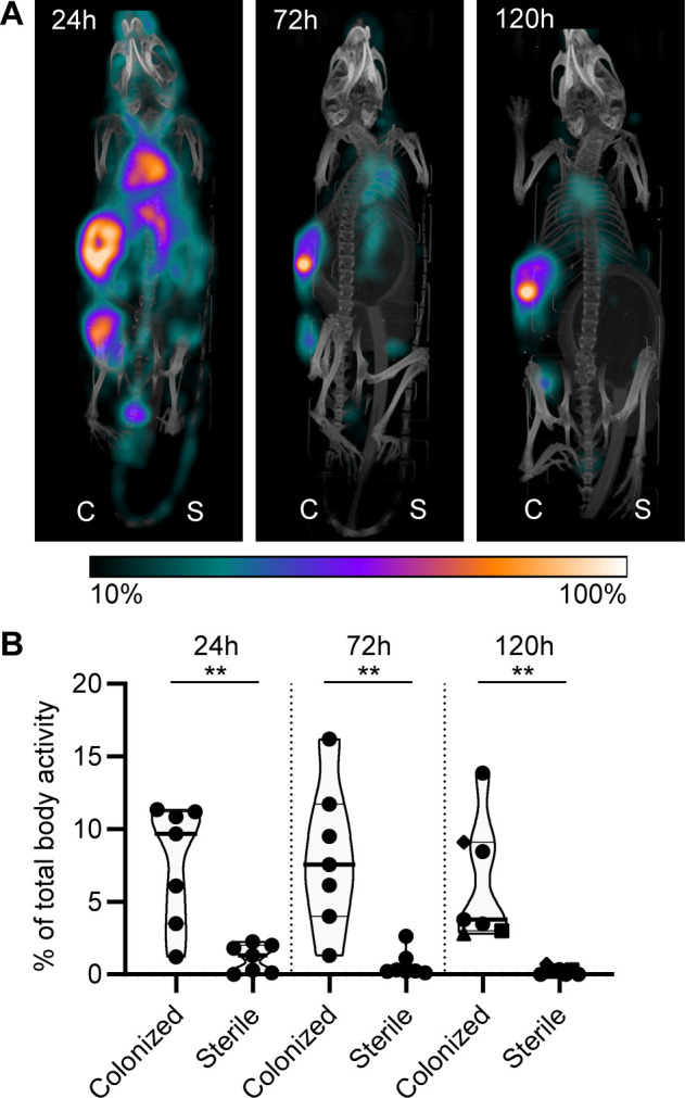
Figure 7.

Figure 7—figure supplement 1. CFU count before implantation and after implantation.
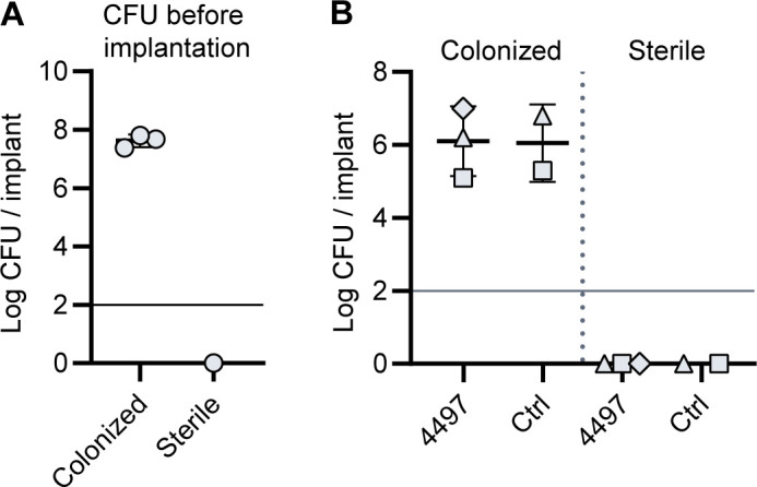
Figure 7—figure supplement 1.

(A) 5 mm PU catheter segments were inoculated with S. aureus LAC. After 48 hr of incubation, catheters were washed and sonicated and viable CFU counts recovered were determined. Each data point represents an independent experiment. (B) Mice received subcutaneous pre-colonized and sterile catheters and 2 days later were injected with [111In]In-4497-IgG1 (n = 3) or [111In]In-palivizumab (n = 2). At time point 120 hr, mice were sacrificed and catheters were removed to determine CFU counts. Horizontal lines indicate detection limit.
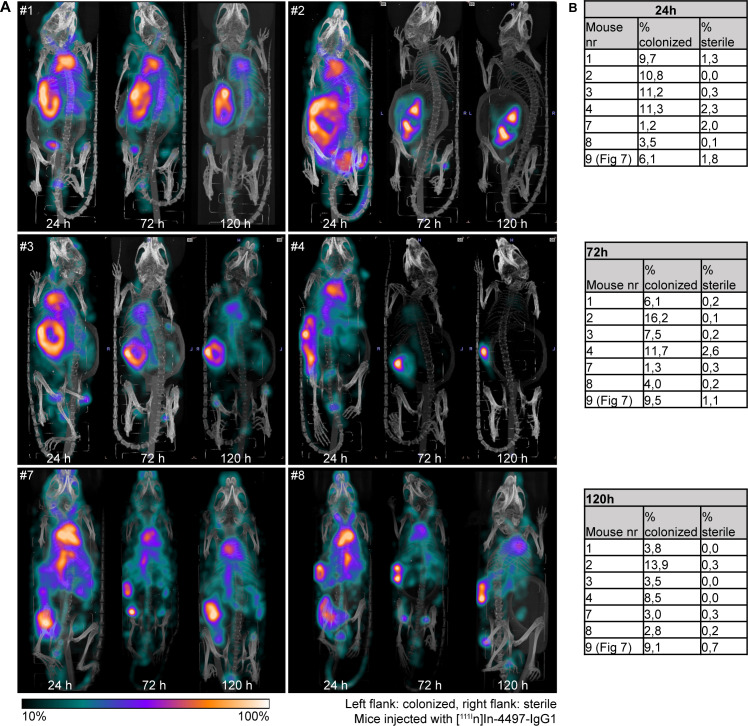
Figure 7—figure supplement 2. Localization of [111In]In-4497-IgG1 to subcutaneous implant pre-colonized with biofilm in a mouse model.

Figure 7—figure supplement 2.

(A) Maximum intensity projections of [111In]In-4497-IgG1 injected in mice subcutaneously bearing pre-colonized (left flank) and sterile (right flank) catheters. Implantation of colonized and sterile implants was randomized; however, for clarity, we here display all colonized implants on the left. (B) Corresponding percentages in regions of interest (ROIs) per mouse.
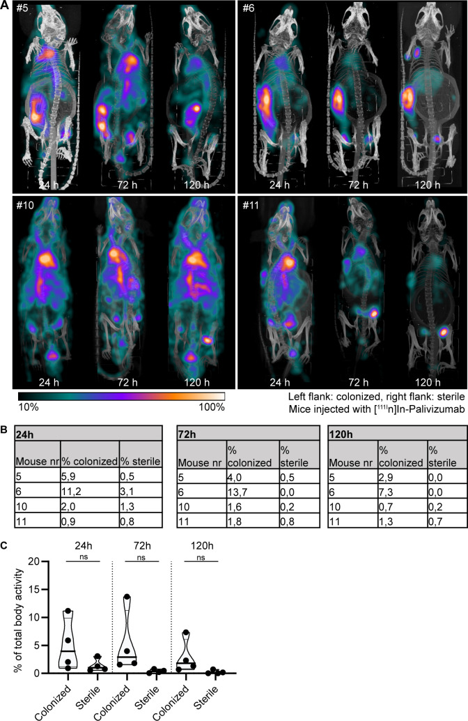
Figure 7—figure supplement 3. Localization of [111In]In-palivizumab to a subcutaneous implant pre-colonized with biofilm.

Figure 7—figure supplement 3.

(A) Maximum intensity projections (corrected for decay) of mice subcutaneously bearing pre-colonized (left flank) and sterile (right flank) catheters. Two days after implantation, mice were injected with 7.5 MBq [111In]In-palivizumab (n = 4) and imaged at 24 hr, 72 hr, and 120 hr after injection. Implantation of colonized and sterile implants was randomized, but for display all colonized implants are shown at the left flank. (B) Corresponding percentages in regions of interest (ROIs) per mouse. (C) The activity detected in regions of interests was expressed as a percentage of total body activity. Each data point represents one mouse. A two-tailed paired t-test was performed to test for differences in activity in sterile versus colonized implants. Exact p-values are displayed in Supplementary file 2.
Figure 7—figure supplement 4. Pilot study for localization of [111In]In-4497-IgG1 to subcutaneous implant-associated biofilm in a mouse model.
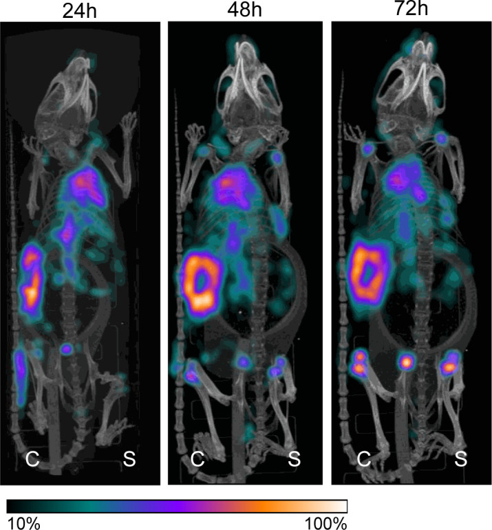
Figure 7—figure supplement 4.

One mouse received subcutaneous pre-colonized and sterile catheters and 2 days later was injected with 4 MBq [111In]In-4497-IgG1. The same mouse was imaged at 24 hr, 48 hr, and 72 hr. Maximum intensity projections of [111In]In-4497-IgG1 injected in mice subcutaneously pre-colonized (left flank) and sterile (right flank) catheters.
Figure 7—animation 1. Localization of [111In]In-4497-IgG1 to subcutaneous implant pre-colonized with biofilm in a mouse model.
3D projections of [111In]In-4497-IgG1 injected in mice subcutaneously bearing pre-colonized and sterile catheters. The same mouse was imaged at 24 hr, 72 hr, and 120 hr.
Figure 7—animation 2. 3D projections of [111In]In-palivizumab injected in mice subcutaneously bearing pre-colonized and sterile catheters.
The same mouse was imaged at 24 hr, 72 hr, and 120 hr.