Sir,
Infantile hemangioma (IH) with minimal or arrested growth (MAG) is a variant of IH increasingly recognized in the literature. IH-MAG usually presents at birth and it should be distinguished from its IH precursor or capillary malformations because of its flat telangiectatic appearance. Mulliken first proposed the term ''reticular'' to identify this type of IH.[1]
Here, we present two cases of IH-MAG associated with cutaneous signs and isolated spinal dysraphism.
Case 1
A firstborn girl presented since birth with an erythematous area with a reticulated background, surrounded by a whitish halo on the lumbar and buttocks region. In correspondence of the intergluteal sulcus, there was a peculiar “skin tag.” In the first few days of life, the detachment of the skin tag occurred. During the following months, the lesion had remained stable. Magnetic resonance imaging was performed, which confirmed the presence of isolated spinal dysraphism, including lipomyelomeningocele.
Case 2
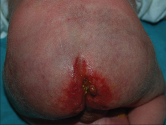
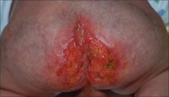
A boy born full-term presented at birth with vascular lesions over the perineal area, the buttocks, and the right lower limb, which started as a bluish-erythematous patch surmounted by red papules. A skin tag was present on the right buttock along the intergluteal sulcus [Figure 1]. After 1 month, numerous ulcers appeared on the buttocks and the posterior region of the thigh. The ulcer at the intergluteal sulcus caused detachment of the skin tag [Figure 2]. Systemic treatment with propranolol at a dose of 2 mg/kg per day was immediately started with a dramatic improvement of the ulceration. The duration of the treatment was 12 months. Magnetic resonance imaging was performed due to the segmental distribution of the lesion, revealing the presence of spinal dysraphism, with lipomyelomeningocele and tethered cord. At 8 months of age, the lipomyelomeningocele was resected and the tethered cord was released.
Figure 1.

Segmental IH-MAG of the perineum and buttocks with skin tag
Figure 2.

Numerous ulcers on the surface of IH-MAG with detachment of the skin tag
IH-MAG appears clinically as a relatively sharply demarcated patch, with a distinctive purple network-like pattern and an irregular distribution of small bright-red papules. The presence of prominent draining veins over the vascular discoloration is also possible. On dermoscopy, focal IH-MAGs present reddish rounded globular vessels, comma- like vessels, and reddish linear vessels.[2] Finally, the histological aspect is similar to capillary malformations, in which the dilated thin walled vessels within the superficial dermis are the main features. However, unlike capillary malformations, IH-MAG is GLUT-1 positive.[1]
IH-MAGs are more likely to be segmental and located on the lower body. Ulceration is common in the lower extremities, occurring in 30% of IH-MAGs and 67% of IH-MAGs with a reticular morphology.[3]
The coexistence of lumbosacral and perineal hemangioma with spinal dysraphism, as well as urogenital and anorectal anomalies, is well-known.[4] The proposed acronyms PELVIS (perineal hemangioma, external genitalia malformations, lipomyelomeningocele, vesicorenal abnormalities, imperforate anus, and skin tag), SACRAL (spinal dysraphism, anogenital, cutaneous, renal and urologic anomalies, associated with an angioma of lumbosacral localization) and LUMBAR (lower body hemangioma-urogenital anomalies-myelopathy-bony deformities-anorectal and arterial malformations-renal anomalies syndrome) underscore that this combination of hemangioma and ventral-caudal structural anomalies is the lower body counterpart of PHACES (posterior fossa brain malformations, hemangioma, arterial lesions, cardiac abnormalities, and eye abnormalities, sternal defects) association.[1]
Our two cases demonstrate the association of bilateral gluteal IH-MAG in a segmental distribution with “skin tags.” A skin tag is a distinct, exophytic, flesh- colored papule usually located on the midline. It has been speculated that it could be rhabdomyomatous mesenchymal hamartoma, composed of overgrowth of mesenchymal structures.[5]
The findings of segmental involvement prompted the evaluation for occult spinal dysraphism, confirmed by magnetic resonance imaging, which revealed the presence in both cases of lipomyelomeningocele.
To our knowledge, the coexistence of IH-MAG, tag-like skin lesions, and spinal dysraphism has not been previously emphasized, since they have been considered as part of syndromic conditions that also included urogenital and anorectal anomalies. Therefore, we believe that, in the presence of IH-MAG of the gluteal region and skin tag, magnetic resonance imaging is required to exclude isolated spinal dysraphism.
Declaration of patient consent
The authors certify that they have obtained all appropriate patient consent forms. In the form the patient(s) has/have given his/her/their consent for his/her/their images and other clinical information to be reported in the journal. The patients understand that their names and initials will not be published and due efforts will be made to conceal their identity, but anonymity cannot be guaranteed.
Financial support and sponsorship
Nil.
Conflicts of interest
There are no conflicts of interest.
Acknowledgments
We are very grateful and would like to acknowledge and thank Professor Ilona Frieden, UCSF Health, who has assisted us with her great expertise and precious suggestions in writing this manuscript submitted for publication.
References
- 1.Mulliken JB, Marler JJ, Burrows PE, Kozakewich HP. Reticular infantile hemangioma of the limb can be associated with ventral-caudal anomalies, refractory ulceration, and cardiac overload. Pediatr Dermatol. 2007;24:356–62. doi: 10.1111/j.1525-1470.2007.00496.x. [DOI] [PubMed] [Google Scholar]
- 2.Oiso N, Kimura M, Kawara S, Kawada A. Clinical, dermoscopic, and histopathologic features in a case of infantile hemangioma without proliferation. Pediatr Dermatol. 2011;28:66–8. doi: 10.1111/j.1525-1470.2010.01363.x. [DOI] [PubMed] [Google Scholar]
- 3.Weitz NA, Bayer ML, Baselga E, Torres M, Siegel D, Drolet BA, et al. The “biker-glove” pattern of segmental infantile hemangiomas on the hands and feet. J Am Acad Dermatol. 2014;71:542–7. doi: 10.1016/j.jaad.2014.04.062. [DOI] [PubMed] [Google Scholar]
- 4.Bouchard S, Yazbeck S, Lallier M. Perineal hemangioma, anorectal malformation, and genital anomaly: A new association? J Pediatr Surg. 1999;34:1133–5. doi: 10.1016/s0022-3468(99)90584-5. [DOI] [PubMed] [Google Scholar]
- 5.Stefanko NS, Davies OMT, Beato MJ, Blei F, Drolet BA, Fairley J, et al. Hamartomas and midline anomalies in association with infantile hemangiomas, PHACE, and LUMBAR syndromes. Pediatr Dermatol. 2020;37:78–85. doi: 10.1111/pde.14006. [DOI] [PubMed] [Google Scholar]


