Abstract
The majority of embolic strokes in patients with nonvalvular atrial fibrillation are caused by thrombi in the left atrial appendage. It is projected that strokes related to atrial fibrillation will markedly increase in the future unless effective mitigation strategies are implemented. Systemic anticoagulation has been known to be highly effective in reducing stroke risk in patients with atrial fibrillation. However, bleeding complications and nonadherence are barriers to effective anticoagulation therapy. Surgical and percutaneous left atrial appendage occlusion devices are nonpharmacologic strategies to mitigate the challenges of drug therapy. We present a contemporary review of left atrial appendage occlusion for stroke prevention in nonvalvular atrial fibrillation. A thorough review of the history of surgical and percutaneous left atrial appendage occlusion devices, recent trials, and US Food and Drug Administration milestones of current left atrial appendage occlusion devices are discussed.
Keywords: atrial fibrillation, left atrial appendage, left atrial appendage occlusion, stroke
Subject Categories: Anticoagulants, Catheter-Based Coronary and Valvular Interventions, Treatment, Ischemic Stroke, Atrial Fibrillation
Nonstandard Abbreviations and Acronyms
- 3D
3‐dimensional
- ACP
Amplatzer Cardiac Plug
- DAPT
dual antiplatelet therapy
- DOAC
direct oral anticoagulant
- DRT
device‐related thrombus
- FDA
Food and Drug Administration
- GA
general anesthesia
- ICE
intracardiac echocardiogram
- LAA
left atrial appendage
- LAAO
left atrial appendage occlusion
- OAC
oral anticoagulation
- TEE
transesophageal echocardiogram
- VKA
vitamin K antagonists
The left atrial appendage (LAA), described as the “most lethal human attachment,” is responsible for >90% of embolic strokes. 1 In 2020, stroke was the fifth leading cause of death in the United States after heart disease, cancer, COVID‐19, and unintentional injury. 2 Atrial fibrillation (AF) is associated with a 4‐ to 5‐fold increased risk of ischemic stroke and accounts for 25% of the 700 000 cerebrovascular accidents that occur in the United States annually. 3 , 4 This translates to an average of $351.2 billion direct and indirect costs in the United States alone. 3 It is projected that strokes related to AF will markedly increase in the future unless effective mitigation strategies are implemented. Historically, the mainstay of treatment for stroke prevention in AF has been oral anticoagulation (OAC). Large, randomized, controlled clinical studies of vitamin K antagonists (VKA), direct thrombin inhibitors, and factor‐Xa inhibitors have established OACs as the standard of care for stroke prevention in AF. 5 , 6 Although systemic anticoagulation has been known to be highly effective in mitigating stroke risk in patients with AF, 7 there are patient, physician, and systemic barriers that make it difficult for many patients to sustain OAC therapy over time. 8 These challenges led to a quest for alternative nonpharmacologic strategies particularly for high‐risk patients who are intolerant to standard therapy.
For decades, OAC therapy for AF was limited to VKAs. Because of its dietary restrictions and need for routine blood monitoring, VKA therapy often leads to poor compliance and frequent measurements outside the therapeutic window. It was hoped that newer direct oral anticoagulants (DOACs) and predictable levels of anticoagulation without blood‐level monitoring would address issues of nonadherence. However, limitations with anticoagulation therapy are present even with DOACs. Significant bleeding, high medication costs, and patient noncompliance are some of the barriers that prevent patients from receiving optimal therapeutic benefit. Although many patients are able to tolerate OACs, there is a large subset of patients who cannot tolerate long‐term anticoagulation. Elderly patients often have unfavorable risk profiles for anticoagulation and high Hypertension, Abnormal Renal/Liver Function, Stroke, Bleeding History or Predisposition, Labile INR, Elderly, Drugs/Alcohol Concomitantly scores. Interestingly, this is the same subset of patients who benefit the most from OAC in terms of stroke risk reduction. This treatment gap has been the impetus for finding an effective and safe nonpharmacologic alternative therapy for stroke prevention.
History of LAA Closure
The history of LAA occlusion (LAAO) or excision can be traced back to the mid‐20th century when cardiac surgical techniques specifically involving the mitral valve were developed. Rheumatic mitral stenosis is notorious for causing embolic strokes especially with concomitant AF. Unsurprisingly, the earliest documented LAA excision procedures were in patients with rheumatic mitral stenosis undergoing cardiac surgery. In 1949, Madden described 2 patients who underwent LAA excision as prophylaxis for recurring atrial thrombi. 9
A meta‐analysis of 23 studies noted that 57% of patients with valvular AF had thrombi localized in the LAA and extended into the left atrial cavity. 10 In contrast, in patients with nonrheumatic AF, 91% of thrombi were localized in the LAA only (P<0.0001). This pivotal observation differentiated the management of rheumatic (valvular) and nonrheumatic (nonvalvular) AF attributed to the site‐specific location of the thrombus that led to embolic stroke. Valvular AF is defined as AF in the setting of moderate to severe mitral valve stenosis from rheumatic heart disease or a mechanical valve. 11 With >90% of thrombi located in the LAA in nonvalvular AF, attention has focused on occlusion or exclusion of the LAA for the prevention of embolic stroke.
Morphology and Function of the LAA
The LAA is a finger‐like projection contiguous with the main body of the left atrium (LA). It is generally regarded as a vestigial remnant of the primordial LA. 12 Knowledge of the LAA shape, size, and spatial relationship with contiguous cardiac structures is vital especially during structural procedures. The LAA has a wide variability in size and shape. In most hearts, the LAA extends between the anterior and lateral walls with its tip pointed antero‐superiorly. In a small percentage of hearts, the LAA is directed laterally and posteriorly or sits in the transverse pericardial sinus. 13
A large study of postmortem hearts examined the variations of LAA lobes. The study demonstrated that 54% of hearts had 2 lobes, 23% had 3 lobes, 20% had 1 lobe, and only 3% had 4 lobes. 14 No significant correlation between sex and age differences in LAA morphologies were found. However, there is a strong association with the number of lobes and the presence of LAA thrombus. In their cohort of patients with AF, most patients with LAA thrombus had ≥3 lobes. In contrast, thrombus was only seen in 0.7% (n=296) with 1 or 2 lobes. 15
LAA Shapes
One of the peculiar features of the LAA is the wide variation of shapes and sizes. In a study using multidetector computed tomography (CT) and cardiac magnetic resonance, LAA morphology was classified into 4 shapes (Figure 1). 13 The most common shape is the “chicken wing.” A prominent bend in the proximal or middle part of the dominant lobe defines this shape. The cactus shape is the next most common LAA morphology. It is characterized by a dominant central lobe with secondary lobes outpouching both superiorly and inferiorly. The third most common is the windsock shape with 1 dominant lobe as the primary structure. Lastly, the cauliflower shape is the least common morphology. It has a variable number of lobes without a dominant lobe. It also has a short length with an irregular orifice. Most importantly, the cauliflower shape is most often associated with embolic events and strokes. 16
Figure 1. Explanted hearts showing different left atrial appendage morphology.
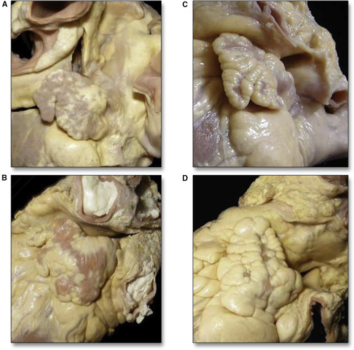
(A) chicken wing, (B) windsock, (C) cauliflower, (D) cactus. Reprinted from Beigel et al 13 with permission. Copyright ©2014, Elsevier.
LAA Function
The LAA has neurohormonal and reservoir functions. It contains specialized endothelial cells involved in the production of natriuretic peptides (ANP [atrial natriuretic peptide] and BNP [B‐type natriuretic peptide]). Compared with the LA, the LAA is more distensible and contains a high concentration of ANP granules. These features allow the LAA to be an ideal decompression chamber when LA pressure increases. The ANP granules also regulate fluid balance. In some studies of surgical removal of the LAA, excision was thought to cause fluid retention in the immediate postoperative period. This observation was apparent in the surgical removal of both the LAA and right atrial appendage during the “cut and sew” Maze procedure. 17 , 18 A current iteration of the procedure only excises the LAA and ameliorates this complication. Experimental studies also demonstrated that mechanical activity of the LAA has no apparent effect on cardiac output, thus closure or excision of the LAA will not have any significant hemodynamic consequence. 18
When the LA undergoes remodeling in patients with chronic AF, the LAA becomes a static pouch. During AF, Doppler flow velocity is reduced and propensity for thrombus formation increases. 12 The pathogenesis of thrombus formation in the LAA has not been fully elucidated. However; the shape of the LAA, relative stasis, and trabeculations appear to play a major role. Several mechanisms occur during AF that promote thrombogenesis as a fulfillment of the Virchow’s triad. Endocardial damage occurs by atrial dilatation, endocardial denudation, and fibroelastic infiltration of the extracellular matrix. 19 Hemostatic and platelet activation, in addition to growth factor changes during AF, promote a hypercoagulable state. Lastly, stasis in the LAA, especially during AF, completes the triad.
Surgical LAA Ligation and Occlusion
The evidence behind surgical closure of the LAA consists mainly of case reports and retrospective case series (Table). Madden published one of the first reports of LAA removal in 2 patients that was later repeated by others. 20 The practice was later abandoned after LAA excision in 8 patients reported a high complication rate that included 3 deaths, 1 paraplegia, and 3 peripheral emboli. 21
Table 1.
Available Literature on Surgical Left Atrial Appendage Occlusion
| Trial name | Year | Publication type | Surgical device used | Control arm | n | Mean follow‐up | Results | ||
|---|---|---|---|---|---|---|---|---|---|
| Primary end point | Closure, % | Conclusions | |||||||
| Johnson et al 1 | 2000 | Observational, retrospective | Excision | … | 437 | Technical feasibility and safety | 100 | Safe, suggested benefit in stroke prevention | |
| Katz et al 25 | 2000 | Observational, retrospective | Endocardial suture | … | 50 | Evaluate postsurgical success with TEE at differed points in time | 64 | Incomplete LAA ligation occurred in 36% of patients irrespective of surgical technique | |
| Garcia‐Fernandez et al 26 | 2003 | Observational, retrospective | Endocardial suture | … | 205 | Occurrence of embolic event | 90 | Positive stroke prevention | |
| Bando et al 75 | 2003 | Observational, retrospective | Endocardial suture | … | 812 | Occurrence of embolic event | Negative for stroke prevention | ||
| Blackshear et al 10 | 2003 | Observational, prospective | Thoracoscopic epicardial purse string | … | 15 | Feasibility of snare loop procedure and stroke incidence | 93 | Positive trend toward stroke prevention | |
| Pennec et al 76 | 2003 | Observational, prospective series |
Endocardial suture Excision |
… | 30 | Safety and efficacy |
70–80 100 |
Negative for stroke prevention Negative outcomes in intra‐atrial approach Positive for stroke prevention |
|
| Schneider et al 77 | 2005 | Observational, prospective series | Endocardial suture | … | 6 | Safety and efficacy | 17 | Incomplete surgical LAA closure may promote rather than reduce the risk of stroke; need for TEE to verify occlusion | |
| Healey et al 24 | 2005 | Randomized controlled |
Epicardial suture Stapler |
No occlusion | 77 | 13 ± 7 mo | Safety and efficacy |
45 72 |
Safe at the time of CABG; positive for stroke prevention (2.6%) |
| Kanderian et al 78 | 2008 | Observational, retrospective |
Excision (52) Suture exclusion (85) Stapler |
… | 137 | Comparison of effectiveness in surgical techniques |
73 23 20 |
There is a high occurrence of unsuccessful surgical LAA closure; of the various techniques, excision appears to be the most successful | |
| Bakhtiary et al 79 | 2008 | Observational, prospective series | Clamp and epicardial suture | … | 259 | Safety, occurrence of stroke | 100 | Positive for stroke prevention | |
| Ailawadi et al, EXCLUDE 28 | 2011 | Observational, prospective series | AtriClip | … | 75 | Safety and efficacy | 95 | Safe and stable on short‐term postprocedural imaging | |
| Whitlock et al, LAAOS II 80 | 2013 | Cross‐sectional | Occlusion | No occlusion | 1889 | 1 y | Safety and efficacy | LAA occlusion can be safely performed at the time of cardiac surgery; positive for stroke prevention | |
| Whitlock et al, LAAOS III 31 | 2021 | Randomized, controlled | Occlusion | No occlusion | 4811 | 3 y | Stroke or systemic embolism | LAA occlusion during cardiac surgery reduced risk of ischemic stroke or systemic embolism in patients with atrial fibrillation | |
CABG indicates coronary artery bypass graft; EXCLUDE, The Exclusion of Left Atrial Appendage with AtriClip Exclusion Device in Patients Undergoing Concomitant Cardiac Surgery; LAA, left atrial appendage; LAAOS II, Left Atrial Appendage Occlusion During Cardiac Surgery to Prevent Stroke; LAAOS III, Left Atrial Appendage Occlusion During Cardiac Surgery to Prevent Stroke; and TEE, transesophageal echocardiogram.
In the 1990s, interest in surgical LAA excision or exclusion resurfaced coincident with the development of the Maze procedure for the treatment AF and the widespread use of transesophageal echocardiogram (TEE). 22 Different techniques of LAA ligation or excision have been developed. Current surgical techniques to exclude the LAA include resection, epicardial stapling, clip application, or endoatrial double‐layer longitudinal suture closure. 23 A surgical stapler for LAA excision was first used at the time of mitral valve surgery. The technique was later supplemented with pericardial buttressing strips in 2005. During the same year, the results of the pilot study LAAOS (Left Atrial Appendage Occlusion Study) were released. This study randomly assigned 77 patients undergoing coronary artery bypass grafting at increased risk for stroke to LAA exclusion using epicardial suture ligation or stapler (n=52) versus control (no LAA exclusion; n=25). 24 TEE was performed after 14 months. Success in the LAA exclusion arm was defined as residual Doppler flow into the appendage or residual “neck” of <1 cm. In the suture exclusion arm, persistent Doppler flow suggested inadequate technical closure. In the staple exclusion arm, the number of subsequent neurological events was attributed to the large residual remnant size (>1 cm) inherent to the surgical technique. However, the stroke rate in both exclusion groups (suture or stapler) was lower than the control group (2.7% versus 5.6%), suggesting benefit despite the differences in technique.
Certain conclusions may be derived from the LAAOS pilot study and other observational studies: (1) successful closure immediately post operation compares well to long‐term observations, suggesting that failure is most likely derived from surgical technique rather than gradual suture/staple dehiscence, 25 (2) incomplete occlusion carries a higher risk than no occlusion in relation to long‐term thromboembolic events, 26 and (3) the LAA orifice is 3‐dimensional (3D) instead of circular, and prosthetic mitral valve material can limit the reach to the most distal edge of the LAA, leading to a large remnant. Other procedural caveats such as shallow suture bites to avoid the left circumflex must be considered. Large remnant sizes >1 cm are inherent to staplers. 27
In response to the multiple difficulties associated with various surgical LAAO techniques, a number of newer surgical devices have been developed. Despite the fact that excision appeared to be more favorable than occlusion, most new devices are designed to occlude rather than excise the LAA. The devices have the ability to sustain a high occlusion pressure compared with suture ligation and stapling. The AtriClip device (Atricure, Dayton, OH; Figure 2), holds the strongest evidence available. This parallel, self‐closing clamp is designed with a cloth covering that exerts uniform pressure at the base of the LAA. The goal is to exert a high occlusion pressure that results in atrophy of the LAA. The AtriClip has the following theoretical advantages over traditional surgical excision: (1) ability to reposition the device, (2) lower risk for tears and bleeding, and (3) decreased left circumflex artery injury. On short‐term follow‐up (3 months), >98% of patients undergoing TEE or CT imaging had complete LAA occlusion after AtriClip. No device‐related adverse events or perioperative mortalities occurred. 28
Figure 2.
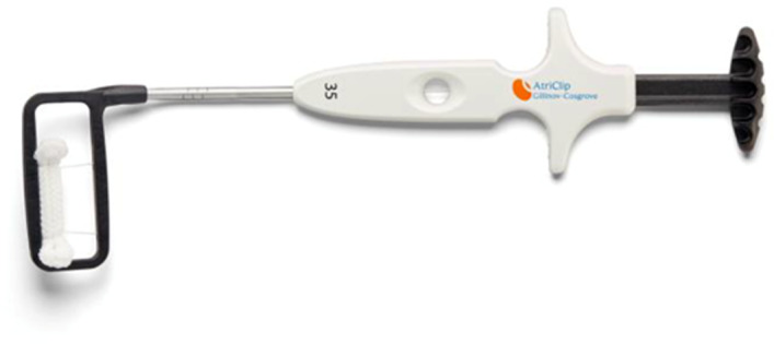
The AtriClip device. Copyright Atricure 2021.
Other surgical LAAO technologies such as the Cardioblate Closure Device (Medtronic, Fridley, MN) and Sierra Ligation System (Aegis Medical Innovations, Inc., Vancouver, BC) are currently investigational. 29 The Tiger Paw System (Maquet Medical Systems, Wayne, NJ) was recalled in 2015 after reports of LAA tears leading to adverse events and death.
A meta‐analysis of patients undergoing surgical LAAO showed significant reductions in stroke at 30 days (0.95% versus 1.9%; P=0.005) and at follow‐up (1.4% versus 4.1%; P=0.01) in patients with AF undergoing surgical LAAO compared with matched controls. In addition, a significant reduction in all‐cause mortality (1.9% versus 5%; P=0.0003) in those who underwent surgical LAAO was observed. 30
Because of the current limited data on surgical LAAO, the 2017 Society of Thoracic Surgeons gave a Class IIa recommendation (Level of Evidence C, “limited data”) for the excision or exclusion of the LAA in concomitant cardiac operations in patients with AF for the prevention of thromboembolism. 23 The recent results of the LAAOS III (Left Atrial Appendage Occlusion During Cardiac Surgery to Prevent Stroke) study may change the recommendation for surgical LAAO in the future.
LAAOS III is a multicenter trial that randomly assigned patients with AF with CHA2D2‐VASc scores ≥2 who were undergoing cardiac surgery for another indication to surgical LAAO (occlusion group) or no LAAO (no‐occlusion group). All patients received the usual postoperative care including oral anticoagulation. At 3 years, 76.8% of the participants continued to receive oral anticoagulation for AF. Stroke or systemic embolism (primary outcome) occurred in 4.8% in the occlusion group and in 7.0% in the no‐occlusion group (hazard ratio, 0.67; 95% CI, 0.53–0.85; P=0.001). 31 The benefit of surgical LAAO on stroke prevention appeared to be an adjunct to oral anticoagulation therapy. However, the study does not support surgical LAAO as a replacement for oral anticoagulation therapy. In contrast to percutaneous LAAO trials, there are still no studies available that randomized surgical closure against oral anticoagulation for stroke prevention.
Percutaneous LAAO Devices
Several percutaneous devices for LAAO are being developed and tested in clinical trials, including the LARIAT device (SentreHeart, Redwood City, CA), the Amulet device (St. Jude Medical, St. Paul, MN), and the WaveCrest device (Biosense Webster, Diamond Bar, CA). The redesigned next‐generation Watchman FLX 2.5 (Boston Scientific, Marlborough, MA), which has been recently approved, is currently available for commercial use. Examples of historical and contemporary percutaneous LAAO devices are shown in Figure 3.
Figure 3. Percutaneous left atrial appendage occlusion devices.
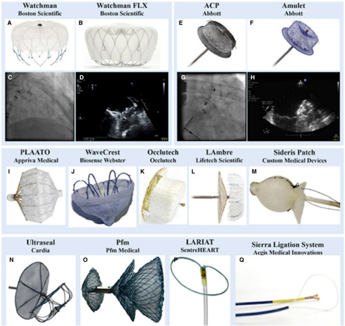
(A) Watchman, (B) Watchman FLX, (C) fluoroscopic image of the Watchman, (D) TEE image showing a deployed Watchman, (E) Amplatzer Cardiac Plug (ACP), (F) Amulet, (G) Fluoroscopic image of the ACP device, (H) transesophageal echocardiogram image showing a deployed Amulet device, (I) PLAATO, (J) WaveCrest, (K) Occlutech, (L) LAmbre, (M) Sideris Patch, (N) Ultraseal, (O) Pfm, (P) LARIAT, (Q) Sierra Ligation System. Reprinted from Asmarats and Rodés‐Cabau 74 with permission. Copyright ©2017 American Heart Association, Inc. ACP indicates Amplatzer Cardiac Plug; and PLAATO, percutaneous left atrial appendage transcatheter occlusion.
Percutaneous Left Atrial Appendage Transcatheter Occlusion
The first percutaneous LAAO device implanted in humans was the Percutaneous Left Atrial Appendage Transcatheter Occlusion (PLAATO) device (ev3 endovascular, Plymouth, MN) (Figure 3I). 32 The device consisted of a self‐expanding nitinol cage enclosed by polytetrafluoroethylene membrane. Early safety and feasibility study were demonstrated in 15 patients with chronic AF at high risk for stroke who were poor candidates for long‐term warfarin therapy. Device deployment was successful in 100% of the patients. No significant adverse events were noted. 32 The European PLAATO study evaluated the safety and efficacy of the device in 180 patients with nonvalvular AF and contraindication to warfarin therapy. 33 The primary end point was LAA closure as determined by TEE after 2 months post procedure and stroke rate at 150 patient‐years. LAA occlusion was successful in 90% of the patients (95% CI, 83.1%–92.9%). However, the trial was inundated with complications, including 2 deaths within 24 hours, 6 cardiac tamponades, and 1 device embolization. The follow‐up phase was halted, and the trial was prematurely discontinued because of financial issues. The PLAATO device is no longer commercially available.
Watchman and Watchman FLX 2.5
The Watchman and Watchman FLX devices (Figure 4) are currently the only US Food and Drug Administration (FDA) approved devices for LAAO in the United States. 34 The Watchman FLX is the next‐generation and current iteration of the Watchman device that gained Conformité Européene marked approval in Europe in March 2019 and US FDA approval in July 21, 2020.
Figure 4. The Watchman (A) and the Watchman FLX device (B).
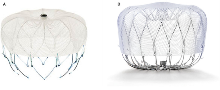
Image provided courtesy of Boston Scientific ©2021 Boston Scientific Corporation or its affiliates. All rights reserved.
The first‐in‐man Watchman device implantation was performed on August 12, 2002, in Europe. The Watchman device consisted of a self‐expanding nitinol 10‐strut frame with a 160‐μm polyethylene terephthalate fabric mesh cap (Figure 4A). The polyethylene terephthalate membrane covers 50% of the proximal outer nitinol frame. This membrane was designed to prevent thrombus embolization from the LAA and to promote endothelialization. The selected device should be larger than the diameter of the LAA ostium to ensure device stability and sufficient compression (8%–20%) against the LAA wall. The length of the device is approximately equal to its width. Fixation anchors, located distal to the distal end of the polyester fabric cap, secure the device within the LAA trabeculae.
The Watchman FLX device (Figure 4B) is the next‐generation Watchman device. This redesigned iteration is fully recapturable and repositionable. It has a shorter device length, 18 struts (versus 10 struts) and an atraumatic closed distal end (versus open end). The Watchman FLX device also has a broader size range (20–35 mm) compared with its predecessor (21–33 mm). This would provide a wider range to accommodate different LAA anatomies. A significant redesign of the device also includes dual row anchors to reduce the risk of embolization and reduced metal exposure. The closed distal end design compared with the open‐ended design of the first Watchman device should theoretically lessen the likelihood of perforation, pericardial effusion, and tamponade.
Amplatzer Cardiac Plug and Amplatzer Amulet Device
Amplatzer Cardiac Plug (ACP; Abbott Vascular, Santa Clara, CA) is a self‐expanding, double‐disc device consisting of a nitinol mesh with polyester fabric (Figure 3E). The length of the device is shorter than the diameter, thereby allowing its use in cases of shorter LAA anatomy. Device size ranges from 16 mm to 30 mm, allowing broader options for different LAA sizes.
The Amulet device is the latest generation of the ACP (Figure 3F). It is based on a design similar to the ACP while incorporating some modifications to allow for easier implantation and reduce periprocedural complications. Long‐term clinical outcomes of the first‐generation and second‐generation Amplatzer devices were similar in terms of safety, efficacy, and clinical benefit. 35 The ACP and Amulet devices received Conformité Européene mark approval in 2008 and 2013, respectively. At this time, the device is for investigational use only in the United States. Several randomized clinical trials evaluating the Amulet device are underway and US FDA approval may be upcoming.
The AMULET Investigational Device Exemption (IDE) trial is a prospective, randomized, multinational trial that is currently ongoing. It is designed to evaluate the safety and effectiveness of the Amplatzer Amulet device in comparison with the Watchman device for stroke prevention in patients with nonvalvular AF. The trial is the first head‐to‐head trial comparing 2 different LAAO devices. A total of 1878 patients will be randomly assigned 1:1 between the Amulet and the Watchman LAAO devices. The 3 primary end points of the trial are the following: (1) composite of procedure‐related complications, or all‐cause death, or major bleeding through 12 months; (2) a composite of ischemic stroke or systemic embolism through 18 months; and (3) effective device LAAO, defined as residual jet around the device ≤5 mm at the 45‐day visit. 36 Initial data from the roll‐in phase appear promising with a low rate of complications and a high procedural success rate. Results from this trial will be helpful in broadening the options available for patients with AF who are high risk for stroke and bleeding. Another upcoming trial (Clinical Trial of Atrial Fibrillation Patients Comparing Left Atrial Appendage Occlusion Therapy to Non‐vitamin K Antagonist Oral Anticoagulants) will randomly assign ≈2650 patients 1:1 to the Amulet device and DOACs at 150 sites worldwide. This trial aims to evaluate the safety and effectiveness of the Amulet device as a viable alternative to DOACs in patients with nonvalvular AF who are at high risk for stroke. Expected completion date of this trial will be in 2024. 37
LARIAT
The LARIAT is a LAAO ligation system that uses a unique technique of combining epicardial and endocardial approaches (Figure 3P). After obtaining percutaneous pericardial access, a magnet‐tipped wire is inserted into the pericardial space. Transseptal access is then performed and a proprietary 20‐mm compliant occlusion balloon catheter with a second magnet‐tipped wire is introduced into the LAA. After the 2 magnets are connected, the LARIAT snare delivery system is advanced over the LAA. This maneuver is guided by the endocardial balloon catheter inside the LAA. The snare is closed and the suture is tightened to ligate the LAA. The US FDA approved the LARIAT in June 2006 for soft tissue approximation but not specifically for prevention of thromboembolism by occluding the LAA.
Aside from the approach, the LARIAT system is unique to other LAAO devices because there is no device left in the endocardial surface of the LAA. Thus, theoretically, anticoagulation is not required post procedure. In large (>40 mm) LAA, which may be prohibitive for both the Watchman or ACP devices, the placement of the LARIAT system may be considered as long as it is anatomically suitable for the device (up to 45 mm [width], 20 mm [height], 70 mm [length]).
In the initial multicenter experience of the device, the rate of acute closure was high (94% device success, defined as suture deployment and <5 mm leak on TEE). 38 However, in the same retrospective study, procedural success (86%) was limited by significant bleeding. There were 14 patients (9.1%) with major bleeds and 16 patients (10.4%) with significant pericardial effusion. 38 Inherent to the technique, the major limitation of the LARIAT device is the need for epicardial access. Most of the complications are related to the pericardial puncture including pericardial effusion, cardiac perforation, and severe pericarditis. Reports of death and major complications including complete LAA detachment and cardiac perforation prompted the US FDA to issue a safety communication in July 2015. Currently, the LARIAT system is generally considered for patients who have LAA anatomy that precludes endovascular LAAO or with absolute contraindications to anticoagulation.
The aMAZE (LAA Ligation Adjunctive to PVI for Persistent or Longstanding Persistent Atrial Fibrillation) study is an ongoing prospective, multicenter, randomized study. The study evaluates the safety and effectiveness of the LARIAT system to ligate the LAA as an adjunct to planned pulmonary vein isolation for the treatment of AF. 39 It is hypothesized that the combination of pulmonary vein isolation and LAA ligation can safely and effectively reduce the incidence of recurrent AF compared with pulmonary vein isolation alone. It is a unique trial because it combines 2 percutaneous procedures with different indications. Enrollment is already completed, and the results are expected in December 2021.
Percutaneous LAAO Device Trials
The LAAO devices have endured a long and arduous journey that culminated with the US FDA approval of the Watchman device in 2015 and the next‐generation Watchman FLX in 2020 (Figure 5). Several other LAAO devices are currently being evaluated in randomized clinical trials and are awaiting US FDA approval. Two prospective, randomized clinical trials evaluating the safety and efficacy of LAAO using the Watchman device were conducted in 2014. PROTECT AF (Watchman Left Atrial Appendage System for Embolic Protection in Patients With Atrial Fibrillation) was the first trial followed by the PREVAIL (Evaluation of the Watchman LAA Closure Device in Patients With Atrial Fibrillation Versus Long‐Term Warfarin Therapy) trial. In both trials, patients were randomized 2:1 to either LAAO using the Watchman device or warfarin. All analyses were by intention to treat. Patients in the LAAO arm used aspirin (81 mg) and warfarin for 45 days, then dual antiplatelet therapy (DAPT) with clopidogrel 75 mg until 6 months, then indefinite aspirin therapy (325 mg). The results of PROTECT AF were ultimately in favor of LAAO, demonstrating both noninferiority and superiority compared with warfarin in the primary end point of stroke, systemic embolism, or cardiovascular mortality. 40 , 41 However, the PROTECT AF trial identified a periprocedural safety hazard particularly cardiac tamponade, pericardial effusion, and procedure‐related strokes. The PREVAIL trial was conducted specifically to further evaluate the safety end point of the Watchman device. This trial mandated at least 25% of new operators be included to further assess the effect of procedural performance. Noninferiority was achieved for a secondary coprimary end point of postprocedure ischemic stroke but did not achieve noninferiority for the first composite coprimary end point of stroke, systemic embolism, or cardiovascular mortality (the same primary end points as PROTECT AF). 42 , 43 Interestingly, there was an unusually low ischemic stroke rate in the warfarin arm (0.73%) in the PREVAIL trial.
Figure 5. Timeline showing important dates of left atrial appendage occlusion trials and US FDA milestones in the United States.
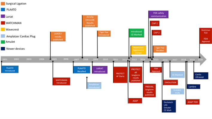
AF indicates atrial fibrillation; ASAP, ASA Plavix Feasibility Study With WATCHMAN Left Atrial Appendage Closure Technology; ASAP TOO, The Assessment of the Watchman Device in Patients Unsuitable for Oral Anticoagulation; CAP, Continued Access to PROTECT AF; CAP2, Continued Access to PREVAIL; CE, conformité européenne; EXCLUDE, Exclusion of Left Atrial Appendage with AtriClip Exclusion Device in Patients Undergoing Concomitant Cardiac Surgery; FDA, Food and Drug Administration; LAA, left atrial appendage; LAAOS I, Left Atrial Appendage Occlusion During Cardiac Surgery to Prevent Stroke; and PLAATO, percutaneous left atrial appendage transcatheter occlusion.
The PROTECT AF trial had a median follow‐up of about 4 years; however, the PREVAIL trial had a rather short follow‐up period with a median of 10 months. Meta‐analysis of patients in both trials followed for 5 years showed that the composite end‐point between groups was similar. Despite ischemic stroke/systemic embolization being numerically higher in LAAO, the difference was not statistically significant (P=0.87). Of note, hemorrhagic stroke, disabling stroke, postprocedure bleeding, and even all‐cause death favored LAAO. 43
Because of the safety concerns from the Watchman trials, the US FDA mandated the continued access registries CAP (Continued Access to PROTECT AF) and CAP2 (Continued Access to PREVAIL). Both are US FDA investigational devices exemption registries that were designed to gain further safety and efficacy data on the Watchman device in both trials. The registries have the same inclusion and exclusion criteria and treatment protocol as PROTECT AF and PREVAIL. The CAP registry included 566 patients with an average follow‐up of 50.1 months, whereas CAP2 included 578 patients with an average of 50.3 months. Both demonstrated similar procedural success rates (94%). The primary end point (composite of stroke, systemic embolism, cardiovascular/unexplained death, and safety) occurred at a rate of 3.05 per 100 patient‐years in CAP, and 4.80 per 100 patient‐years in CAP2. Total stroke rates were significantly lower in both registries (CAP, 78% reduction; CAP2, 69% reduction). 44 Both registries contain the largest follow‐up data of patients with the Watchman device. They reaffirm the safety and effective therapy of LAAO for patients with nonvalvular AF.
A rigid anticoagulation protocol (VKA for 45 days, then DAPT for 6 months, and then lifelong aspirin) was part of the trial design in PROTECT AF and PREVAIL. Patients with contraindication to warfarin were excluded in these trials. However, in real‐world populations, many patients cannot tolerate any anticoagulation because of bleeding. After LAAO, these patients end up using only DAPT, single antiplatelet therapy, or nothing at all. EWOLUTION (Evaluating Real‐World Clinical Outcomes in Atrial Fibrillation Patients Receiving the WATCHMAN Left Atrial Appendage Closure Technology) is the largest prospective real‐world registry on Watchman. The study showed that at hospital discharge after device implant, patients were using VKA (16%), DOAC (11%), DAPT (60%), single antiplatelet therapy (7%), or no anticoagulation (6%) at all. After a 2 year follow‐up, the patients (85%) who discontinued their DAPT and DOAC not surprisingly showed a 46% lower major bleeding rate compared with historic controls. However, the overall stroke rate remained low and in line with the results of the PROTECT AF, PREVAIL, and the CAP registries. 45
The next‐generation Watchman FLX device was evaluated in a prospective, nonrandomized multicenter trial to establish safety and effectiveness. PINNACLE FLX (The Protection Against Embolism for Non‐valvular AF Subjects: Investigational Evaluation of the WATCHMAN FLX™ LAA Closure Technology) is a single‐arm investigational device exemption trial that eventually enrolled 400 patients. 46 The primary effectiveness end point (100%; P<0.0001) was the incidence of effective closure (peridevice flow of ≤5 mm) at 12 months assessed by TEE. The primary safety end point (0.5%; P<0.0001) was the occurrence of death, ischemic stroke, systemic embolism, or device‐related or procedure‐related events requiring cardiac surgery within 7 days post procedure. Moreover, there were no device embolizations or pericardial effusions requiring cardiac surgery. 47
Several devices are also currently being evaluated in randomized trials in comparison with the Watchman device. A randomized, multicenter clinical trial (AMPLATZER Amulet LAA Occluder Investigational Device Exemption Trial) comparing the Amulet device to Watchman device (1:1) is set to be completed in 2024. 48 The WAVECREST 2 (WaveCrest Vs. Watchman TranssEptal LAA Closure to Reduce AF‐Mediated Stroke 2) is a prospective, multicenter, randomized trial designed to demonstrate the safety and effectiveness of the WaveCrest device in comparison to the Watchman device (control arm). 49 The target completion date will be in 2028.
Preprocedural Imaging
The success of the LAAO procedure hinges on the 3D appreciation of LAA anatomy and its surrounding structures. Multimodality imaging is essential in assessing LAA anatomy, site‐specific transseptal puncture, device selection, and positioning. 50 A thorough preprocedure analysis of LAA dimensions and potential anatomical pitfalls is mandatory. This can be accomplished by cardiac CT or TEE. Recently, adjunctive measures such as 3D printing of the LAA provide additional information to the operators. 50
TEE has remained the gold standard in LAAO preprocedural assessment. The presence of LAA or LA thrombus, determination of LAA dimensions and morphology, and feasibility of LAAO implant are the primary objectives of preprocedural imaging. The LAA is evaluated by using different TEE omni plane angles (0°, 45°, 90°, and 135°). Based on these views, the maximal and minimal diameters of the ostium, depth of the LAA, and perimeter are noted. The device size is chosen based on the manufacturer’s sizing guide of the selected device.
The oval‐shaped ostium of the LAA exposes a major flaw of TEE in LAAO procedures. The use of preprocedural TEE systematically underestimates the LAA ostium. This underestimation may lead to erroneous device size selection. Experience from transcatheter aortic valve replacement showed that the aortic annulus sizing by echocardiography resulted in underestimation of the diameter of the annulus. CT‐based sizing provides a more accurate assessment of aortic annulus size and is now standard in selecting transcatheter aortic valve replacement valve sizes. 51 Compared with the aortic annulus, the LAA is an even more complex structure. Despite a thorough evaluation using multiple TEE omni plane angles, the true LAA diameter may still be underestimated. 52 Moreover, TEE requires a skilled echocardiographer and has a wide interoperator variability.
CT is widely used for preprocedural assessment in LAAO. It is less invasive than TEE with less interoperator variability. A standard CT analysis includes an assessment of the LAA anatomy in the sagittal, axial, and coronal planes. The perimeter, maximal, and minimal diameters of the LAA ostium and landing zone are standard measurements in cardiac CT analysis of the LAA.
Preprocedural cardiac CT may also be used in evaluating for the presence or absence of LAA thrombus. However, filling defects in the LAA may just represent slow contrast mixing attributed to low flow velocities and may be mistaken for an LAA thrombus. 53 This usually triggers a confirmatory secondary imaging either with TEE or ICE. A delayed imaging acquisition, in which a second scan is acquired after a time delay, has been studied to increase the diagnostic accuracy (sensitivity 100% and specificity 99%) in detecting LAA thrombus. 54
A study was done comparing preprocedural TEE and multislice CT in aiding device selection for LAAO. The use of multislice CT resulted in 83% correct LAAO device selection compared with 57% using TEE (P<0.01). 52 The study also showed that the best parameter to guide Amulet and Watchman FLX (closed‐ended devices) is the perimeter‐derived mean diameter. On the other hand, maximal diameter was the best parameter when using an open‐ended device such as the Watchman.
A small case‐control study demonstrated the potential value of 3D printing of the LAA for device sizing. It also demonstrated the potential impact on reducing device leak. Patient‐specific 3D printed models of the LAA were made from preoperative CT images. Proposed device sizing based on 3D modeling was compared with the size of the device that was implanted using a standard approach. The results demonstrated that there was an agreement of device size in 35% of the time. However, compared with the 3D printed model, 55% of the devices were underestimated. In addition, there was a significantly higher prevalence of device leak in the subset of patients where the device size was underestimated (P=0.019). 55
Procedural Techniques
Although the procedural techniques for most LAAO devices are similar (except for the LARIAT), the steps for the Watchman procedure will be primarily discussed here. The LAAO procedure is performed via a femoral venous approach using standard transseptal techniques. Right femoral venous access is preferred because it facilitates transseptal access. Hemostasis is commonly achieved with either manual compression, “figure‐of‐8” suture, or preclosure technique using the 6‐F Perclose ProGlide Suture‐Mediated Closure System (Abbott Vascular, Temecula, CA). An 8‐French transseptal needle, dilator, and sheath assembly are advanced via the femoral vein to access the LA. Transseptal puncture is carried out in the usual manner (details of transseptal puncture are available elsewhere). 56 The long axis of the LAA is oriented anteriorly, and its ostium is perpendicular to this axis. Successful engagement of the LAAO device access sheath will therefore be facilitated by a posterior and inferior puncture in the fossa ovalis. 57 Bi‐caval (90°) and short‐axis (45°) TEE views are used for obtaining an optimal site‐specific transseptal puncture. Intracardiac echocardiogram (ICE) may also be used to guide transseptal access. Anatomic landmarks on fluoroscopy alone can also sometimes be used by experienced operators for transseptal access; however, echo guidance is strongly recommended to ensure safe site‐specific puncture and access to the LA. Intravenous heparin is administered before or immediately following transseptal puncture to maintain an activated clotting time of >300 seconds. There are no large studies available on specific activated clotting time goals for transseptal puncture. However, data on patients undergoing LA ablation demonstrated that an increased intensity of anticoagulation (activated clotting time >300 seconds) prevented thrombus formation. 57 , 58 LAA size is dependent on preload and volume status of the patient. Patients are fasting before LAAO procedures, thus intraprocedural measurements may differ from preprocedural imaging measurements (TEE or CT). Intraprocedural volume loading (LA pressure >12 mm Hg) with a saline bolus may be considered to accurately measure the LAA before final device size selection. 59
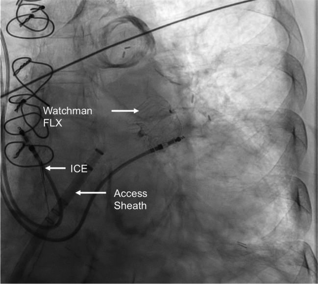
Once transseptal access is obtained, LA access is secured with a long (260‐cm) J‐tipped stiff 0.035‐inch wire after which the transseptal sheath is exchanged for the 14‐French device access sheath (for the Watchman and Watchman FLX devices). The access sheath is available in a variety of curves. The double curve configuration is the most commonly used as it allows easier access to superiorly directed distal lobes. A 5‐French or 6‐French pigtail is positioned inside the LAA thru the access sheath. Cine angiograms are performed in multiple projections to ascertain the LAA anatomy and measurements to guide precise device sizing (Figure 6). Caudal projections allow better visualization of the mid‐distal LAA for the Watchman device, whereas the right anterior oblique and cranial projections are better in visualizing the ostium and proximal LAA for the ACP. 29 Watchman device sizing is determined by the maximum LAA ostium diameter (measured from the left circumflex artery to 1–2 cm within the pulmonary vein ridge at 0°, 45°, 90°, and 135° TEE angles) and depth (from ostium to the tip of LAA). After appropriate device size selection, the device and device delivery catheter ensemble are prepped and meticulously deaired before insertion into the access sheath. After locking the access sheath and delivery system together, the device is ready to be deployed. Preferably under apnea for enhanced stability, device deployment is accomplished by slowly unsheathing the device under fluoroscopy. TEE and cine‐angiographic evaluations are then performed to assess for adequate compression, seal, and stability of the device. The Watchman FLX device can be partially or fully recaptured and redeployed if the initial deployment is not acceptable. The device is then released by using the deployment knob under fluoroscopy (Figure 7).
Figure 6. Contrast injection using a pigtail catheter and Watchman access sheath showing a multilobed left atrial appendage (LAA).
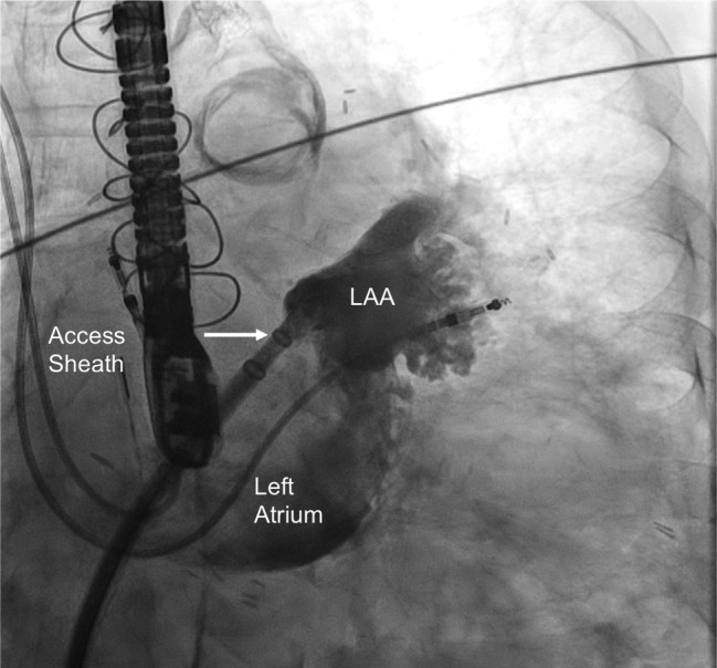
Figure 7. Fluoroscopic image of a Watchman FLX device released using intracardiac echo guidance (ICE).
The ACP and Amulet devices are implanted through 9‐French to 13‐French and 12‐French to 14‐French sheaths, respectively. There are 3 ACP/Amulet delivery sheaths available, of which the TorqVue 45°×45° is used most often (>95% of cases). It has a 3D distal tip, allowing anterior and superior angulation for coaxial positioning at the landing zone. The procedural steps are similar to those previously described for the Watchman device. Device sizing is based on the widest diameter of the landing zone. An oversizing of 3 to 5 mm for the ACP and 2 to 4 mm for the Amulet is generally recommended. TEE measurements at both the short axis (30°–60°) and long axis (120°–150°) of the landing zone and orifice (from the left circumflex artery to the pulmonary vein ridge) are used for LAA evaluation. The landing zone is measured at 10 mm and 12 to 15 mm from the orifice for the ACP and Amulet devices, respectively. A right anterior oblique (30°) and cranial (10°–20°) angiographic view is recommended during device deployment. 29
Intraprocedural Imaging
Intraprocedural echo imaging guidance is essential for a successful LAAO device implant.
TEE traditionally has been the gold standard for imaging guidance during LAAO. Familiarity of structural proceduralists with imaging views has made TEE the imaging modality of choice in LAAO procedures. The TEE probe is reusable and thus defrays the cost of the procedure. However, a major disadvantage of using TEE is the requirement for general anesthesia (GA). The patient is exposed not only to the risks of GA but also to the risks (albeit small) of the TEE procedure itself. Esophageal perforation is a very rare but catastrophic and potentially fatal complication of TEE. A recent prospective study investigated injuries during TEE‐guided structural cardiac interventions. 60 An esophagogastroduodenoscopy was performed before and after the structural procedures to evaluate new esophageal lesions. The study demonstrated that 86% of patients have new injuries, with 40% of them labeled as complex lesions. Longer procedure time (for each 10‐minute increment in procedure time; odds ratio [OR], 1.27; 95% CI, 1.01–1.59) and suboptimal image quality (OR, 4.93; 95% CI, 1.10–22.02) were identified as independent factors associated with an increased risk of complex lesions. Despite TEE being the gold standard during LAAO closures, imagers should be aware of the potential complications and take necessary precautions.
ICE is the mainstay of intraprocedural imaging guidance for congenital heart disease interventions, specifically atrial septal defect and patent foramen ovale closure. ICE is an alternative imaging technique that may obviate some of the disadvantages of TEE. ICE is a viable and safe alternative for intraprocedural imaging during LAAO given its simplicity, single‐operator use, and avoidance of GA. The steep learning curve in using ICE on top of the already complex LAAO procedure prolongs procedure time for the early adaptor. Also, deploying during induced apnea may be impossible if the patient is not under GA. However, the procedure length is attenuated by the faster turnover of the catheterization laboratory mostly because of faster anesthesia recovery. 61
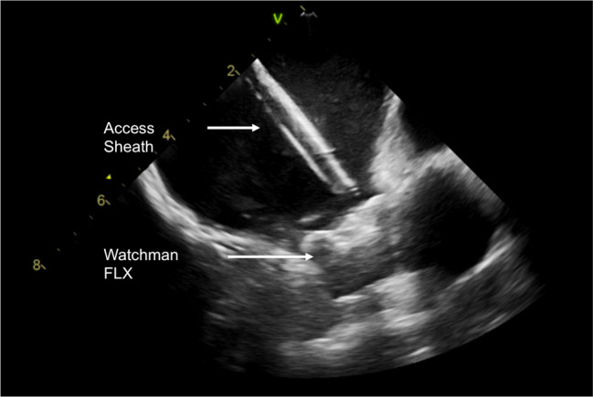
There is no significant difference in LAAO device complications whether TEE or ICE is used. 62 However, additional venous access is required for the ICE catheter, which potentially increases vascular complications. Also, unlike the TEE probe, most ICE systems are single‐use catheters that add to the cost of the procedure. The ICE catheter can be positioned in the right atrium, LA, right ventricular outflow tract, left upper pulmonary vein, or left pulmonary artery. 63 The right atrial position of the ICE catheter is ideal for transseptal puncture but is often inadequate for visualization of the LAA because of the distance from the transducer. A single or double transseptal puncture is required to enter the LA for optimal imaging of the LAA. Once inside the LA, imaging of the LAA is comparable or may even be superior to TEE (Figure 8). Technical success with ICE compared with TEE was also comparable. LAA ostium size and landing zone measurements by ICE correlated well with TEE in a study using the ACP device. 64
Figure 8. Intracardiac echocardiogram image of a deployed Watchman FLX device (27 mm).
Complications of LAAO
In general, complications from LAAO are related to (1) vascular access, (2) device implantation, (3) and antithrombotic therapy. Complications related to vascular access and antithrombotic therapy are common but are not unique to the LAAO procedure. Intraprocedural complications, specifically related to device implantation, will be focused here. Although the PROTECT AF trial established noninferiority of the Watchman device compared with warfarin therapy for the prevention of stroke, systemic embolism and cardiovascular death were significantly higher compared with warfarin. Although ischemic stroke and bleeding were also present, the safety events noted in the Watchman group in the PROTECT AF trial were largely procedure‐related events. However, subsequent analysis of both the PROTECT AF and CAP demonstrated that these safety events decrease with greater operator experience. 65
Pericardial Effusion
Postprocedural pericardial effusion with tamponade physiology was a significant safety event in the PROTECT AF trial. Pericardial effusion was noted to be serious if patient hospitalization was extended or hemodynamic compromise was present. 65 Serious pericardial effusions were seen in 5.2% (28/542) in PROTECT AF, whereas only 2.2% (10/460) in CAP. This was a relative reduction of 58% (P=0.014). 65 In the PROTECT AF cohort, the majority of the effusions were considered serious and required either percutaneous or surgical drainage. Effusions were detected within 24 hours in 89% of the cases. The exact mechanism of pericardial effusion has yet to be elucidated. A root cause analysis showed that the majority of effusions had no single definitive cause identified and are likely multifactorial. 65 Several notable steps or maneuvers during the procedure have been hypothesized to cause effusions. These include the device deployment process, manipulation of the delivery system in the LAA, a guidewire or adjunctive device in the LAA, and initial transseptal puncture. 65 However, with increasing operator experience and procedural changes, such as requiring the use of a pigtail catheter inside the LAA to position the access sheath, the rate of pericardial effusions has dramatically decreased. Early detection is key so that prompt percutaneous or surgical intervention can be initiated. It remains unclear if a routine echocardiogram post procedure is needed to detect pericardial effusions. Further studies are needed to mandate the necessity of routine echocardiograms, until then, postprocedure echocardiography is at the operator’s discretion. In addition, the next‐generation Watchman FLX device with its innovative closed design may offer a greater safety margin during the implantation procedure, resulting in fewer pericardial effusions and tamponade.
Procedure‐Related Stroke
Procedure‐related stroke is a known complication of any left‐sided cardiac procedure. This occurs presumably because of thrombus formation and embolization (thrombus or air) to the cerebral circulation. In the PROTECT AF trial, a low incidence of stroke was seen (n=5, 0.9%). On the other hand, no strokes were seen in the CAP registry (P=0.039). 65 Interestingly, the cause of the strokes was clearly identified as air embolism from the transseptal access sheath as opposed to thrombus formation. Prevention of procedure‐related stroke can be mitigated by judicious flushing, meticulous inspection of any air bubbles in the system, and confirmation of therapeutic anticoagulation during the procedure. There is no standard procedural anticoagulation strategy during LAAO. Some operators administer (1) full anticoagulation (100 units/kg of heparin) upfront after vascular access, (2) full anticoagulation (100 units/kg of heparin) after successful transseptal puncture, or (3) 50% of the heparin dose after vascular access then 50% after successful transseptal puncture.
Device Embolization
Device embolization is a rare but catastrophic complication of LAAO. An incidence of 0.6% and 0.7% of patients enrolled were noted in the PROTECT AF and PREVAIL trials, respectively. 41 , 42 In the CAP registry, 0.2% of patients had a reported device embolization. There were no Watchman device embolizations noted in the CAP2 registry. 44 , 65 A systematic review from 20 studies reporting device embolization of the Watchman device and the ACP noted 31 cases of embolization (13 Watchman devices and 18 ACP devices). 66 Embolization was noted acutely or periprocedurally in 65% of the cases reported. Late device embolization was noted to be rare. The devices embolized into the aorta, left atrial cavity, and left ventricle. The embolized devices in the left ventricle were associated with a higher rate of surgical retrieval compared with percutaneous retrieval (88% versus 17%; P=0.0019). Embolized devices in the aorta and LA can be retrieved successfully by percutaneous techniques via snares but with much difficulty. After Conformité Européene marked approval in Europe, the initial roll out of the Watchman FLX was halted because of unexplained high rates (6 of 207 implants) of device embolization. 67 The current iteration of the Watchman FLX has dual row anchors, which was designed for optimal device engagement to reduce the risk of embolization. The PINNACLE FLX study showed no cases of device embolization using the Watchman FLX device. 47
Device‐Related Thrombus
Device‐related thrombus (DRT) is a known complication detected by TEE after implantation of a LAAO device. The overall incidence of DRT is 3.9% in an analysis of 30 studies. 68 Despite the potential clinical impact of DRT, it only carries a small risk of stroke. In the PROTECT‐AF cohort, DRT was found in 4.2% (20 of 478) of the patients but only 3 developed an ischemic stroke. This translates to an annual stroke rate of only 0.3% per 100 patient‐years. 65 A prospective analysis after implantation of the ACP or Watchman device after a 12‐month follow‐up showed that varying durations of DAPT were not shown to be associated with DRT rates. 69 Several patient‐related and procedure‐related factors were noted to be associated with DRT. Low ejection fraction, history of thromboembolism, deep implantation of the LAAO device, and large occluders were noted to increase the rates of DRT. 69 Treatment consists of anticoagulation with either low‐molecular‐weight heparin or OAC. Complete resolution of the thrombus was achieved in 95% of cases using low‐molecular‐weight heparin or OAC with a varied treatment duration (median 45 days). 68
Future Directions
As operators become more adept in ICE and LAAO, eliminating GA would shift the paradigm to an even more minimally invasive approach. This is parallel to the early experience in transcatheter aortic valve replacement wherein GA was first exclusively used and then shifted rapidly to moderate sedation. Avoidance of GA may dramatically lessen turnover time, TEE‐related complications, and hospital stay while accelerating patient recovery.
Despite the advancements of periprocedural imaging, device size selection remains a challenge mostly because of the marked variability of the size and shape of the LAA. Accurate device size is critical in preventing residual leaks and avoiding complications. The current iteration of LAAO devices adapts to the variable LAA sizes by having a wide selection of device sizes. However, the LAA shape remains a variable that is not addressed by existing devices, although the Watchman FLX partially compensates for the variable LAA morphologies by avoiding a deep implantation. A device specifically tailored to the size and shape of the LAA via 3D printing may be the future of LAAO procedures.
Three‐dimensional printing technology can offer a vital advantage by allowing the implanters to physically “deploy” the LAAO device in the patient’s simulated LAA before the procedure. This facilitates not only precise device size selection but also aid in selection of catheters that will successfully navigate the patient’s unique LAA anatomy. The completeness of LAA occlusion by the device can be inspected beforehand to minimize device leak. In addition, 3D printed models allow implanters to visualize the fossa ovalis and assess the ideal transseptal puncture. This information is crucial especially in patients with septal occluders and patent foramen ovale. 70 Three‐dimensional printing technology may also provide an impact on training new and aspiring implanters and optimizing the techniques of current implanters.
Currently, only the Watchman and Watchman FLX devices are approved by the US FDA for LAAO. The LARIAT, Amulet, and WaveCrest devices are currently undergoing clinical trials. Once these devices are approved by the US FDA, the LAAO registry will most probably initiate the evaluation of the safety and effectiveness of these newer devices over time. Once these are commercially available, there will be broader treatment options to accommodate a wider range of LAA anatomy.
All of the early trials compared LAAO devices with a vitamin K antagonist. DOACs are now widely used as anticoagulation therapy for AF. Upcoming trials are now comparing LAAO devices with DOACs for the prevention of stroke. The Left Atrial Appendage Closure Versus Direct Oral Anticoagulants in High‐Risk Patients With Atrial Fibrillation trial compared the use of DOACs versus LAAO in patients with nonvalvular AF who are at high risk for stroke and increased risk of bleeding. These patients had a history of bleeding requiring hospitalization or intervention, cardioembolic event while on oral anticoagulant, and/or a CHA2D2‐VASc score of ≥3, and a Hypertension, Abnormal Renal/Liver Function, Stroke, Bleeding History or Predisposition, Labile INR, Elderly, Drugs/Alcohol Concomitantly score of ≥2. 71 LAAO demonstrated noninferiority to OACs in preventing AF‐related cardiovascular, neurological, and bleeding events. However, the trial highlighted persistent safety concerns regarding LAAO. A complication rate of 4.5% were noted in the trial including a procedure‐related death and device‐related death.
The results of the pivotal trials were conclusive; however, the increased adverse event rates have diluted their initial impact. The recurring theme of the safety with LAAO mandates the refinement in both the device technology and operator skills. As with any invasive procedure, increased operator experience significantly improves the safety of the procedure. 65 The NCDR (National Cardiovascular Data Registry) LAAO registry enrolled >38 000 patients with a higher stroke and bleeding risk compared with patients in the landmark trials. It is hopeful that despite having an older cohort with more comorbidities, the registry showed lower adverse event rates compared with the early trials. 72
As with any emerging transcatheter therapy, cost and reimbursement is also a significant issue in LAAO procedures. Despite the encouraging results from recent trial data, reimbursement for LAAO can be challenging. The cost‐effectiveness of LAAO (Watchman) in comparison with OAC and warfarin for stroke prevention has recently been demonstrated. 73 Professional societies should continue to engage third‐party payers to recognize the clinical and economic benefits of LAAO. Cost‐effective data analysis should also be considered when formulating policy and clinical practice guidelines for stroke prevention in AF.
Meticulous screening is mandatory before proceeding to LAAO. LAAO is unique to other structural heart interventions because it is performed prophylactically in patients who are stable and “asymptomatic.” Thus, the patient would not have any relief or noticeable symptomatic benefit after undergoing LAAO. Serious complications from an elective and prophylactic procedure including death, tamponade, and stroke are therefore unacceptable and cannot be tolerated. Further analysis of the registry data may help guide the selection criteria for patients who would benefit from LAAO or anticoagulation alone.
Data from the early pivotal trials were initially concerning because of safety events. Encouraging safety and efficacy data from subsequent trials, replicated by registries in real‐world populations, have allayed these concerns. Increasing operator experience, evolving device technology, and upcoming trials of new LAAO devices are reassuring aspects of the future of LAAO. These will ensure the delivery of a safe and effective nonpharmacologic strategy to prevent stroke in patients with nonvalvular AF.
Disclosures
None.
This manuscript was sent to N.A. Mark Estes III, MD, Guest Editor, for review by expert referees, editorial decision, and final disposition.
For Disclosures, see page 15.
REFERENCES
- 1. Johnson WD, Ganjoo AK, Stone CD, Srivyas RC, Howard M. The left atrial appendage: our most lethal human attachment! Surgical implications. Eur J Cardiothorac Surg. 2000;17:718–722. doi: 10.1016/S1010-7940(00)00419-X [DOI] [PubMed] [Google Scholar]
- 2. Ahmad FB, Cisewski JA, Minino A, Anderson RN. Provisional mortality data—United States, 2020. MMWR Morb Mortal Wkly Rep. 2021;70:519–522. doi: 10.15585/mmwr.mm7014e1 [DOI] [PMC free article] [PubMed] [Google Scholar]
- 3. Benjamin EJ, Muntner P, Alonso A, Bittencourt MS, Callaway CW, Carson AP, Chamberlain AM, Chang AR, Cheng S, Das SR, et al. American Heart Association Council on Epidemiology and Prevention Statistics Committee and Stroke Statistics Subcommittee . Heart disease and stroke statistics‐2019 update: a report from the American Heart Association. Circulation. 2019;139:e56–e528. doi: 10.1161/CIR.0000000000000659 [DOI] [PubMed] [Google Scholar]
- 4. Virani SS, Alonso A, Aparicio HJ, Benjamin EJ, Bittencourt MS, Callaway CW, Carson AP, Chamberlain AM, Cheng S, Delling FN, et al. American Heart Association Council on Epidemiology and Prevention Statistics Committee and Stroke Statistics Subcommittee . Heart disease and stroke statistics‐2021 update: a report from the American Heart Association. Circulation. 2021;143:e254–e743. doi: 10.1161/CIR.0000000000000950 [DOI] [PubMed] [Google Scholar]
- 5. Hindricks G, Potpara T, Dagres N, Arbelo E, Bax JJ, Blomstrom‐Lundqvist C, Boriani G, Castella M, Dan GA, Dilaveris PE, et al. 2020 ESC guidelines for the diagnosis and management of atrial fibrillation developed in collaboration with the European Association of Cardio‐Thoracic Surgery (EACTS). Eur Heart J. 2021;42:373–498. doi: 10.1093/eurheartj/ehaa612 [DOI] [PubMed] [Google Scholar]
- 6. Ruff CT, Giugliano RP, Braunwald E, Hoffman EB, Deenadayalu N, Ezekowitz MD, Camm AJ, Weitz JI, Lewis BS, Parkhomenko A, et al. Comparison of the efficacy and safety of new oral anticoagulants with warfarin in patients with atrial fibrillation: a meta‐analysis of randomised trials. Lancet. 2014;383:955–962. doi: 10.1016/S0140-6736(13)62343-0 [DOI] [PubMed] [Google Scholar]
- 7. Hart RG, Pearce LA, Aguilar MI. Meta‐analysis: antithrombotic therapy to prevent stroke in patients who have nonvalvular atrial fibrillation. Ann Intern Med. 2007;146:857–867. doi: 10.7326/0003-4819-146-12-200706190-00007 [DOI] [PubMed] [Google Scholar]
- 8. Bungard TJ, Ghali WA, Teo KK, McAlister FA, Tsuyuki RT. Why do patients with atrial fibrillation not receive warfarin? Arch Intern Med. 2000;160:41–46. doi: 10.1001/archinte.160.1.41 [DOI] [PubMed] [Google Scholar]
- 9. Madden JL. Resection of the left auricular appendix; a prophylaxis for recurrent arterial emboli. J Am Med Assoc. 1949;140:769–772. doi: 10.1001/jama.1949.02900440011003 [DOI] [PubMed] [Google Scholar]
- 10. Blackshear JL, Odell JA. Appendage obliteration to reduce stroke in cardiac surgical patients with atrial fibrillation. Ann Thorac Surg. 1996;61:755–759. doi: 10.1016/0003-4975(95)00887-X [DOI] [PubMed] [Google Scholar]
- 11. January CT, Wann LS, Calkins H, Chen LY, Cigarroa JE, Cleveland JC Jr, Ellinor PT, Ezekowitz MD, Field ME, et al. 2019 AHA/ACC/HRS focused update of the 2014 AHA/ACC/HRS guideline for the management of patients with atrial fibrillation: a report of the American College of Cardiology/American Heart Association task force on clinical practice guidelines and the heart rhythm society in collaboration with the society of thoracic surgeons. Circulation. 2019;140:e125–e151. doi: 10.1016/j.jacc.2019.01.011 [DOI] [PubMed] [Google Scholar]
- 12. Al‐Saady NM, Obel OA, Camm AJ. Left atrial appendage: structure, function, and role in thromboembolism. Heart. 1999;82:547–554. doi: 10.1136/hrt.82.5.547 [DOI] [PMC free article] [PubMed] [Google Scholar]
- 13. Beigel R, Wunderlich NC, Ho SY, Arsanjani R, Siegel RJ. The left atrial appendage: anatomy, function, and noninvasive evaluation. JACC Cardiovasc Imaging. 2014;7:1251–1265. doi: 10.1016/j.jcmg.2014.08.009 [DOI] [PubMed] [Google Scholar]
- 14. Veinot JP, Harrity PJ, Gentile F, Khandheria BK, Bailey KR, Eickholt JT, Seward JB, Tajik AJ, Edwards WD. Anatomy of the normal left atrial appendage: a quantitative study of age‐related changes in 500 autopsy hearts: implications for echocardiographic examination. Circulation. 1997;96:3112–3115. doi: 10.1161/01.CIR.96.9.3112 [DOI] [PubMed] [Google Scholar]
- 15. Yamamoto M, Seo Y, Kawamatsu N, Sato K, Sugano A, Machino‐Ohtsuka T, Kawamura R, Nakajima H, Igarashi M, Sekiguchi Y, et al. Complex left atrial appendage morphology and left atrial appendage thrombus formation in patients with atrial fibrillation. Circ Cardiovasc Imaging. 2014;7:337–343. doi: 10.1161/CIRCIMAGING.113.001317 [DOI] [PubMed] [Google Scholar]
- 16. Di Biase L, Santangeli P, Anselmino M, Mohanty P, Salvetti I, Gili S, Horton R, Sanchez JE, Bai R, Mohanty S, et al. Does the left atrial appendage morphology correlate with the risk of stroke in patients with atrial fibrillation? Results from a multicenter study. J Am Coll Cardiol. 2012;60:531–538. doi: 10.1016/j.jacc.2012.04.032 [DOI] [PubMed] [Google Scholar]
- 17. Cox JL, Boineau JP, Schuessler RB, Kater KM, Lappas DG. Five‐year experience with the maze procedure for atrial fibrillation. Ann Thorac Surg. 1993;56:814–823. discussion 823–814. [DOI] [PubMed] [Google Scholar]
- 18. Cox JL. Mechanical closure of the left atrial appendage: is it time to be more aggressive? J Thorac Cardiovasc Surg. 2013;146:1018–1027.e1012. [DOI] [PubMed] [Google Scholar]
- 19. Watson T, Shantsila E, Lip GY. Mechanisms of thrombogenesis in atrial fibrillation: Virchow's triad revisited. Lancet. 2009;373:155–166. doi: 10.1016/S0140-6736(09)60040-4 [DOI] [PubMed] [Google Scholar]
- 20. Odell JA, Blackshear JL, Davies E, Byrne WJ, Kollmorgen CF, Edwards WD, Orszulak TA. Thoracoscopic obliteration of the left atrial appendage: potential for stroke reduction? Ann Thorac Surg. 1996;61:565–569. [DOI] [PubMed] [Google Scholar]
- 21. Leonard FC, Cogan MA. Failure of ligation of the left auricular appendage in the prevention of recurrent embolism. N Engl J Med. 1952;246:733–735. doi: 10.1056/NEJM195205082461903 [DOI] [PubMed] [Google Scholar]
- 22. Cox JL, Ad N, Palazzo T. Impact of the maze procedure on the stroke rate in patients with atrial fibrillation. J Thorac Cardiovasc Surg. 1999;118:833–840. doi: 10.1016/S0022-5223(99)70052-8 [DOI] [PubMed] [Google Scholar]
- 23. Badhwar V, Rankin JS, Damiano RJ, Gillinov AM, Bakaeen FG, Edgerton JR, Philpott JM, McCarthy PM, Bolling SF, Roberts HG, et al. The society of thoracic surgeons 2017 clinical practice guidelines for the surgical treatment of atrial fibrillation. Ann Thorac Surg. 2017;103:329–341. doi: 10.1016/j.athoracsur.2016.10.076 [DOI] [PubMed] [Google Scholar]
- 24. Healey JS, Crystal E, Lamy A, Teoh K, Semelhago L, Hohnloser SH, Cybulsky I, Abouzahr L, Sawchuck C, Carroll S, et al. Left Atrial Appendage Occlusion Study (LAAOS): results of a randomized controlled pilot study of left atrial appendage occlusion during coronary bypass surgery in patients at risk for stroke. Am Heart J. 2005;150:288–293. doi: 10.1016/j.ahj.2004.09.054 [DOI] [PubMed] [Google Scholar]
- 25. Katz ES, Tsiamtsiouris T, Applebaum RM, Schwartzbard A, Tunick PA, Kronzon I. Surgical left atrial appendage ligation is frequently incomplete: a transesophageal echocardiograhic study. J Am Coll Cardiol. 2000;36:468–471. doi: 10.1016/s0735-1097(00)00765-8 [DOI] [PubMed] [Google Scholar]
- 26. Garcia‐Fernandez MA, Perez‐David E, Quiles J, Peralta J, Garcia‐Rojas I, Bermejo J, Moreno M, Silva J. Role of left atrial appendage obliteration in stroke reduction in patients with mitral valve prosthesis: a transesophageal echocardiographic study. J Am Coll Cardiol. 2003;42:1253–1258. doi: 10.1016/S0735-1097(03)00954-9 [DOI] [PubMed] [Google Scholar]
- 27. Gillinov AM, Pettersson G, Cosgrove DM. Stapled excision of the left atrial appendage. J Thorac Cardiovasc Surg. 2005;129:679–680. doi: 10.1016/j.jtcvs.2004.07.039 [DOI] [PubMed] [Google Scholar]
- 28. Ailawadi G, Gerdisch MW, Harvey RL, Hooker RL, Damiano RJ Jr, Salamon T, Mack MJ. Exclusion of the left atrial appendage with a novel device: early results of a multicenter trial. J Thorac Cardiovasc Surg. 2011;142:1002–1009, 1009 e1001. [DOI] [PubMed] [Google Scholar]
- 29. Saw J, Lempereur M. Percutaneous left atrial appendage closure: procedural techniques and outcomes. JACC Cardiovasc Interv. 2014;7:1205–1220. doi: 10.1016/j.jcin.2014.05.026 [DOI] [PubMed] [Google Scholar]
- 30. Tsai YC, Phan K, Munkholm‐Larsen S, Tian DH, La Meir M, Yan TD. Surgical left atrial appendage occlusion during cardiac surgery for patients with atrial fibrillation: a meta‐analysis. Eur J Cardiothorac Surg. 2015;47:847–854. doi: 10.1093/ejcts/ezu291 [DOI] [PubMed] [Google Scholar]
- 31. Whitlock RP, Belley‐Cote EP, Paparella D, Healey JS, Brady K, Sharma M, Reents W, Budera P, Baddour AJ, Fila P, et al. Left atrial appendage occlusion during cardiac surgery to prevent stroke. N Engl J Med. 2021;384:2081–2091. doi: 10.1056/NEJMoa2101897 [DOI] [PubMed] [Google Scholar]
- 32. Sievert H, Lesh MD, Trepels T, Omran H, Bartorelli A, Della Bella P, Nakai T, Reisman M, DiMario C, Block P, et al. Percutaneous left atrial appendage transcatheter occlusion to prevent stroke in high‐risk patients with atrial fibrillation: early clinical experience. Circulation. 2002;105:1887–1889. doi: 10.1161/01.CIR.0000015698.54752.6D [DOI] [PubMed] [Google Scholar]
- 33. Bayard YL, Omran H, Neuzil P, Thuesen L, Pichler M, Rowland E, Ramondo A, Ruzyllo W, Budts W, Montalescot G, et al. PLAATO (Percutaneous Left Atrial Appendage Transcatheter Occlusion) for prevention of cardioembolic stroke in non‐anticoagulation eligible atrial fibrillation patients: results from the European PLAATO Study. EuroIntervention. 2010;6:220–226. doi: 10.4244/EIJV6I2A35 [DOI] [PubMed] [Google Scholar]
- 34. U.S. Food & Drug Administration Watchman left atrial appendage closure device with delivery system and Watchman FLX left atrial appendage closure device with delivery system–p130013/s035. 2020. https://www.accessdata.fda.gov/scripts/cdrh/cfdocs/cfpma/pma.cfm?id=P130013. Accessed January 4, 2021.
- 35. Kleinecke C, Cheikh‐Ibrahim M, Schnupp S, Fankhauser M, Nietlispach F, Park JW, Brachmann J, Windecker S, Meier B, Gloekler S. Long‐term clinical outcomes of Amplatzer Cardiac Plug versus Amulet occluders for left atrial appendage closure. Catheter Cardiovasc Interv. 2020;96:E324–E331. doi: 10.1002/ccd.28530 [DOI] [PubMed] [Google Scholar]
- 36. Lakkireddy D, Windecker S, Thaler D, Søndergaard L, Carroll J, Gold MR, Guo H, Brunner KJ, Hermiller JB, Diener H‐C, et al. Rationale and design for AMPLATZER Amulet Left Atrial Appendage Occluder IDE randomized controlled trial (Amulet IDE trial). Am Heart J. 2019;211:45–53. doi: 10.1016/j.ahj.2018.12.010 [DOI] [PubMed] [Google Scholar]
- 37. Amplatzer amulet LAAO vs. NOAC (CATALYST). 2020. Clinical Trials. https://clinicaltrials.gov/ct2/show/NCT04226547. Accessed January 5, 2021.
- 38. Price MJ, Gibson DN, Yakubov SJ, Schultz JC, Di Biase L, Natale A, Burkhardt JD, Pershad A, Byrne TJ, Gidney B, et al. Early safety and efficacy of percutaneous left atrial appendage suture ligation: results from the U.S. transcatheter LAA ligation consortium. J Am Coll Cardiol. 2014;64:565–572. doi: 10.1016/j.jacc.2014.03.057 [DOI] [PMC free article] [PubMed] [Google Scholar]
- 39. aMAZE study: LAA ligation adjunctive to PVI for persistent or longstanding persistent atrial fibrillation (aMAZE) . 2020. https://clinicaltrials.gov/ct2/show/NCT02513797. Accessed May 8, 2021. Identifier: NCT02513797.
- 40. Reddy VY, Sievert H, Halperin J, Doshi SK, Buchbinder M, Neuzil P, Huber K, Whisenant B, Kar S, Swarup V, et al. Percutaneous left atrial appendage closure vs warfarin for atrial fibrillation: a randomized clinical trial. JAMA. 2014;312:1988–1998. doi: 10.1001/jama.2014.15192 [DOI] [PubMed] [Google Scholar]
- 41. Holmes DR, Reddy VY, Turi ZG, Doshi SK, Sievert H, Buchbinder M, Mullin CM, Sick P, Investigators PA . Percutaneous closure of the left atrial appendage versus warfarin therapy for prevention of stroke in patients with atrial fibrillation: a randomised non‐inferiority trial. Lancet. 2009;374:534–542. doi: 10.1016/S0140-6736(09)61343-X [DOI] [PubMed] [Google Scholar]
- 42. Holmes DR Jr, Kar S, Price MJ, Whisenant B, Sievert H, Doshi SK, Huber K, Reddy VY. Prospective randomized evaluation of the watchman left atrial appendage closure device in patients with atrial fibrillation versus long‐term warfarin therapy: the prevail trial. J Am Coll Cardiol. 2014;64:1–12. doi: 10.1016/j.jacc.2014.04.029 [DOI] [PubMed] [Google Scholar]
- 43. Reddy VY, Doshi SK, Kar S, Gibson DN, Price MJ, Huber K, Horton RP, Buchbinder M, Neuzil P, Gordon NT, et al. 5‐year outcomes after left atrial appendage closure: from the prevail and protect AF trials. J Am Coll Cardiol. 2017;70:2964–2975. doi: 10.1016/j.jacc.2017.10.021 [DOI] [PubMed] [Google Scholar]
- 44. Holmes DR Jr, Reddy VY, Gordon NT, Delurgio D, Doshi SK, Desai AJ, Stone JE Jr, Kar S. Long‐term safety and efficacy in continued access left atrial appendage closure registries. J Am Coll Cardiol. 2019;74:2878–2889. doi: 10.1016/j.jacc.2019.09.064 [DOI] [PubMed] [Google Scholar]
- 45. Boersma LV, Ince H, Kische S, Pokushalov E, Schmitz T, Schmidt B, Gori T, Meincke F, Protopopov AV, Betts T, et al. Evaluating real‐world clinical outcomes in atrial fibrillation patients receiving the WATCHMAN left atrial appendage closure technology: final 2‐year outcome data of the EWOLUTION trial focusing on history of stroke and hemorrhage. Circ Arrhythm Electrophysiol. 2019;12:e006841. doi: 10.1161/CIRCEP.118.006841 [DOI] [PubMed] [Google Scholar]
- 46. Investigational device evaluation of the Watchman FLX LAA closure technology (PINNACLE FLX) . https://clinicaltrials.gov/ct2/show/NCT02702271. Accessed February 20, 2021.
- 47. Kar S, Doshi SK, Sadhu A, Horton R, Osorio J, Ellis C, Stone J, Shah M, Dukkipati SR, Adler S, et al. Primary outcome evaluation of a next generation left atrial appendage closure device: results from the PINNACLE FLX trial. Circulation. 2021;143:1754–1762. doi: 10.1161/CIRCULATIONAHA.120.050117 [DOI] [PubMed] [Google Scholar]
- 48. Amplatzer amulet LAA occluder trial (Amulet IDE) . https://clinicaltrials.gov/ct2/show/NCT02879448. Accessed February 20, 2021.
- 49. WaveCrest vs. Watchman transseptal LAA closure to reduce af‐mediated stroke 2 (WaveCrest 2) . https://clinicaltrials.gov/ct2/show/NCT03302494. Accessed January 5, 2021.
- 50. Iriart X, Ciobotaru V, Martin C, Cochet H, Jalal Z, Thambo JB, Quessard A. Role of cardiac imaging and three‐dimensional printing in percutaneous appendage closure. Arch Cardiovasc Dis. 2018;111:411–420. doi: 10.1016/j.acvd.2018.04.005 [DOI] [PubMed] [Google Scholar]
- 51. Kempfert J, Van Linden A, Lehmkuhl L, Rastan AJ, Holzhey D, Blumenstein J, Mohr FW, Walther T. Aortic annulus sizing: echocardiographic versus computed tomography derived measurements in comparison with direct surgical sizing. Eur J Cardiothorac Surg. 2012;42:627–633. doi: 10.1093/ejcts/ezs064 [DOI] [PubMed] [Google Scholar]
- 52. Chow DH, Bieliauskas G, Sawaya FJ, Millan‐Iturbe O, Kofoed KF, Sondergaard L, De Backer O. A comparative study of different imaging modalities for successful percutaneous left atrial appendage closure. Open Heart. 2017;4:e000627. doi: 10.1136/openhrt-2017-000627 [DOI] [PMC free article] [PubMed] [Google Scholar]
- 53. Korsholm K, Berti S, Iriart X, Saw J, Wang DD, Cochet H, Chow D, Clemente A, De Backer O, Moller Jensen J, et al. Expert recommendations on cardiac computed tomography for planning transcatheter left atrial appendage occlusion. JACC Cardiovasc Interv. 2020;13:277–292. doi: 10.1016/j.jcin.2019.08.054 [DOI] [PubMed] [Google Scholar]
- 54. Romero J, Husain SA, Kelesidis I, Sanz J, Medina HM, Garcia MJ. Detection of left atrial appendage thrombus by cardiac computed tomography in patients with atrial fibrillation: a meta‐analysis. Circ Cardiovasc Imaging. 2013;6:185–194. doi: 10.1161/CIRCIMAGING.112.000153 [DOI] [PubMed] [Google Scholar]
- 55. Conti M, Marconi S, Muscogiuri G, Guglielmo M, Baggiano A, Italiano G, Mancini ME, Auricchio F, Andreini D, Rabbat MG, et al. Left atrial appendage closure guided by 3D computed tomography printing technology: a case control study. J Cardiovasc Comput Tomogr. 2019;13:336–339. doi: 10.1016/j.jcct.2018.10.024 [DOI] [PubMed] [Google Scholar]
- 56. Kenny D. Transseptal basics: what you need to know to ensure safe puncture as this technique reemerges. Cardiac Interventions Today. 2012. [Google Scholar]
- 57. Alkhouli M, Rihal CS, Holmes DR Jr. Transseptal techniques for emerging structural heart interventions. JACC Cardiovasc Interv. 2016;9:2465–2480. doi: 10.1016/j.jcin.2016.10.035 [DOI] [PubMed] [Google Scholar]
- 58. Ren JF, Marchlinski FE, Callans DJ, Gerstenfeld EP, Dixit S, Lin D, Nayak HM, Hsia HH. Increased intensity of anticoagulation may reduce risk of thrombus during atrial fibrillation ablation procedures in patients with spontaneous echo contrast. J Cardiovasc Electrophysiol. 2005;16:474–477. doi: 10.1046/j.1540-8167.2005.40465.x [DOI] [PubMed] [Google Scholar]
- 59. Spencer RJ, DeJong P, Fahmy P, Lempereur M, Tsang MYC, Gin KG, Lee PK, Nair P, Tsang TSM, Jue J, et al. Changes in left atrial appendage dimensions following volume loading during percutaneous left atrial appendage closure. JACC Cardiovasc Interv. 2015;8:1935–1941. doi: 10.1016/j.jcin.2015.07.035 [DOI] [PubMed] [Google Scholar]
- 60. Freitas‐Ferraz AB, Bernier M, Vaillancourt R, Ugalde PA, Nicodème F, Paradis J‐M, Champagne J, O’Hara G, Junquera L, del Val D, et al. Safety of transesophageal echocardiography to guide structural cardiac interventions. J Am Coll Cardiol. 2020;75:3164–3173. doi: 10.1016/j.jacc.2020.04.069 [DOI] [PubMed] [Google Scholar]
- 61. Hemam ME, Kuroki K, Schurmann PA, Dave AS, Rodriguez DA, Saenz LC, Reddy VY, Valderrabano M. Left atrial appendage closure with the watchman device using intracardiac vs transesophageal echocardiography: procedural and cost considerations. Heart Rhythm. 2019;16:334–342. doi: 10.1016/j.hrthm.2018.12.013 [DOI] [PMC free article] [PubMed] [Google Scholar]
- 62. Berti S, Pastormerlo LE, Santoro G, Brscic E, Montorfano M, Vignali L, Danna P, Tondo C, Rezzaghi M, D'Amico G, et al. Intracardiac versus transesophageal echocardiographic guidance for left atrial appendage occlusion: the LAAO Italian Multicenter Registry. JACC Cardiovasc Interv. 2018;11:1086–1092. doi: 10.1016/j.jcin.2018.05.008 [DOI] [PubMed] [Google Scholar]
- 63. Alkhouli M, Hijazi ZM, Holmes DR Jr, Rihal CS, Wiegers SE. Intracardiac echocardiography in structural heart disease interventions. JACC Cardiovasc Interv. 2018;11:2133–2147. doi: 10.1016/j.jcin.2018.06.056 [DOI] [PubMed] [Google Scholar]
- 64. Berti S, Paradossi U, Meucci F, Trianni G, Tzikas A, Rezzaghi M, Stolkova M, Palmieri C, Mori F, Santoro G. Periprocedural intracardiac echocardiography for left atrial appendage closure: a dual‐center experience. JACC Cardiovasc Interv. 2014;7:1036–1044. doi: 10.1016/j.jcin.2014.04.014 [DOI] [PubMed] [Google Scholar]
- 65. Reddy VY, Holmes D, Doshi SK, Neuzil P, Kar S. Safety of percutaneous left atrial appendage closure: results from the Watchman Left Atrial Appendage System for Embolic Protection in Patients with AF (protect AF) clinical trial and the continued access registry. Circulation. 2011;123:417–424. doi: 10.1161/CIRCULATIONAHA.110.976449 [DOI] [PubMed] [Google Scholar]
- 66. Aminian A, Lalmand J, Tzikas A, Budts W, Benit E, Kefer J. Embolization of left atrial appendage closure devices: a systematic review of cases reported with the watchman device and the Amplatzer Cardiac Plug. Catheter Cardiovasc Interv. 2015;86:128–135. doi: 10.1002/ccd.25891 [DOI] [PubMed] [Google Scholar]
- 67. Neale T New‐generation watchman FLX device pulled from European shelves over embolism concerns. TCT MD. 2016. https://www.tctmd.com/news/new‐generation‐watchman‐flx‐device‐pulled‐european‐shelves‐over‐embolism‐concerns. Accessed February 28, 2021.
- 68. Lempereur M, Aminian A, Freixa X, Gafoor S, Kefer J, Tzikas A, Legrand V, Saw J. Device‐associated thrombus formation after left atrial appendage occlusion: a systematic review of events reported with the Watchman, the Amplatzer Cardiac Plug and the Amulet. Catheter Cardiovasc Interv. 2017;90:E111–E121. doi: 10.1002/ccd.26903 [DOI] [PubMed] [Google Scholar]
- 69. Pracon R, Bangalore S, Dzielinska Z, Konka M, Kepka C, Kruk M, Kaczmarska‐Dyrda E, Petryka‐Mazurkiewicz J, Bujak S, Solecki M, et al. Device thrombosis after percutaneous left atrial appendage occlusion is related to patient and procedural characteristics but not to duration of postimplantation dual antiplatelet therapy. Circ Cardiovasc Interv. 2018;11:e005997. doi: 10.1161/CIRCINTERVENTIONS.117.005997 [DOI] [PubMed] [Google Scholar]
- 70. Kaafarani M, Saw J, Daniels M, Song T, Rollet M, Kesinovic S, Lamorgese T, Kubiak K, Qi Z, Pantelic M, et al. Role of CT imaging in left atrial appendage occlusion for the WATCHMAN device. Cardiovasc Diagn Ther. 2020;10:45–58. doi: 10.21037/cdt.2019.12.01 [DOI] [PMC free article] [PubMed] [Google Scholar]
- 71. Osmancik P, Herman D, Neuzil P, Hala P, Taborsky M, Kala P, Poloczek M, Stasek J, Haman L, Branny M, et al. Left atrial appendage closure versus direct oral anticoagulants in high‐risk patients with atrial fibrillation. J Am Coll Cardiol. 2020;75:3122–3135. doi: 10.1016/j.jacc.2020.04.067 [DOI] [PubMed] [Google Scholar]
- 72. Freeman JV, Varosy P, Price MJ, Slotwiner D, Kusumoto FM, Rammohan C, Kavinsky CJ, Turi ZG, Akar J, Koutras C, et al. The NCDR left atrial appendage occlusion registry. J Am Coll Cardiol. 2020;75:1503–1518. doi: 10.1016/j.jacc.2019.12.040 [DOI] [PMC free article] [PubMed] [Google Scholar]
- 73. Reddy VY, Akehurst RL, Armstrong SO, Amorosi SL, Beard SM, Holmes DR Jr. Time to cost‐effectiveness following stroke reduction strategies in AF: warfarin versus NOACs versus LAA closure. J Am Coll Cardiol. 2015;66:2728–2739. doi: 10.1016/j.jacc.2015.09.084 [DOI] [PubMed] [Google Scholar]
- 74. Asmarats L, Rodés‐Cabau J. Percutaneous left atrial appendage closure. Circ Cardiovasc Interv. 2017;10. e005359 [DOI] [PubMed] [Google Scholar]
- 75. Bando K, Kobayashi J, Hirata M, Satoh T, Niwaya K, Tagusari O, Nakatani S, Yagihara T, Kitamura S. Early and late stroke after mitral valve replacement with a mechanical prosthesis: risk factor analysis of a 24‐year experience. J Thorac Cardiovasc Surg. 2003;126:358–364. [DOI] [PubMed] [Google Scholar]
- 76. Pennec PY, Jobic Y, Blanc JJ, Bezon E, Barra JA. Assessment of different procedures for surgical left atrial appendage exclusion. Ann Thorac Surg. 2003;76:2168–2169. doi: 10.1016/s0003-4975(03)00738-0. PMID: 14667677 [DOI] [PubMed] [Google Scholar]
- 77. Schneider B, Stollberger C, Sievers HH. Surgical closure of the left atrial appendage – a beneficial procedure? Cardiology. 2005;104(3):127–132. doi: 10.1159/000087632. Epub 2005 Aug 22. PMID: 16118490 [DOI] [PubMed] [Google Scholar]
- 78. Kanderian AS, Gillinov AM, Pettersson GB, Blackstone E, Klein AL. Success of surgical left atrial appendage closure. Assessment by transesophageal echocardiography. J Am Coll Cardiol. 2008;52(11):924–929. doi: 10.1016/j.jacc.2008.03.067 [DOI] [PubMed] [Google Scholar]
- 79. Bakhtiary F, Kleine P, Martens S, Dzemali O, Dogan S, Keller H, Ackermann H, Zierer A, Ozaslan F, Wittlinger T, et al. Simplified technique for surgical ligation of the left atrial appendage in high‐risk patients. J Thorac Cardiovasc Surg. 2008;135(2):430–431. doi: 10.1016/j.jtcvs.2007.08.057. PMID: 18242281 [DOI] [PubMed] [Google Scholar]
- 80. Whitlock RP, Vincent J, Blackall MH, Hirsh J, Fremes S, Novick R, Devereaux PJ, Teoh K, Lamy A, Connolly SJ, et al. Left atrial appendage occlusion study II (LAAOS II). Can J Cardiol. 2013;29(11):1443–1447. doi: 10.1016/j.cjca.2013.06.015. Epub 2013 Sep 20. PMID: 24054920 [DOI] [PubMed] [Google Scholar]


