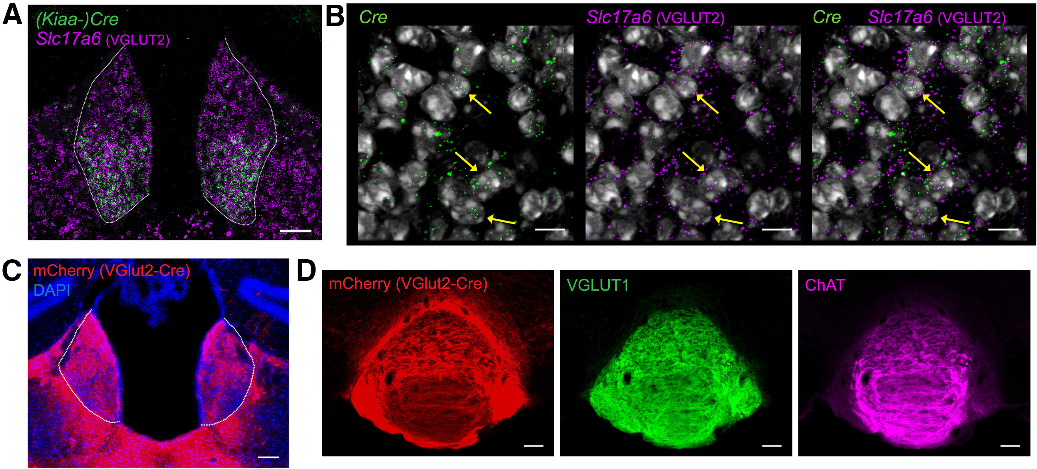
Figure 3.

VGLUT2-expressing projections from MHb to IPN. A, Fluorescent in situ hybridization from KiaaCre mouse showing Cre and Slc17a6 expression in the MHb (outlined); scale bar: 100 μm. B, High-resolution image showing expression of Cre, Slc17a6 (VGLUT2), and DAPI in MHb of KiaaCre mouse; scale bar: 10 μm. Yellow arrows indicate some of the cells containing both Cre and Slc17a6 mRNA. C, Image of MHb from Slc17a6Cre (VGLUT2) mouse injected with AAV1-Ef1α-DIO-ChR2:mCherry bilaterally into the MHb (outlined); scale bar: 100 μm. D, Immunohistochemistry of IPN from Slc17a6Cre (VGLUT2-Cre) mouse injected bilaterally with AAV1-Ef1α-DIO-ChR2:mCherry in MHb. VGLUT2Cre MHb terminals in IPN represented by mCherry expression, stained with VGLUT1 and ChAT; scale bar: 100 μm.
