Abstract
The aim of this review to determine recommendations for classification and radiological diagnosis of thoracolumbar spine fractures. Recommendation was made through a literature review of the last 10 years. The statements created by the authors were discussed and voted on during 2 consensus meetings organized by the WFNS (World Federation Neurosurgical Societies) Spine Committee. The literature review was yielded 256 abstracts, of which 32 were chosen for full-text analysis. Thirteen papers evaluated the reliability of a classification system by our expert members and were also chosen in this guideline analysis. This literature review-based recommendation provides the classification and radiologic diagnosis in thoracolumbar spine fractures that can elucidate the management decision-making in clinical practice.
Keywords: Thoracolumbar spine fracture, Classifications, Therapy recommendation, Radiological diagnosis
INTRODUCTION
The thoracolumbar spine is the most often fractured part of the spine [1]. Wang et al. [1] in 2012, recorded that 54.9% of patients had injuries in the thoracolumbar spine in a comprehensive series of 3,142 patients at high-risk spinal fractures. The thoracolumbar junction is marked by significant biomechanical strain due to its location between the rigid thoracic and dynamic lumbar spines [2]. For more than 8 decades, trauma and spine surgeons have been perplexed by a broadly agreed thoracolumbar fracture classification. Persistent occurrences refer to the seriousness of an injury [3]. A description of spine fractures is essential to create a common language for diagnostic purposes [4]. To promote the creation of a globally accepted treatment algorithm and to make treatment guidelines for a wide variety of thoracolumbar injuries [5]. Encouraging constant communication between practicing doctors in order to develop a common terminology for identifying an injury configuration when a patient requires surgery, and directing care with a comparative assessment of the type and severity of injury [5]. This clinical guideline was developed to enhance patient care by recommending a description and diagnosis for thoracolumbar spine fractures [6].
MATERIALS AND METHODS
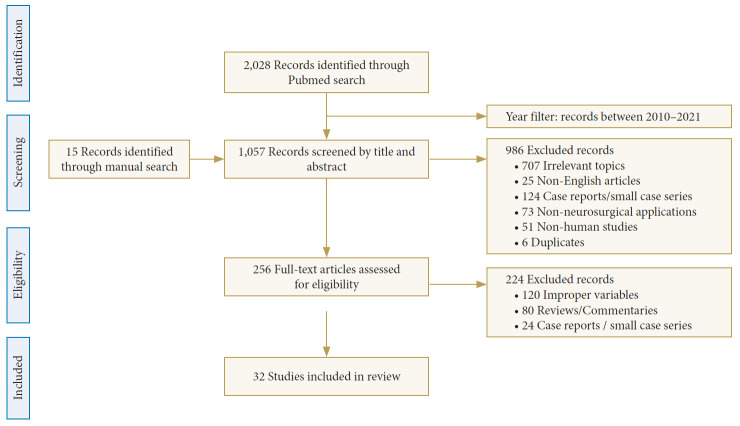
The guidelines for thoracolumbar spine fracture classification were proposed by World Federation of Neurosurgical Societies (WFNS) members following a study of the literature on the classification and radiological analysis of thoracolumbar spine fractures. We conducted a review of the literature from 2010 to 2020 using the keywords “thoracolumbar fracture and classification” and “thoracolumbar fracture and radiology”; PubMed and MEDLINE reported 1,057 findings. Additionally, we conducted a manual search and discovered 15 journals. We excluded articles written in non-English languages, case notes, and low-quality case series. Then, for this analysis, we reviewed 32 papers. Fig. 1 depicts the flowchart of the literature findings. The WFNS Spine Committee reviewed current evidence on the recognition and radiological evaluation of thoracolumbar fractures in order to reach consensus at 2 consensus meetings. The first meeting was held in Peshawar in December 2019 with the attendance and involvement of WFNS Spine Committee members. On June 12, 2020, a virtual meeting was held.
Fig. 1.
Flowchart of the literature search we used.
Both meetings were designed to discuss a preformulated survey from detailed literature reviews and comments about the current state of evidence in order to elicit recommendations in a thorough polling session.
To ensure a high degree of validity, we administered the questionnaire using the Delphi system. To achieve consensus, participants individually voted on their degree of agreement or disagreement on each item using a Likert-type scale ranging from 1 to 5 (1=firmly oppose, 2=disagree, 3=fairly agree, 4=agree, 5=strongly agree). The results are expressed as a percentage of respondents who responded with a 1 or 2 (disagreement) or a 3, 4, or 5 for each object (agreement). Consensus was reached as the total of the points of dispute or compromise reached 66%. Each consensus point was established in detail, including the strength of the proof, the recommendation score, and the consensus standard.
RESULTS
The literature search yielded 2,028 abstracts, 32 of which were selected for full-text review. Our task force participants tested the effectiveness of a classification scheme in 13 articles, which were all included in this research guideline.
DISCUSSION
1. Thoracolumbar Spine Fractures Classifications
1) Goal of classification
The classification of thoracolumbar fractures aims to promote better coordination between treating doctors, to provide a standard vocabulary for describing injury configurations, and to provide a consistent measure of the injury’s form and severity. Additionally, the designation would serve as a clear guide for management, regardless of whether the patient requires surgery [4].
Finally, the classification's primary objective is to facilitate the implementation of a globally agreed therapeutic algorithm that can offer treatment guidelines for a broad range of thoracolumbar injuries [7].
2) Requirements for an ideal classification
A reasonable spinal fracture classification system should be detailed, intuitive, and easy to use. Additionally, the definition must be consistent in describing the injury to be tested using a variety of techniques. Additionally, it must forecast the result and have a moderate to fair degree of reliability [3].
The majority of current classifications do not meet the above requirements. While others are oversimplified, some are much too inclusive and nuanced for everyday use. In a functional level, these classifications fail to correctly indicate whether an injury is stable or unstable [3].
3) History and evolution of classification system
The definition of thoracolumbar fractures dates all the way back to1929, when Boehler proposed his accident types. He described 5 distinct categories of thoracolumbar injuries by fusing anatomical details of the fracture with information about the cause of injury [8,9].
Watson-Jones suggested a new classification scheme based on Boehler's study in 1938 [10]. Watson-Jones was the first to emphasize the importance of maintaining the continuity of the posterior ligamentous complex (PLC) for spinal cohesion, as well as the principles of anatomic reduction and radiographic orientation [8]. Nicoll [11] sought to better characterize equilibrium in 1949 by using an anatomical classification. In contrast to his contemporaries, Nicoll believed that the main determinant of equilibrium was the integrity of interspinous ligament (ISL) [12].
Holdsworth extended Nicoll’s scheme to include the whole spine more than 25 years later. It was a breakthrough when the “panel idea” was introduced [13]. Holdsworth divided the spine into 2 major columns; anterior and posterior [12]. This definition emphasized the integrity of the ISL and emphasized the importance of the posterior column in maintaining spinal stabilization [8]. In1968, Kelly and Whitesides [14] worked to improve Holdsworth’s classification system. They asserted that burst fractures are intrinsically fragile [8].
With the introduction of computed tomography (CT) technology in the early 1980s, it became essential to revise previous classification schemes. Denis [15] updated Holdsworth’s “column principle” in 1983 by substituting 3 columns for two. The middle column consisted of the vertebral body’s posterior portion, which included the annulus fibrosus and the posterior longitudinal ligament.
McAfee et al. [16] improved and extended the Denis scale in order to further describe the enigmatic property of uncertainty. This classification scheme places a focus on the PLC as the primary determinant of fracture stability. McAfee suggested that the failure of the middle column could be calculated and that this aspect affected injury stability.
Ferguson and Allen [17] rejected the “panel definition” in 1984 and suggested a classification scheme based on injury function and loss patterns that included anterior and posterior spinal “elements.” Their suggested classification scheme for thoracolumbar injuries was an extension to their more commonly accepted cervical injury classification, which is a strictly mechanical classification [8].
The Arbeitsgemeinschaft für Osteosynthesefragen (AO) classification scheme was established in 1994 as a result of a multicenter review involving over 1,445 thoracolumbar injuries. Initially, the AO classification was extended to extremity fractures, with injuries classified on a progressive severity scale ranging from type A to type C. As was the case with Ferguson and Allen [17], the severity of injury is determined by a variety of factors, including technical integrity and the likelihood of neurologic injury [18].
A1 was the least severe fracture, while C3 was the most severe. There were a total of 53 distinct trends. Magerl et al. [18] abandoned Denis’s 3-column theory in favor of Holdsworth’s 2-column theory while establishing the AO classification. The AO classification attempted to provide a systematic scheme that encompassed all fractures. Nonetheless, the scheme proved to be confusing beyond the most elementary classification stage and exhibited only modest inter- and intraobserver reliability [19,20]. Additionally, the AO classification lacked a concrete description of stability and failed to characterize neurologic deficits. The AO classification and magnetic resonance imaging (MRI) results of instability based on posterior ligamentous integrity had a low correlation [21].
The thoracolumbar injury classification and severity score (TLICS) was established in 2002 by the Spine Trauma Study Group to help determine which considerations to include when performing surgery on thoracolumbar injuries. Additionally, this multinational community of spine surgeons sought to find commonalities in care algorithms perceived to be driven by the thoracolumbar injury characteristics. The TLICS was developed as an injury severity scale with the aim of informing treatment and predicting outcome. In general, TLICS could be used to systematically classify accidents and guide care, following the requirements for a classification system [22].
The TLICS is focused on 3 main characteristics of injury: the cause of injury, the PLC’s dignity, and neurologic condition. An analysis of imaging research, including plain radiographs, CT, and MRI, is used to conclude the cause of damage and the state of the PLC. It was the first to use data from MRI. A cumulative score is derived from the severity scores assigned to these 3 groups and can be used to coordinate care [8].
While TLICS remains the most robust grading scale to date, further work should be done to improve the system’s accuracy and validity [1]. One significant criticism leveled at this designation is that its authenticity has never been independently checked by a panel other than the creators [19,23].
In 2012, the AOSpine Knowledge Forum Trauma committee revised the classification scheme and created a single classification system for spinal column trauma. The upper cervical spine, the subaxial cervical spine, the thoracolumbar spine, and the sacrum are also included. The morphological component of the system is focusing on the hierarchical AO fracture classification system, which divides fractures into 3 major groups (A, B, and C), each of which has many subtypes depending on the severity of the injury. Neurologic status at the time of initial admission is often graded using a simple and understandable procedure that is consistent in all spine areas. To improve the granularity of the underlying patient status and to integrate critical additional clinical facts, a system of supplementary modifiers was developed by consensus of the world's largest multispecialty spine society through a series of consensus-based confirmation studies led by the AOSpine Trauma Knowledge Forum. In addition to the basic alphanumeric injury description scheme, patient-specific modifiers take into account important injury and disease features that can affect the choice of operative versus conservative care and outcome. These are critical factors when treating trauma patients, as modifiers assist in coordination between surgeons and help to consolidate care habits. Validation tests conducted on the subaxial cervical spine and thoracolumbar spine suggest their inter- and intraobserver reliability [7,24].
4) Recent literature thoracolumbar fracture classifications
The TLICS classification, established in 2002 by the Spine Trauma Study Group, was widely used by physicians and radiologists to classify thoracolumbar fractures in order to develop an effective therapeutic approach [25]. This has been the most often used classification method for patients with thoracolumbar injuries until recently [6]. TLICS attempted to quantify the severity of injuries and aid in the development of care guidelines [26] as well as serve as the best possible measure of surgical versus nonsurgical management [4].
Initial research with TLICS demonstrated a high degree of reliability and variability in accident classification and recovery plans. However, some of the experiments were written by the TLICS system’s developers [22,23,27]. Both subgroups’ interrater kappa statistics were within the spectrum of modest to significant reproducibility (0.45–0.74). It possesses an adequate degree of reliability and validation for classifying thoracolumbar fractures and prescribing effective therapeutic measures [28], and reliable when used by experts [26].
TLICS also has some problems, despite its high reliability and validity. Worldwide implementation is restricted due to the understanding of its creators’ treatment prejudices and the fact that they do not correctly reflect widely recognized treatment algorithms in many parts of the world [27,29,30].
The PLC score was the least reliable component [26] since it necessitates the use of an MRI that is restricted in functional emergency applications. The condition of the PLC is indeterminate. Neurological scoring can be impossible during spinal shock. Additionally, there is a need to determine the indirect radiographic parameters of an unstable spine [3]. TLICS does not prescribe surgical stabilization in cases of rotational damage (score of 3), with intact PLC and neurology. However, this kind of injury is classified as more unstable than distraction injuries in the AO classification (type C) [8].
Additionally, there is a question for burst fractures. For instance, an L1 fracture with compression and burst fracture injury (1 and 1), an intact PLC (0 point), and a normal neurologic status (0 points). At the TLICS level 2, the classification takes into account nonoperative management. However, the analysis of Joaquim and Patel [27] shows that this situation results in a 20% reduction of height with a 19° kyphotic angulation. Additionally, considering the availability of 3 randomized trials, there is confusion about the optimal treatment of patients with moderate incidence burst fractures (TLICS score 4, the integrity of the PLC is indeterminate) [6].
Some other recent classification system for TL fractures is the Latest AOSpine Thoracolumbar Classification System, which was revealed in 2012. Verheyden et al. [2] discuss how this classification scheme establishes accurate terms for spinal criteria and therapeutic mechanisms, which is essential for clinical consultation and medication recommendations. Additionally, they demonstrate that the device can aid in successful diagnosis by accurately assessing the fracture morphology. In the other hand, this diagnostic scheme highlights critical morphological modifiers for decision-making, such as sagittal or coronal symmetry disruption, the extent of vertebral body destruction, spinal canal stenosis, and intervertebral disc lesion.
Gamanagatti et al. [4] argue that this is the most comprehensive and realistic classification scheme available, reflecting a progressive progression of morphological injury that allows for the determination of the degree of instability. Additionally, the categorization is precise and scientifically relevant; a higher level implies greater seriousness, and therefore greater uncertainty, as well as the style of management and prognosis. This system is straightforward, convenient, and easy to use due to a basic algorithm designed for radiologists/clinicians [4]. Additionally, the inter- and intraobserver reliability of this classification scheme has been shown. It means that the case presentations were interpreted consistently [31].
Furthermore, there is a reasonable recommendation for therapy. Following a study of the most recent trials with the highest level of data, it was determined that the clinical guidelines in this classification scheme were beneficial for everyday practice. However, morphological modifiers could be discussed in future research to determine their impact on daily decision-making and result. Additionally, more differentiated evidence-based therapy algorithms should be available on this framework in the future [2].
Despite favorable reviews of the current AOSpine Thoracolumbar Classification System and its morphological modifiers, some condemn the system for being too inclusive, nuanced, and inefficient for normal clinical practice. Additionally, it made no recommendations for therapy, and their reproducibility is a major issue [3,7]. Finally, Table 1 summarizes the 2 most recent and widely used thoracolumbar fracture classification schemes. The description of thoracolumbar fractures is recommended at the conclusion of the article.
Table 1.
Comparison of TLICS and the new AOSpine classification systems for thoracolumbar fractures
| Features | TLICS | New AO |
|---|---|---|
| Correct terminology | ± | + |
| Accurate morphology diagnosis | - | ++ |
| PLC injury determination | ± | + |
| Burst fracture determination | ± | + |
| Assessment of severity | + | ++ |
| Recommendation for therapy | ++ | + |
| Inter-/intraobserver reliability | + | + |
| Validity in the interpretation | + | + |
| Ease of use in clinical practice | Easy | Complex |
TLICS, thoracolumbar injury classification and severity score; AO, Arbeitsgemeinschaft für Osteosynthesefragen; PLC, posterior ligamentous complex; ±, fair; +, strong; -, none; ++, very strong.
2. Radiological Diagnosis of Thoracolumbar Spine Fractures
1) Radiological evaluation
Anteroposterior and lateral radiographs are also common and necessary radiographic examinations. The above examination should include spinal orientation, any rotation or translation, kyphosis, vertebral height loss, and widened interpedicular or interspinous distances [32].
CT is also a vital modality for evaluating thoracolumbar fractures, especially in emergency situations. It contains additional information about the fracture and the degree of canal compromise. However, about 25% of burst fractures are mistaken as stress fractures based solely on radiographs [32].
MRI may also be used to evaluate thoracolumbar fractures, but it is not recommended in emergency situations. In these cases, MRI provides useful knowledge about nonbony structures and soft tissues, especially about spinal cord or root injury. It can detect and quantify cord edema and hemorrhage, as well as epidural hematoma. Additionally, it can test intervertebral disc and PLC damage and detect the existence of noncontiguous injuries (23,8%) by evaluating the whole spine [32].
2) Radiological factors on diagnosis of thoracolumbar spine fractures
Numerous radiological influences influence thoracolumbar fracture diagnosis, medical considerations, and outcome. Both of these factors can occur individually or in combination with other factors, depending on the mechanism of the trauma.
(1) Vertebral body height
(1) Vertebral body heightIn the event of a vertebral body fracture, the height of the vertebral body can decrease. It is most often found in the anterior portion of the vertebral body and is graded as A1–4 by the AOSpine classification [33].
Numerous calculations may be used to quantify it. The first equation is the most straightforward. The anterior vertebral body compression percentage (AVBC) is specified as the ratio of the anterior vertebral body height (AVH) to the posterior vertebral body height (PVH) [34].
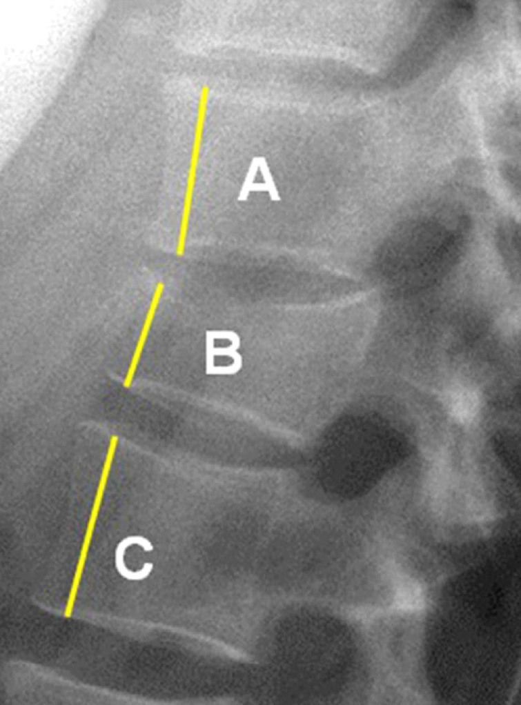
The second equation is used to calculate the loss of AVH. It is calculated by dividing the difference between the mean height of the anterior vertebral body above and below the broken vertebrae by the height of the anterior vertebral body inside the fractured vertebrae (Fig. 2). The equation as follows:
Fig. 2.

Measurements used in the calculation of anterior vertebral body height loss. Measure from the lateral x-ray film the height of the most anterior corpus vertebra (straight line): (A) the height of normal superior vertebra; (B) the height of the fractured vertebral body; (C) the height of the normal inferior vertebra. Reprinted from Ruiz Santiago et al., Quant Imaging Med Surg 2016;6:772-84 [35].
These calculations have clinical significance in determining the kyphotic lesion. If the AVH decreases by more than 50%, the resulting segment is kyphotic [35].
(2) PLC and interspinous distance
The PLC is a group of spine components comprised of the supraspinous ligament, the ISL, the facet capsule ligament, and the ligament flavum [36]. This complex works in conjunction with the surrounding fascia and muscles to withstand bending and compressive stresses, thus stabilizing the spine by preventing repetitive motion. In a case of thoracolumbar fracturing, disruption of PLC integrity results in instability and kyphotic deformity [37-40]. It is critical in determining stabilization and subsequent medication selection.
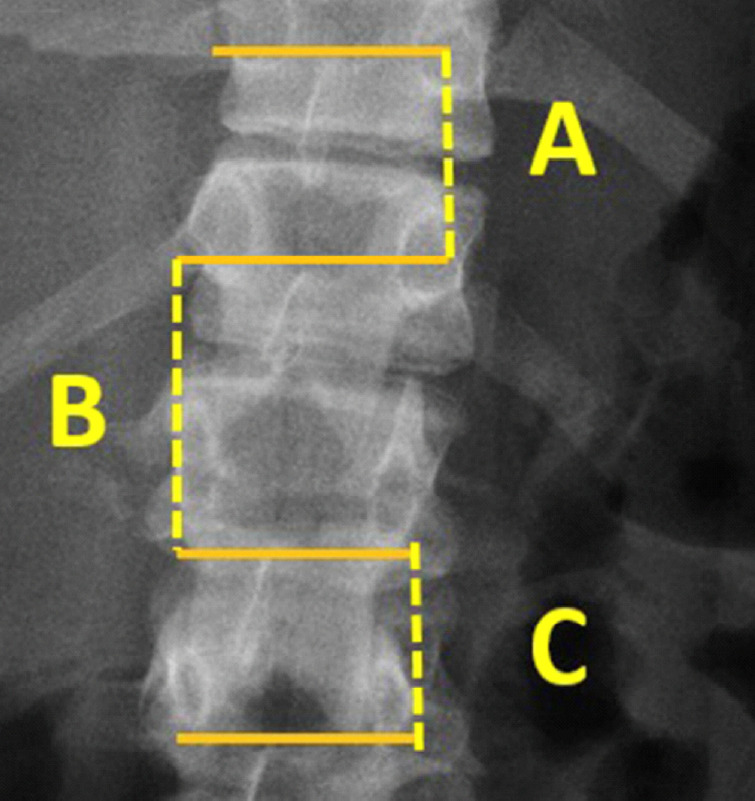
PLC evaluation is critical because its deficiency has a direct effect on the seriousness of the fracture [41]. More than 3.5 mm of vertebral translation is strongly associated with PLC damage. Variations of up to 7 mm in the interspinous gap are possible. However, technically, an expansion of the interspinous gap of more than 20% indicates an unstable PLC that requires surgical care. However, it should be checked by an MRI evaluation [42,43]. The interspinous distance equation is depicted in Fig. 3 [35]:
Fig. 3.

Measurements used in the calculation of interspinous distance in anterorposterior x-ray film, Make a horizontal straight line right on the upper edge of each spinous process, then the distance between the adjacent horizontal lines is the interspinous process distance: (A) Interspinous distance of normal superior vertebra; (B) Interspinous distance of a fractured vertebra; (C) Interspinous distance of normal inferior vertebra. Reprinted from Ruiz Santiago et al., Quant Imaging Med Surg 2016;6:772-84 [35].
Thake note that the PLC injury is very unpredictable, and associated brain injury necessitates surgery [44]. Due to the critical nature of the PLC evaluation, PCL integrity can necessitate a shift in care from conservative to surgical management [32,45].
CT scan findings are correlated with clinical neurological findings about the extent and degree of spinal cord trauma, as well as the presence and severity of PLC injury in this situation [32,45]. Apart from CT scans, such radiological observations, such as vertebral body translation on simple x-ray, are also accurate indicators of PLC damage (AO type C). Without vertebral body fracture, focal kyphosis is also a good indicator of PLC injury. Disruption of the PLC portion is also the most accurate predictor of PLC injury on T1 sagittal segment MRI [44]. Moreover, there is a chance of development of kyphosis if a PLC lesion is discovered in conjunction with a burst fracture, whether complete or incomplete [34].
(3) Facet joints/pedicles
The stiffness of the facet joints and pedicles also has an impact on the treatment options for thoracolumbar injuries. Bilateral facet joint-pedicle dislocation-fracture is synonymous with thoracolumbar AO type B2 damage [33].
(4) Intervertebral disc space
Most often, anterior disc space expansion occurs with or without avulsion and/or shear chip fracturing of the anterior endplate. It is associated with disability caused by hyperextension (AO type B3 on thoracolumbar fractures classification). Increased disc space width signals an unstable injury that can necessitate surgical intervention [33,44,45].
(5) Spinal canal diameter
Details regarding the degree of canal stenosis should be used in burst fractures. Since the degree of stenosis in neurological injury differs between vertebral stages, it can be expressed as a percentage. However, a sagittal-to-transverse diameter ratio of 0.40 is strongly correlated with neurological damage [46].
The spinal canal diameter ratio can be easily determined using CT scan axial cuts. Given that B is the spinal canal diameter ratio at the level of injury, A and C are the spinal canal diameter ratios of surrounding tissues vertebrae, respectively, above and below the level of injury [46]. The equation as it follows (Fig. 4) [35].
Fig. 4.
Measurements used in the calculation of canal narrowing percent in axial computed tomography scan of the vertebral body. The ratio of each spinal canal is the division of the sagittal diameter (s) by the transverse diameter (t). (A) Canal ratio of the normal superior vertebra; (B) Canal ratio of the fractured vertebra; (C) Canal ratio of the normal inferior vertebra. Reprinted from Ruiz Santiago et al., Quant Imaging Med Surg 2016;6:772-84 [35].
(6) Displacement - dislocation
Vertebral displacement or dislocation is a form of thoracolumbar fracture that is extremely unstable. Since it involves the whole vertebral column, it is graded as the most severe level of damage on the AOSpine classification of thoraco-lumbar fractures (AO Spine type C – translational injuries). Indeed, surgical intervention is needed [4,33].
(7) Spinal lordosis - kyphosis
This may be the product of incomplete correction, which results in kyphosis progressing late. Kyphosis resulted in constant back pain, and its correction improved clinical symptoms in posttraumatic kyphosis [46,47].
In spinal surgery, it is necessary to restore the usual range of thoracolumbar curvature in order to prevent further improper loading of the spine, which may result in another secondary complication [48]. As a result, the surgery’s objective is to achieve a fair adjustment and appropriate alignment. Cobb’s 7° are the desired practical result. Cobb’s 20° correction deficiency has a clear association with extreme back pain [49]. Cobb’s angle greater than 10.5° immediately after operation was significantly correlated with an adverse radiographic result, which is associated with a worse clinical outcome [50].
(8) Statements about radiological diagnosis of thoracolumbar spine fractures
As a result of the preceding examination, multiple radiological causes may be identified with the diagnosis of thoracolumbar fractures and can be considered when planning surgery and assessing the surgical outcome. A reduction of more than 50% of the AVH results in the onset or development of kyphotic. PLC evaluation is critical because its deficiency has a direct effect on the seriousness of the fracture. Widening of the interspinous gap by 20% or more is considered a symptom of an unhealthy PLC and should be checked via MRI. Stable disc lesions include those depicted in AO type B-3.1 and those caused by hyperextension damage with an anterior subluxation. Details regarding the degree of canal stenosis should be used in burst fractures. A ratio of sagittal-to-transverse diameters of 0.40 is strongly correlated with neurological damage. Cobb’s angle greater than 10.5° directly after operation is associated with adverse radiological effects and a weak clinical result. Cobb’s 20° correction deficiency has a clear association with extreme back pain. Table 2 contains a summary of the study.
Table 2.
Review statements: radiological factors which affect the clinical symptoms
| 1. | A decrease in anterior vertebral height of more than 50% should be considered as a risk in its course of causing kyphotic development. |
| 2. | Widening the distance between spinous processes by more than 20% can be a sign of posterior ligamentous complex instability and should be confirmed by magnetic resonance imaging. |
| 3. | In burst fractures, the degree of stenosis of the canal should be measured. A sagittal-transverse diameter ratio < 0.40 may be associated with neurological injury. |
| 4. | A Cobb angle of > 10.5° immediately after surgery may lead to unfavorable radiological results. Besides, loss of Cobb angle correction ≥ 20° can be associated with severe back pain. |
CONCLUSION
Besides being critical for physician-physician communication, terminology and clinical algorithms may also determine the therapy, so it is crucial to examine fracture anatomy carefully. The current AO and TLICS classifications for thoracolumbar fractures have demonstrated their validity and utility in clinical practice. The AOSpine classification scheme for thoracolumbar spinal injuries is complicated. Nonetheless, it contains the majority of details on the degree of vertebral body injury, canal stenosis, intervertebral disk lesion, and neurological trauma.
Numerous radiological considerations have an impact on the diagnosis, the need of treatment, and the kind of surgery that can be done. As a result, the option of x-ray, CT scan, or MRI imaging is highly dependent on the nature of the observation. The radiological evaluation would determine whether or not the surgical objectives were met.
WFNS SPINE COMMITTEE RECOMMENDATIONS
Classification of Thoracolumbar Fractures
• Both the current AO and TLICS classifications are valid and can be used in clinical practice for traumatic thoracolumbar fractures.
• Recent research indicates that, considering its complexity, the current AO classification could be more useful in treating thoracolumbar fractures.
Radiological Diagnosis of Thoracolumbar Fractures
• If a CT scan/MRI scan is not available, anteroposterior and normal lateral radiographs may be obtained.
• Although CT is still critical for trauma evaluation, it cannot accurately demonstrate the disco-ligamentous complex; therefore, MRI should be considered.
• MRI is the most often used advanced imaging technique. It is the preferred treatment for discoligamentous disorders, spinal cord abnormalities, and other pathologies involved with spinal trauma.
Acknowledgments
The authors would like to thank their respected institution for the support: Department of Neurosurgery, Faculty of Medicine - Universitas Airlangga, Surabaya, Indonesia; Airlangga University Hospital Surabaya, Surabaya; LPDP Indonesia; Dr. Soetomo General Academic Hospital, Surabaya, Indonesia; Ege University Neurosurgery Department, Izmir, Turkey; Department of Neurosurgery, Royal Victoria Hospital, Belfast Health and Social Care Trust, Belfast, Northern Ireland, UK.
Footnotes
The authors have nothing to disclose.
REFERENCES
- 1.Wang H, Zhang Y, Xiang Q, et al. Epidemiology of traumatic spinal fractures: experience from medical university–affiliated hospitals in Chongqing, China, 2001–2010. J Neurosurg Spine. 2012;17:459–68. doi: 10.3171/2012.8.SPINE111003. [DOI] [PubMed] [Google Scholar]
- 2.Verheyden AP, Spiegl UJ, Ekkerlein H, et al. Treatment of fractures of the thoracolumbar spine: recommendations of the spine section of the German Society for Orthopaedics and Trauma (DGOU) Glob Spine J. 2018;8(2 Suppl):34S–45S. doi: 10.1177/2192568218771668. [DOI] [PMC free article] [PubMed] [Google Scholar]
- 3.Azam MQ, Sadat-Ali M. The concept of evolution of thoracolumbar fracture classifications helps in surgical decisions. Asian Spine J. 2015;9:984–94. doi: 10.4184/asj.2015.9.6.984. [DOI] [PMC free article] [PubMed] [Google Scholar]
- 4.Gamanagatti S, Rathinam D, Rangarajan K, et al. Imaging evaluation of traumatic thoracolumbar spine injuries: Radiological review. World J Radiol. 2015;7:253–65. doi: 10.4329/wjr.v7.i9.253. [DOI] [PMC free article] [PubMed] [Google Scholar]
- 5.Tjoumakaris SI, Harrop JS. In: Spine and spinal cord trauma: evidence based management. Vaccaro AR, Fehlings MG, Dvorak MF, editors. New York: Thieme; 2011. American Spinal Injury Association Neurological Examination and Neurological Assessment of Spinal Cord Injury. [Google Scholar]
- 6.Nataraj A, Jack AS, Ihsanullah I, et al. Outcomes in thoracolumbar burst fractures with a thoracolumbar injury classification score (TLICS) of 4 treated with surgery versus initial conservative management. Clin Spine Surg. 2018;31:E317–21. doi: 10.1097/BSD.0000000000000656. [DOI] [PubMed] [Google Scholar]
- 7.Vaccaro AR, Schroeder GD, Kepler CK, et al. The surgical algorithm for the AOSpine thoracolumbar spine injury classification system. Eur Spine J. 2016;25:1087–94. doi: 10.1007/s00586-015-3982-2. [DOI] [PubMed] [Google Scholar]
- 8.Sethi MK, Schoenfeld AJ, Bono CM, et al. The evolution of thoracolumbar injury classification systems. Spine J. 2009;9:780–8. doi: 10.1016/j.spinee.2009.04.003. [DOI] [PubMed] [Google Scholar]
- 9.Böhler L. Die techniek der knochenbruchbehandlung im grieden und im kriege. Verlag von Wilheim Maudrich. Vienna: Verlag von Wilheim Maudrich; 1930. [Google Scholar]
- 10.Watson-Jones R. The results of postural reduction of fractures of the spine. J Bone Jt Surg. 1938;20:567–86. [Google Scholar]
- 11.Nicoll EA. Fractures of the dorso-lumbar spine. J Bone Joint Surg Br. 1949;31B:376–94. [PubMed] [Google Scholar]
- 12.Srinivasan P. A review of thoracolumbar spine fracture classifications. Indian Spine J. 2018;1:71–8. [Google Scholar]
- 13.Holdsworth F. Fractures, dislocations, and fracture-dislocations of the spine. J Bone Joint Surg Am. 1970;52:1534–51. [PubMed] [Google Scholar]
- 14.Kelly RP, Whitesides TE. Treatment of lumbodorsal fracturedislocations. Ann Surg. 1968;167:705–17. doi: 10.1097/00000658-196805000-00009. [DOI] [PMC free article] [PubMed] [Google Scholar]
- 15.Denis F. The three column spine and its significance in the classification of acute thoracolumbar spinal injuries. Spine (Phila Pa 1976) 1983;8:817–31. doi: 10.1097/00007632-198311000-00003. [DOI] [PubMed] [Google Scholar]
- 16.McAfee PC, Yuan HA, Fredrickson BE, et al. The value of computed tomography in thoracolumbar fractures. An analysis of one hundred consecutive cases and a new classification. J Bone Joint Surg Am. 1983;65:461–73. [PubMed] [Google Scholar]
- 17.Ferguson RL, Allen BL. A mechanistic classification of thoracolumbar spine fractures. Clin Orthop Relat Res. 1984;189:77–88. [PubMed] [Google Scholar]
- 18.Magerl F, Aebi M, Gertzbein SD, et al. A comprehensive classification of thoracic and lumbar injuries. Eur Spine J. 1994;3:184–201. doi: 10.1007/BF02221591. [DOI] [PubMed] [Google Scholar]
- 19.Bono CM, Vaccaro AR, Hurlbert RJ, et al. Validating a newly proposed classification system for thoracolumbar spine trauma: looking to the future of the thoracolumbar injury classification and severity score. J Orthop Trauma. 2006;20:567–72. doi: 10.1097/01.bot.0000244999.90868.52. [DOI] [PubMed] [Google Scholar]
- 20.Wood KB, Khanna G, Vaccaro AR, et al. Assessment of two thoracolumbar fracture classification systems as used by multiple surgeons. J Bone Joint Surg Am. 2005;87:1423–9. doi: 10.2106/JBJS.C.01530. [DOI] [PubMed] [Google Scholar]
- 21.Oner FC, van Gils AP, Dhert WJ, et al. MRI findings of thoracolumbar spine fractures: a categorisation based on MRI examinations of 100 fractures. Skeletal Radiol. 1999;28:433–43. doi: 10.1007/s002560050542. [DOI] [PubMed] [Google Scholar]
- 22.Vaccaro AR, Lehman RA, Jr, Hurlbert RJ, et al. A new classification of thoracolumbar injuries: the importance of injury morphology, the integrity of the posterior ligamentous complex, and neurologic status. Spine (Phila Pa 1976) 2005;30:2325–33. doi: 10.1097/01.brs.0000182986.43345.cb. [DOI] [PubMed] [Google Scholar]
- 23.Raja Rampersaud Y, Fisher C, Wilsey J, et al. Agreement between orthopedic surgeons and neurosurgeons regarding a new algorithm for the treatment of thoracolumbar injuries: a multicenter reliability study. J Spinal Disord Tech. 2006;19:477–82. doi: 10.1097/01.bsd.0000211237.14211.21. [DOI] [PubMed] [Google Scholar]
- 24.Divi SN, Schroeder GD, Oner FC, et al. AOSpine-Spine Trauma Classification System: the value of modifiers: a narrative review with commentary on evolving descriptive principles. Global Spine J. 2019;9(1 Suppl):77S–88S. doi: 10.1177/2192568219827260. [DOI] [PMC free article] [PubMed] [Google Scholar]
- 25.Park HJ, Lee SY, Park NH, et al. Modified thoracolumbar injury classification and severity score (TLICS) and its clinical usefulness. Acta Radiol. 2016;57:74–81. doi: 10.1177/0284185115580487. [DOI] [PubMed] [Google Scholar]
- 26.Lenarz CJ, Place HM, Lenke LG, et al. Comparative reliability of 3 thoracolumbar fracture classification systems. J Spinal Disord Tech. 2009;22:422–7. doi: 10.1097/BSD.0b013e31818a38cd. [DOI] [PubMed] [Google Scholar]
- 27.Joaquim AF, Patel AA. Thoracolumbar spine trauma: Evaluation and surgical decision-making. J Craniovertebr Junction Spine. 2013;4:3–9. doi: 10.4103/0974-8237.121616. [DOI] [PMC free article] [PubMed] [Google Scholar]
- 28.Rihn JA, Anderson DT, Harris E, et al. A review of the TLICS system: a novel, user-friendly thoracolumbar trauma classification system. Acta Orthop. 2008;79:461–6. doi: 10.1080/17453670710015436. [DOI] [PubMed] [Google Scholar]
- 29.Reinhold M, Audigé L, Schnake KJ, et al. AO spine injury classification system: a revision proposal for the thoracic and lumbar spine. Eur Spine J. 2013;22:2184–201. doi: 10.1007/s00586-013-2738-0. [DOI] [PMC free article] [PubMed] [Google Scholar]
- 30.Schnake KJ, Stavridis SI, Kandziora F. Five-year clinical and radiological results of combined anteroposterior stabilization of thoracolumbar fractures. J Neurosurg Spine. 2014;20:497–504. doi: 10.3171/2014.1.SPINE13246. [DOI] [PubMed] [Google Scholar]
- 31.Kepler CK, Vaccaro AR, Schroeder GD, et al. The thoracolumbar AOSpine injury score. Global Spine J. 2016;6:329–34. doi: 10.1055/s-0035-1563610. [DOI] [PMC free article] [PubMed] [Google Scholar]
- 32.Rajasekaran S, Kanna RM, Shetty AP. Management of thoracolumbar spine trauma: an overview. Indian J Orthop. 2015;49:72–82. doi: 10.4103/0019-5413.143914. [DOI] [PMC free article] [PubMed] [Google Scholar]
- 33.Vaccaro AR, Oner C, Kepler CK, et al. AOSpine thoracolumbar spine injury classification system: fracture description, neurological status, and key modifiers. Spine (Phila Pa 1976) 2013;38:2028–37. doi: 10.1097/BRS.0b013e3182a8a381. [DOI] [PubMed] [Google Scholar]
- 34.Curfs I, Grimm B, van der Linde M, et al. Radiological prediction of posttraumatic kyphosis after thoracolumbar fracture. Open Orthop J. 2016;10:135–42. doi: 10.2174/1874325001610010135. [DOI] [PMC free article] [PubMed] [Google Scholar]
- 35.Ruiz Santiago F, Tomás Muñoz P, Moya Sánchez E, et al. Classifying thoracolumbar fractures: role of quantitative imaging. Quant Imaging Med Surg. 2016;6:772–84. doi: 10.21037/qims.2016.12.04. [DOI] [PMC free article] [PubMed] [Google Scholar]
- 36.Li Y, Shen Z, Huang M, et al. Stepwise resection of the posterior ligamentous complex for stability of a thoracolumbar compression fracture: an in vitro biomechanical investigation. Medicine (Baltimore) 2017;96:e7873. doi: 10.1097/MD.0000000000007873. [DOI] [PMC free article] [PubMed] [Google Scholar]
- 37.Vaccaro AR, Lee JY, Schweitzer KM, Jr, et al. Assessment of injury to the posterior ligamentous complex in thoracolumbar spine trauma. Spine J. 2006;6:524–8. doi: 10.1016/j.spinee.2006.01.017. [DOI] [PubMed] [Google Scholar]
- 38.Radcliff K, Su BW, Kepler CK, et al. Correlation of posterior ligamentous complex injury and neurological injury to loss of vertebral body height, kyphosis, and canal compromise. Spine (Phila Pa 1976) 2012;37:1142–50. doi: 10.1097/BRS.0b013e318240fcd3. [DOI] [PubMed] [Google Scholar]
- 39.Johnson GM, Zhang M. Regional differences within the human supraspinous and interspinous ligaments: a sheet plastination study. Eur Spine J. 2002;11:382–8. doi: 10.1007/s00586-001-0378-2. [DOI] [PMC free article] [PubMed] [Google Scholar]
- 40.Oner FC, van Gils AP, Faber JA, et al. Some complications of common treatment schemes of thoracolumbar spine fractures can be predicted with magnetic resonance imaging: prospective study of 53 patients with 71 fractures. Spine (Phila Pa 1976) 2002;27:629–36. doi: 10.1097/00007632-200203150-00012. [DOI] [PubMed] [Google Scholar]
- 41.Machino M, Yukawa Y, Ito K, et al. The complement of the load-sharing classification for the thoracolumbar injury classification system in managing thoracolumbar burst fractures. J Orthop Sci. 2013;18:81–6. doi: 10.1007/s00776-012-0319-4. [DOI] [PubMed] [Google Scholar]
- 42.Radcliff K, Kepler CK, Rubin TA, et al. Does the load-sharing classification predict ligamentous injury, neurological injury, and the need for surgery in patients with thoracolumbar burst fractures?: Clinical article. J Neurosurg Spine. 2012;16:534–8. doi: 10.3171/2012.3.SPINE11570. [DOI] [PubMed] [Google Scholar]
- 43.Neumann P, Wang Y, Kärrholm J, et al. Determination of inter-spinous process distance in the lumbar spine. Evaluation of reference population to facilitate detection of severe trauma. Eur Spine J. 1999;8:272–8. doi: 10.1007/s005860050172. [DOI] [PMC free article] [PubMed] [Google Scholar]
- 44.Vaccaro AR, Lim MR, Hurlbert RJ, et al. Surgical decision making for unstable thoracolumbar spine injuries: results of a consensus panel review by the Spine Trauma Study Group. J Spinal Disord Tech. 2006;19:1–10. doi: 10.1097/01.bsd.0000180080.59559.45. [DOI] [PubMed] [Google Scholar]
- 45.Rajasekaran S, Vaccaro AR, Kanna RM, et al. The value of CT and MRI in the classification and surgical decision-making among spine surgeons in thoracolumbar spinal injuries. Eur Spine J. 2017;26:1463–9. doi: 10.1007/s00586-016-4623-0. [DOI] [PubMed] [Google Scholar]
- 46.Vaccaro AR, Nachwalter RS, Klein GR, et al. The significance of thoracolumbar spinal canal size in spinal cord injury patients. Spine (Phila Pa 1976) 2001;26:371–6. doi: 10.1097/00007632-200102150-00013. [DOI] [PubMed] [Google Scholar]
- 47.Zeng J, Gong Q, Liu H, et al. Complete fracture-dislocation of the thoracolumbar spine without neurological deficit: a case report and review of the literature. Medicine (Baltimore) 2018;97:e0050. doi: 10.1097/MD.0000000000010050. [DOI] [PMC free article] [PubMed] [Google Scholar]
- 48.Khoueir P, Oh BC, Wang MY. Delayed posttraumatic thoracolumbar spinal deformities: diagnosis and management. Neurosurgery. 2008;63(3 Suppl):117–24. doi: 10.1227/01.NEU.0000320385.27734.CB. [DOI] [PubMed] [Google Scholar]
- 49.Xu BS, Tang TS, Yang HL. Long-term results of thoracolumbar and lumbar burst fractures after short-segment pedicle instrumentation, with special reference to implant failure and correction loss. Orthop Surg. 2009;1:85–93. doi: 10.1111/j.1757-7861.2009.00022.x. [DOI] [PMC free article] [PubMed] [Google Scholar]
- 50.Seo DK, Kim CH, Jung SK, et al. Analysis of the risk factors for unfavorable radiologic outcomes after fusion surgery in thoracolumbar burst fracture: what amount of postoperative thoracolumbar kyphosis correction is reasonable? J Korean Neurosurg Soc. 2019;62:96–105. doi: 10.3340/jkns.2017.0214. [DOI] [PMC free article] [PubMed] [Google Scholar]


