ABSTRACT
African swine fever (ASF) is currently causing a major pandemic affecting the swine industry and protein availability from Central Europe to East and South Asia. No commercial vaccines are available, making disease control dependent on the elimination of affected animals. Here, we show that the deletion of the African swine fever virus (ASFV) E184L gene from the highly virulent ASFV Georgia 2010 (ASFV-G) isolate produces a reduction in virus virulence during the infection in swine. Of domestic pigs intramuscularly inoculated with a recombinant virus lacking the E184L gene (ASFV-G-ΔE184L), 40% experienced a significantly (5 days) delayed presentation of clinical disease and, overall, had a 60% rate of survival compared to animals inoculated with the virulent parental ASFV-G. Importantly, all animals surviving ASFV-G-ΔE184L infection developed a strong antibody response and were protected when challenged with ASFV-G. As expected, a pool of sera from ASFV-G-ΔE184L-inoculated animals lacked any detectable antibody response to peptides partially representing the E184L protein, while sera from animals inoculated with an efficacious vaccine candidate, ASFV-G-ΔMGF, strongly recognize the same set of peptides. These results support the potential use of the E184L deletion for the development of vaccines able to differentiate infected from vaccinated animals (DIVA). Therefore, it is shown here that the E184L gene is a novel ASFV determinant of virulence that can potentially be used to increase safety in preexisting vaccine candidates, as well as to provide them with DIVA capabilities. To our knowledge, E184L is the first ASFV gene product experimentally shown to be a functional DIVA antigenic marker.
IMPORTANCE No commercial vaccines are available to prevent African swine fever (ASF). The ASF pandemic caused by the ASF virus Georgia 2010 (ASFV-G) strain is seriously affecting pork production in a contiguous geographical area from Central Europe to East Asia. The only effective experimental vaccines are viruses attenuated by deleting ASFV genes associated with virus virulence. Therefore, identification of such genes is of critical importance for vaccine development. Here, we report the discovery of a novel determinant of ASFV virulence, the E184L gene. Deletion of the E184L gene from the ASFV-G genome (ASFV-G-ΔE184L) produced a reduction in virus virulence, and importantly, animals surviving infection with ASFV-G-ΔE184L were protected from developing ASF after challenge with the virulent parental virus ASFV-G. Importantly, the virus protein encoded by E184L is highly immunogenic, making a virus lacking this gene a vaccine candidate that allows the differentiation of infected from vaccinated animals (DIVA). Here, we show that unlike what is observed in animals inoculated with the vaccine candidate ASFV-G-ΔMGF, ASFV-G-ΔE184L-inoculated animals do not mount a E184L-specific antibody response, indicating the feasibility of using the E184L deletion as the antigenic marker for the development of a DIVA vaccine in ASFV.
KEYWORDS: ASF, ASFV, African swine fever, DIVA, E184L
INTRODUCTION
African swine fever virus (ASFV) is a large and structurally complex virus that is currently causing a disease pandemic affecting swine production and wild boar population. The initial outbreak outside Africa occurred in the Republic of Georgia in 2007, and then the first outbreak in China, the world’s largest pig-producing country, in 2018 caused the rapid spread of ASF throughout all of southeast Asia. Currently, ASFV is causing outbreaks in Central and Eastern Europe and throughout Asia. In 2020, ASFV spread to Papua New Guinea, India, and Germany. In 2021, ASF spread to Malaysia and to domestic swine farms in Germany. Recently, in August 2021, the outbreaks of ASF detected in the Dominican Republic, extended the ASF pandemic to the Western Hemisphere, causing additional concerns for a continued worldwide spread. As a result of the continued outbreaks of ASF in affected countries, the disease is causing devastating economic losses in swine production, as well as a shortage in worldwide protein availability. The ASFV strains causing this pandemic phylogenetically are all descendants of the highly virulent isolate identified during the initial 2007 outbreak in the Republic of Georgia, ASFV Georgia 2010 (ASFV-G), making this initial 2007 outbreak outside Africa the source of the current pandemic.
ASFV is an enveloped virus containing a double-stranded DNA genome of approximately 180 to 190 kbp. The ASFV genome encodes approximately 150 to 160 open reading frames (ORFs) (1). The functions of most ASFV proteins encoded within these ORFs are unknown or have been predicted using only functional genomics (1, 2), and very few of the resulting viral proteins have had any experimental function described.
Currently, there is no vaccine to prevent ASF. Consequently, the control of the disease relies on the quarantine and elimination of affected animals. Several experimental live attenuated vaccines have been shown to induce protection against infection with historical virulent virus strains (3, 4) and against the current pandemic strain (5–10). Generally, animals inoculated with attenuated viruses containing genetically engineered deletions of virus genes involved in the process of virulence are protected against infection with the homologous virulent parental virus (3–10). Therefore, the identification and genetic manipulation of virus genes associated with virulence is necessary for the rational design of genetically modified virus strains to be used as live attenuated ASFV vaccine candidates.
Here, we report the identification of a novel determinant of ASFV virulence, the E184L gene. An ASFV-G recombinant virus without the E184L gene, ASFV-G-ΔE184L, has reduced virulence when inoculated in swine, and animals surviving the infection are protected against challenge with the virulent parental virus. We also demonstrated that E184L is a highly immunogenic protein, as evidenced during inoculation with the vaccine candidate ASFV-G-ΔMGF; this immune response is completely absent in ASFV-G-ΔE184L-infected animals. Therefore, deletion of the E184L gene can act as an antigenic marker that could be used to develop vaccines that allow the differentiation of infected and vaccinated animals (DIVA).
RESULTS
Conservation of E184L gene across different ASFV isolates.
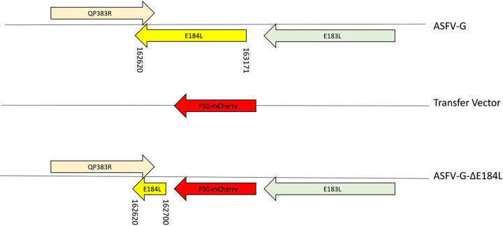
ASFV E184L gene encodes a 184-amino acid protein and is positioned on the negative strand between nucleotide positions 163174 and 162620 of the ASFV-G genome (Fig. 1). The translated product of the ASFV E184L gene is a 22-kDa protein of unknown function (11) expressed during the virus replication cycle in pigs (12), inducing a strong antibody response (13, 14).
FIG 1.
Diagram indicating the position of the E184L open reading frame in the African swine fever virus strain Georgia 2010 (ASFV-G) genome. The donor plasmid with the homologous arms to ASFV-G and the mCherry under the control of the p30 promoter in the orientation are indicated. The final genomic changes were introduced to develop ASFV-G-ΔE184L where the sequence of the donor plasmid mCherry reporter is introduced to replace the ORF of E184L as indicated. The numbers refer to the nucleotide positions of the ORF of E184L in ASFV-G or the residual portion of E184L that remains in ASFV-G-ΔE184L.
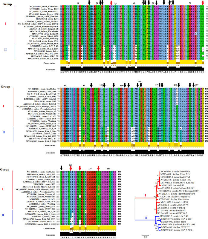
To assess the nucleotide and amino acid homology among different isolates of ASFV representing the genetic diversity of gene E184L, multiple pairwise comparisons were performed using the algorithm ClustalW. In general, the average identities at the nucleotide and amino acid levels were calculated to be 95.65 and 92.67%, respectively. However, we found that there is a disparate range of identity at the nucleotide (90.42 to 99.80%) and amino acid (83.60 to 99.45%) levels, indicating the moderated conservation of the E184L protein among some ASFV isolates. Overall, no differences at nucleotide or amino acid levels were found within isolates belonging to the epidemic Eurasia lineage.
To get more insights about the disparate levels of identity at the amino acid level, a phylogenetic analysis was performed by neighbor joining using the Jones-Taylor Thornton (JTT) model and 1,000 bootstrap replicates to assess the confidence probability of the analysis. The results indicated that based on their amino acid differences, multiple ASFV isolates can be classified in two main phylogenetic groups (Fig. 2). Furthermore, pairwise calculations using the p-distance model (P < 0.05) showed that the average levels of identity within groups I and II were 98.86 and 97.89%, respectively, with the average identity between groups as low as 84.20%.
FIG 2.
Multiple sequence alignment of the indicated ASFV isolates of viral protein E184L. A total of 21 protein sequences representing the genetic diversity of gene E184L of ASFV in the GenBank database were used to conduct this alignment. The alignment contains 148 conserved and 36 variable sites. Among the variable sites, conservation scores are displayed based on the biological properties of each amino acid, with the lower scores associated with more divergent replacements. Symbols indicate residue conservation (*) or replacement for an amino acid with similar properties (+). Based on phylogenetic analysis (conducted on Mega version 10.0.5), protein sequences were classified in two different groups. The numbers in the branches represent bootstrap values. Additionally, black and red arrows represent relevant residues evolving under negative or positive selection, respectively. Alignment was conducted on the Jalview software version 2.11.1.3, using the ClustalW algorithm.
No homology was found among 19 protein families when E184L protein was evaluated using the program Pfam 34.0 (15). Interestingly, evolutionary analysis performed by the algorithms fixed-effects likelihood (FEL) model (16) and the mixed effects model of evolution (MEME) (17) indicated that negative selection was the main force driving the evolution of E184L (dN/dS = 0.323). A total of 22 amino acid residues appeared to evolve under negative selection (P = 0.1), showing the relevance of the conservation of these amino acids during the evolution of E184L (Fig. 2). Conversely, five residues were found evolving under positive selection (P = 0.1), indicating the importance of these residues in the promotion of the divergence of E184L protein (Fig. 2). No evidence of recombination was found using the genetic algorithm for recombination detection (GARD) (18).
E184L is a late-transcribed gene.
To determine whether the E184L gene is transcribed during the infectious cycle, a time-course experiment was performed to analyze the kinetics of RNA transcription in primary swine macrophages infected with ASFV strain Georgia. Swine macrophage cultures were infected with a multiplicity of infection (MOI) of 1 with ASFV-G, and cell lysate samples were taken at 4, 6, 8, and 24 hours postinfection (hpi). The presence of E184L RNA was detected by real-time PCR (RT-PCR) as described in Material and Methods. Transcription of E184L was detected starting at 6 hpi and remained stable until 24 hpi (Fig. 3). The pattern of expression of the well-characterized ASFV early protein p30 (CP204L) and the late protein p72 (B646L) has been previously described and is used here as a representation of early- and late-transcription profiles. Expression of E184L practically overlaps with that of the p72 gene. This result agrees with recently published work describing the expression profile for all ASFV genes by next generation sequencing (NGS), characterizing E184L as a late-transcribed gene. Therefore, the ASFV E184L gene encodes a protein that is expressed late in the virus replication cycle.
FIG 3.
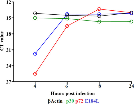
Expression profile of the E184L gene of ASFV during infection in porcine macrophages. qRT-PCR analysis was performed to assess the profile expression of E184L at different hours postinfection. As controls for this analysis, genes encoding p30 (early expression) and p72 (late expression) were used for this experiment. The housekeeping β-actin was used as an endogenous control for the analysis. CT, cycle threshold.
Development of the E184L gene deletion mutant in the ASFV-Georgia isolate.
Although it is known that the E184L gene is expressed during infection in pigs and induces a strong antibody response, the biological function of the gene remains completely unknown. To investigate the function of the E184L gene during virus infection in cell cultures and its impact on disease phenotype, a recombinant virus harboring a deletion of the E184L gene was developed (ASFV-G-ΔE184L) from the parental highly virulent ASFV Georgia 2010 (ASFV-G). The E184L gene was replaced by a cassette containing the fluorescent reporter gene, mCherry, under the ASFV p30 promoter (Fig. 1). The recombinant ASFV-G-ΔE184L was purified by limiting dilution based on the presence of fluorescent activity.
To evaluate the accuracy of the genetic modification introduced in ASFV-G-ΔE184L, as well as the integrity of the virus genome, the full genomic sequence of the recombinant virus was obtained using NGS. Subsequent sequencing reads were analyzed using CLC genomics workbench 21 (Qiagen), and reads that aligned to the ASFV-G sequence were used for analysis. Two separate NGS sequencing runs were performed to provide sequencing reads with a minimum depth of 30 across the ASFV-G genome, and a depth of over 200 was achieved on either side of the E184L deletion. Variant detection is performed to identify any point mutations that could have occurred in the genome. Comparison of ASFV-G-ΔE184L and ASFV-G genomic sequences showed a deletion of 471 nucleotides (covering nucleotide positions 162704 and 163174) corresponding with the deletion of the E184L gene (Fig. 1). Additionally, the ASFV-G-ΔE184L genome possesses an insertion of 3,944 nucleotides, consistent with the introduction of the p30mCherry cassette substituting the E184L gene. No unwanted additional genomic modifications were found in the rest of the ASFV-G-ΔE184L genome. No E184L gene sequences were detected by NGS, indicating the purity of the recombinant virus stock.
Replication of ASFV-G-ΔE184L in primary swine macrophages.
The impact of the removal of the E184L gene from the genome of ASFV-G was assessed by a growth kinetics study using primary swine macrophage cultures, the main cell type targeted by ASFV during infection in swine. The kinetics of replication of ASFV-G-ΔE184L were compared with that of the parental ASFV-G in multistep growth curves (Fig. 4). Primary cultures of swine macrophages were infected (MOI of 0.01), and samples were collected at 2, 24, 48, 72, and 96 h postinfection (hpi). Analysis of the results indicates that ASFV-G-ΔE184L exhibited a replication kinetic significantly diminished compared to that of the parental ASFV-G. ASFV-G-ΔE184L titers are between 10- and 100-fold lower than those of ASFV-G, depending on the time point considered. Therefore, deletion of the E184L gene moderately diminished the capability of the virus to replicate in swine macrophage cultures.
FIG 4.

In vitro growth characteristics of parental ASFV-G and recombinant ASFV-G-ΔE184, ASFV-G-ΔMGF, and ASFV-G-ΔMGF/ΔE184L. Primary swine macrophage cell cultures were infected (MOI = 0.01) with each of the viruses, and virus yield was titrated at the indicated times postinfection. The data represent means from three independent experiments. The sensitivity of virus detection was ≥1.8 log10 HAD50/mL. Significant differences in viral yields between ASFV-G-ΔE184L versus ASFV-G (black asterisks) and between ASFV-G-ΔMGF versus ASFV-G-ΔMGF/ΔE184L (red asterisks) are shown at specific time points. Statistical analysis was conducted by the unpaired t test using the two-stage step-up (Benjamini, Krieger, and Yekutieli) method, assuming individual variance for each time point. P values of <0.05 were considered statistically significant. TCID50, 50% tissue culture infective dose.
Assessment of ASFV-G-ΔE184L virulence in swine.
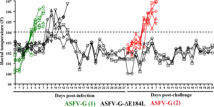
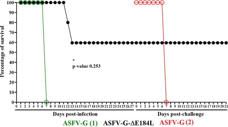
Evaluating the impact of the removal of the E184L gene from the ASFV-G genome on virus virulence in swine was assessed by experimentally infecting domestic pigs with ASFV-G-ΔE184L, for comparison with animals infected with parental virulent ASFV-G. Groups of five 80- to 90-pound pigs were intramuscularly (i.m.) inoculated with a 102 50% hemadsorption dose (HAD50) of either ASFV-G-ΔE184L or ASFV-G and observed for 28 days. All five animals infected with ASFV-G had increased body temperature (>104°F) by day 4 or 5 postinfection, followed by development and rapid progression of clinical ASF signs, with all animals euthanized in extremis by 7 days postinfection (pi) due to the severity of the disease (Table 1 and Fig. 5 and 6). The five animals infected with ASFV-G-ΔE184L presented with a heterogenous response. All animals had increased body temperature (>104°F) by day 10 or 11 pi. Two of those animals started showing clinical signs of the disease (anorexia, depression, skin lesions, and, later, incoordination) that evolved over 2 to 3 days to a more severe form of the disease with the animals being euthanized by day 13 or 14 pi. The remaining three animals in the group did not show any clinical sign of the disease aside from the initial rise in body temperature, remaining clinically normal until day 28 pi (Fig. 5 and 6). Therefore, deletion of the E184L gene resulted in an attenuation of the ASFV-G strain, with 40% of infected animals experiencing a significantly delayed disease onset before developing lethal ASF disease and 60% of animals exhibiting a late and transient rise of body temperature with no additional clinical signs. Furthermore, the latest was statistically supported (P < 0.05) when survival curves between groups of pigs infected with ASFV-G or ASFV-G-ΔE184L were compared by two independent statistical analyses.
TABLE 1.
Swine survival and fever response following infection with a 102 HAD50 of ASFV-G-ΔE184L or parental ASFV-G
| Fever |
|||||
|---|---|---|---|---|---|
| Virus (HAD50) | No. of survivors/total | Mean time to death (days ± SD) | Time to onset (days ± SD) | Duration (days ± SD) | Maximum daily temp (°F ± SD) |
| ASFV-G | 0/5 | 7 ± 0a | 4.6 ± 0.55 | 2.4 ± 0.55 | 105.52 ± 0.79 |
| ASFV-G-ΔE184L | 3/5 | 13.5 ± 0.71a,b | 10.5 ± 0.71b | 3 ± 0b | 105.45 ± 0.07 |
aAll animals were euthanized for humanitarian reasons following the corresponding IACUC protocol.
bData refer to the only two animals in the group developing disease. The other three animals remained clinically normal during the observational period.
FIG 5.
Kinetics of body temperature values in pigs i.m. inoculated with 102 HAD50 of either ASFV-G-ΔE184L or ASFV-G [ASFV-G (1)] before and after the challenge with 102 HAD50 of ASFV-G [ASFV-G (2)]. Each curve represents values from an individual animal in each group.
FIG 6.
Kinetics of mortality in pigs i.m. inoculated with 102 HAD50 of either ASFV-G-ΔE184L or ASFV-G [ASFV-G (1)] before and after the challenge with 102 HAD50 of ASFV-G [ASFV-G (2)]. The asterisk (*) represents the statistical difference in the survival curves between groups of pigs during the first stage of the experiment (postinfection).
The level of virus replication, as represented by the viremia values, was analyzed in both groups of animals. As expected, the animals infected with ASFV-G had high titers (105.5 to 108 HAD50/mL) by day 4 pi, which rapidly increased (to around 108.5 HAD50/mL) by day 7 pi, when all animals were euthanized (Fig. 7). Conversely, ASFV-G-ΔE184L-infected animals had viremia kinetics that paralleled the development of clinical signs. Animals had viremia titers ranging between 102.5 and 106.5 HAD50/mL by day 4 pi, increasing to titer values of 105.5 to 107.5 HAD50/mL by day 7 pi. Viremia titers progressively decreased until day 28 pi, reaching titer values between 104.5 and 107 HAD50/mL. The two euthanized animals had final titers of 107 HAD50/mL. Therefore, the disappearance of ASFV virulence caused by deletion of the E184L gene is accompanied by a reduced but stable virus replication presenting long viremias with relatively moderate disease.
FIG 7.
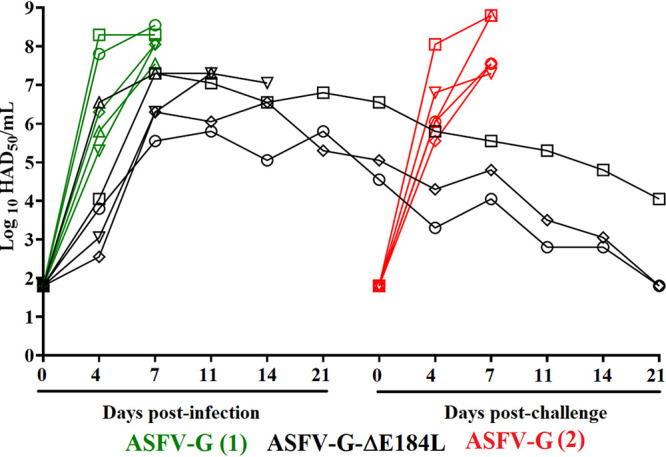
Viremia titers detected in pigs i.m. inoculated with a 102 50% hemadsorption dose (HAD50) of either ASFV-G-ΔE184L or ASFV-G [ASFV-G (1)] before and after the challenge with 102 HAD50 of ASFV-G [ASFV-G (2)]. Each curve represents values from an individual animal in each group. The sensitivity of virus detection was ≥1.8 log10 HAD50/mL.
Protective efficacy of ASFV-G-ΔE184L against challenge with ASFV-G.
Infection with attenuated strains of ASFV consistently protected animals against the disease caused by the virulent parental virus. The ability of ASFV-G-ΔE184L to induce protection against disease caused by parental ASFV-G was assessed in the animals surviving the ASFV-G-ΔE184L infection. The three animals were challenged i.m. 28 days after ASFV-G-ΔE184L infection with 102 HAD50 of ASFV-G. An additional group of five naive animals were challenged under the same conditions as a control group.
Animals in the control group started displaying ASF-related clinical signs 4 to 5 days postchallenge (pc), with quick progression to severe disease; all animals were euthanized by day 7 pc (Table 2 and Fig. 5 and 6). Conversely, all three animals infected with ASFV-G-ΔE184L did not display any clinical signs of ASF during the 21-day observational period. Therefore, ASFV-G-ΔE184L infection induced protection against development of ASF when challenged with the highly virulent parental strain ASFV-G.
TABLE 2.
Swine survival and fever response in ASFV-G-ΔE184L-infected animals with a 102 HAD50 of ASFV-G virus 28 days later
| Virus (102 HAD50) | Fever |
||||
|---|---|---|---|---|---|
| No. of survivors/total | Mean time to death (days ± SD) | Time to onset (days ± SD) | Duration (days ± SD) | Maximum daily temp (°F ± SD) | |
| Mock | 0/5 | 7 ± 0a | 4.2 ± 0.45 | 2.8 ± 0.45 | 105.98 ± 0.94 |
| ASFV-G-ΔE184L | 3/3 | 103.06 ± 0.29 | |||
aAll animals were euthanized due to humanitarian reasons following the corresponding IACUC protocol.
After challenge, virus titers in the control animals were high (ranging between 106.5 and 107 HAD50/mL) by day 4 pi, increasing (ranging from 107.5 to 108 HAD50/mL) by day 7 pi, when all animals were euthanized (Fig. 7). After challenge with ASFV-G, none of the three ASFV-G-ΔE184L-infected animals developed viremia values higher than titers present at the time of challenge. The titers in two of the animals progressively decreased until the end of the observational period (21 days after challenge) when no detectable titers were found in the animals, whereas the one remaining animal had a viremia titer of 104.5 HAD50/mL at the end of the experiment.
Antibody response in animals infected with ASFV-G-ΔE184L.
Although the immune mechanisms producing protection in animals infected with attenuated strains of virus are still surrounded by controversy, our experience working with several vaccine candidates is that the only parameter consistently associated with protection is the presence of ASFV-specific circulating antibodies (6, 7, 9, 10, 19, 20). Therefore, we tried to associate the presence of anti-ASFV circulating antibodies in ASFV-G-ΔE184L-infected animals with protection against challenge. A robust virus-specific antibody response, detected using a direct enzyme-linked immunosorbent assay (ELISA) developed in-house, was detected in the sera of all three animals (Fig. 8). Antibody response, mediated by IgG isotype, was detected in all the animals by day 11 pi, remaining high until the day of the challenge. Therefore, in agreement with previous reports, a close association exists between the presence of anti-ASFV antibodies at the moment of challenge and protection of the animals.
FIG 8.

Anti-ASFV antibody titers detected by ELISA in pigs i.m. inoculated with 102 HAD50 of ASFV-G-ΔE184L. Each point represents a value from an individual animal.
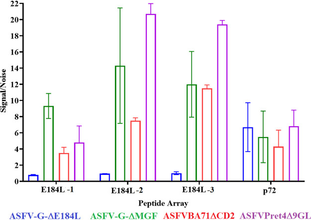
The E184L protein has been shown to elicit a strong immune response when used as an individual protein (13) and, more importantly, during viral infection (14). Therefore, the E184L gene product is potentially a target for the development of a DIVA test to discriminate between animals immunized with a recombinant vaccine lacking the E184L gene from those infected with wild-type ASFV. In this study, we assessed the potential of E184L as a DIVA marker. The response of a pool of sera from animals immunized with vaccine candidates ASFV-G-ΔMGF (7), ASFV-BA71-ΔCD2 (4), and ASFV-Pret4-Δ9GL (19) to a set of 15-mer partially overlapping peptides representing the carboxyl end of the translated sequence of the E184L gene was evaluated in a peptide array assay and compared to that raised in the ASFV-G-ΔE184L-infected animals. Synthetic peptides representing partial amino acid sequences of highly immunogenic ASFV protein p54 were included as positive controls. The results demonstrate that pooled sera from ASFV-G-ΔMGF-, ASFV-Ba71-ΔCD2-, or ASFV-Pret4-Δ9GL- and ASFV-G-ΔE184L-infected animals strongly recognized peptides representing amino acid sequences of the control antigen p54 (Fig. 9). In addition, pooled sera from ASFV-G-ΔMGF-, ASFV-Ba71-ΔCD2-, or ASFV-Pret4-Δ9GL-infected animals also strongly recognized the E184L peptides. Conversely, and as expected, the pooled sera from ASFV-G-ΔE184L-infected animals failed to recognize any of the peptides representing the E184L amino acid sequence. These results, together with the positive response found after vaccination with ASFV-G-ΔMGF, ASFV-Ba71-ΔCD2, or ASFV-Pr4-Δ9GL (Fig. 9), affirm the potential use of the E184L gene as the molecular basis of a DIVA marker in the ASFV genome.
FIG 9.
Antibody response to ASFV E184L protein or p54 in pool of sera of animals inoculated with either ASFV-G-ΔE184L, ASFV-G-ΔMGF, ASFVPret4-Δ9GL, or ASFv-Ba71ΔCD2 detected by peptide array. The results are presented as signal/noise OD values (and their SD) of each pool sera related to OD of a pool of preimmune sera. E184L peptides 1, 2, and 3 represent amino acid residues at positions 163 to 177, 167 to 181, and 170 to 184 of the E184L protein, respectively.
Development of an ASFV vaccine candidate harboring E184L deletion as potential DIVA marker.
Deletion of E184L does not lead to a complete attenuation of ASFV-G; therefore, to be used as a potential DIVA marker, removal of the gene should be performed in a ASFV vaccine candidate with a minimal safety profile. To test the DIVA functionality of E184L we deleted the gene from the vaccine candidate ASFV-G-ΔMGF (7). The ΔMGF deletion was constructed using a p72Gus reporter gene cassette as described previously (7), allowing for the same strategy and methodology to develop the resulting recombinant virus, ASFV-G-ΔMGF/ΔE184L, which was already described for ASFV-G-ΔE184L. The E184L gene was replaced by the p30mCherry cassette provoking the same modifications in the ASFV-G-ΔMGF/ΔE184L genome as those described in ASFV-G-ΔE184L (Fig. 1).
The effect of the E184L gene deletion on the replication ability of ASFV-G-ΔMGF was assessed by a growth kinetics study in primary swine macrophages of the ASFV-G-ΔMGF/ΔE184L compared with that of ASFV-G, and the parentals ASFV-G-ΔMGF and ASFV-G-ΔE184L in multistep growth curves (Fig. 4). Interestingly, ASFV-G-ΔMGF/ΔE184L exhibited a replication kinetic almost indistinguishable from that of ASFV-G-ΔE184L and approximately 10-fold lower kinetics than those of ASFV-G and ASFV-G-ΔMGF. Therefore, deletion of the E184L gene produced a moderate decrease in the ability of the resulting virus (ASFV-G-ΔE184L and ASFV-G-ΔMGF/ΔE184L) to replicate compared with the replication ability of the corresponding parental virus.
The potential use of ASFV-G-ΔMGF/ΔE184L as a DIVA marker experimental vaccine was tested in domestic pigs in comparison with the efficacy of the parental ASFV-G-ΔMGF vaccine candidate. Two groups of five 80- to 90-pound pigs were i.m. inoculated with either 104 HAD50 or 106 HAD50 of ASFV-G-ΔMGF/ΔE184L, while a third group was inoculated with 104 HAD50 of ASFV-G-ΔMGF. All animals were observed for 28 days with no development of clinical disease.
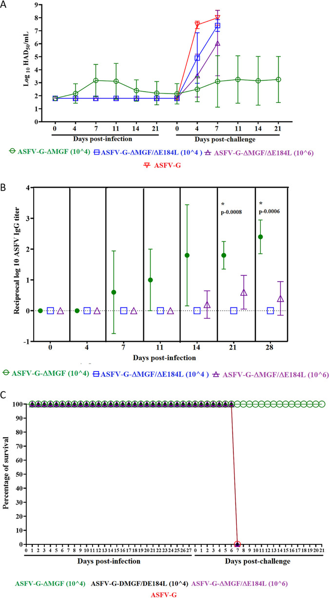
Analysis of the viremia titers in all groups showed that, as already reported (7), animals infected with ASFV-G-ΔMGF displayed a heterogenous pattern with three animals presenting low to medium (102.5 to 105.5 HAD50) titers between days 4 and 28 pi and the other two not producing detectable titers at all during the prechallenge period (Fig. 10A). Interestingly, in both groups of animals inoculated with ASFV-G-ΔMGF/ΔE184L, no viremia titers were detected in any of the animals at any time point tested.
FIG 10.
(A) Viremia titers detected in pigs i.m. inoculated with either 104 or 106 HAD50 of ASFV-G-ΔMGF/ΔE184L or 104 HAD50 of ASFV-G-ΔMGF before and after the challenge with 102 HAD50 of ASFV-G (ASFV-G 2). Each curve represents average values and corresponding SDs from each animal group. The sensitivity of virus detection was ≥1.8 log10 HAD50/ml. (B) Anti-ASFV antibody titers detected by ELISA in pigs i.m. inoculated with either 104 or 106 HAD50 of ASFV-G-ΔMGF/ΔE184L or 104 HAD50 of ASFV-G-ΔMGF. Each point represents average values and corresponding SDs from each animal group. Asterisks represent time points at which statistically significant differences were found between groups. (C) Kinetics of mortality in pigs i.m. inoculated with either 104 or 106 HAD50 of ASFV-G-ΔMGF/ΔE184L or 104 HAD50 of ASFV-G-ΔMGF before and after the challenge with 102 HAD50 of ASFV-G.
According to the viremia kinetics, antibody titers developed in ASFV-G-ΔMGF-infected animals starting at day 11 pi, increasing through days 14 to 21 pi, reaching the highest titers by the day of challenge (Fig. 10B). Conversely, no detectable antibody titers were found at any time point in animals inoculated with ASFV-G-ΔMGF/ΔE184L with the exception of very late, low titers in two animals receiving 106 HAD50. Similar results have been previously observed when making additional deletions to vaccine strains where the lack of virus replication in vivo resulted in the inability to produce antibodies to ASFV, resulting in a loss of vaccine efficacy (20).
All three groups were challenged at 28 days pi with virulent ASFV-G along with five naive pigs used as the control group. Control animals developed the expected clinical disease with a rise in body temperature by day 4 pc with a rapid progression in severity until all animals were euthanized by day 7 pc (Fig. 10C). After the challenge, as described earlier (7), animals infected with ASFV-G-ΔMGF remained clinically normal throughout the observational period. On the other hand, all animals inoculated with ASFV-G-ΔMGF/ΔE184L developed a clinical disease undistinguishable from that experienced by the control animals and were euthanized by day 7 pc. Again, viremia profiles followed presentation of clinical signs (Fig. 10A). The control group developed high titers (107.05 to 107.8 HAD50) by day 4 pc, reaching 107.05 to 108.55 HAD50 by the time they were euthanized. After the challenge, all animals infected with 104 HAD50 of ASFV-G-ΔMGF except one (which remained aviremic) developed viremia titers ranging from 102.55 to 106.55 HAD50 at different times pc. Animals receiving 104 HAD50 of ASFV-G-ΔMGF/ΔE184L ranged from 104.55 to 107.55 HAD50 by day 4 pc, reaching high titers (107.3 to 108.5 HAD50). Those animals inoculated with 106 HAD50 of ASFV-G-ΔMGF/ΔE184L displayed a range of titers (from undetected to 105.05 HAD50) by day 4 pc, reaching high titers (105.8 to 108.5 HAD50) in all euthanized animals at day 7 pc.
DISCUSSION
The use of experimental vaccines based on the use of live attenuated strains is a practical approach to the development of an effective ASF vaccine. Attenuated virus strains can be developed using different approaches, from the use of natural attenuated field isolates (21) to attenuation by adapting virulent field isolates to growth in cell cultures (22, 23) or attenuation by genetic manipulation, deleting viral genes associated with virulence (3–10, 24–28). The latter appears effective and perhaps is a safer methodology compared to the use of naturally attenuated isolates. In the referenced examples, genetic manipulation causing deletions of single genes or a group of genes produced attenuated virus strains that induce protection against the virulent parental virus. Here, we present the identification of a previously uncharacterized ASFV gene, E184L, as a novel viral genetic determinant of virulence. Deletion of E184L partially attenuates ASFV-G in swine, when used at doses of 102 HAD50. Interestingly, only six other genetic modifications have been shown to decrease virulence in the highly virulent ASFV Georgia isolate or its derivative isolates. In our laboratory, we showed that deletion of the 9GL and UK genes or a deletion of a group of six genes from the MGF360 and 530 or the I177L gene induced a complete virus attenuation when inoculated i.m. in a dose range of 103 to 106 HAD (6, 7, 10). We also have shown that the deletion of I177L when inoculated i.m. was 100% effective at doses as low as 102 HAD (10). This low-dose potency was maintained when an additional deletion in the left variable region was introduced to adapt the ASFV-G-ΔI177L experimental vaccine to cell culture (26). More recently, results using virulent field isolates from China have shown that deletion of the MGF110-9L or MGF505-7R gene induces complete or partial attenuation when i.m. tested at 10 HAD (24, 29), whereas deletion of a group of genes, L7L to L11L (27), in a recombinant virus i.m. inoculated at doses of 103 to 106 HAD induces partial attenuation. Therefore, deletion of the E184L gene constitutes the seventh genetic modification, leading to a decrease in virulence of the Georgia 2007 virus or its derivatives. Importantly, along with the surviving animals infected with ASFV-G-ΔE184L, only recombinant viruses lacking I177L, 9GL, 9GL/UK, CD2/UK, MGF, L7L to L11L, or MGF/CD2 genes, in the context of the Georgia 2007 strain or its derivative isolates, have been used as experimental vaccines to protect against the corresponding virulent parental virus (5–10, 27, 28).
Because residual virulence remained using the ASFV-G-ΔE184L recombinant virus, it is clear that the individual deletion of E184L cannot be used in the development of an attenuated virus strain but must occur in combination with other gene deletions to achieve complete attenuation. The deletion of E184L in the context of a vaccine candidate genome would potentially serve to both increase vaccine safety and provide a negative antigenic marker in the vaccine candidate. The recognized immunogenicity of the E184L protein product (13) constitutes a critical characteristic for its potential use as an antigenic marker to develop a DIVA-compatible vaccine. Our results demonstrated that E184L deletion can be used as a negative antigenic marker in a potential live attenuated vaccine. Animals surviving inoculation with ASFV-G-ΔE184L mounted a vigorous ASFV-specific antibody response while failing to recognize the E184L protein product. To our knowledge, this constitutes the first report describing a specific ASFV protein functioning as an antigenic DIVA marker in pigs vaccinated with an experimental attenuated ASFV vaccine that confers protection against virulent challenge. Additional studies using E184L as a potential target marker to develop a DIVA test would likely include the detection of the E184L antibody response mediated by both IgG (as tested in this study) and IgM to ensure the earliest possible detection of the E184L-specific response. Additionally, the reactivity of E184L-specific antibodies from other genotypes of ASFV will also need to be tested for validation of any future DIVA test using E184L as a viral antigen.
We attempted to present a proof of concept of E184L gene deletion in a genome of the experimental attenuated vaccine candidate ASFV-G-ΔMGF. Deletion of E184L clearly affects the ability of ASFV-G-ΔMGF/ΔE184L to replicate in vivo. Interestingly, this replication deficiency does not appear to be as drastic in primary macrophage cell cultures, indicating the existence of other to-be-determined host factors involved in the decreased replication of ASFV-G-ΔMGF/ΔE184L in pigs. The reduced replication resulting from multiple gene deletions from the virus genome is not an uncommon event. Subsequent deletion of genes from a virus genome already containing deletion of genes associated with virulence usually decreases the ability of the novel recombinant virus to replicate, particularly in inoculated swine. Some examples of this phenomenon are the deletion of six genes of the MGF360/505 family or the CD2-like gene from the genome of the ASFV-G-Δ9GL virus (20, 30), the deletion of the NL or UK genes in naturally attenuated OURT T88/3 (36), the deletion of CD2-like gene in HLJ/18-6GD (28), the deletion of UK in CD2-deleted virus (8), the deletion of UK from ASFV-G-Δ9GL (6), and the deletion of NL and UK genes from ASFV-G-Δ9GL (31). The outcome of these combined deletions is unpredictable, in some cases producing a desirable increase of virus attenuation (6, 8, 28) and in others, as in the case of ASFV-G-ΔMGF/ΔE184L, producing a decreased efficacy in inducing protection by the modified vaccine candidate harboring the novel deletion (20, 30–32). Therefore, deleting the E184L gene from the genome of a vaccine candidate to gain DIVA functionality will need to be clinically evaluated to ensure the resulting virus still efficaciously protects animals. The decrease in replication associated with deletion of E184L could also restrict the large-scale production of any DIVA vaccine candidate with E184L deleted.
We believe the results presented here demonstrate that deletion of the E184L gene should be considered a valid approach to produce a vaccine candidate with DIVA capability and to potentially increase vaccine safety. To our knowledge, E184L is the first ASFV gene candidate functionally tested as a negative antigenic marker for the development of live attenuated ASF vaccines with serological DIVA capability. Therefore, the addition of the deletion of E184L could be included in a ASFV vaccine candidate to produce a potential DIVA ASF vaccine.
MATERIALS AND METHODS
Cell culture and viruses.
Culture of primary swine macrophages was performed as described elsewhere (33). Briefly, blood mononuclear leukocytes were separated over a Ficoll-Paque density gradient (Pharmacia, Piscataway, NJ). Monocyte/macrophage cells were cultured in plastic Primaria tissue culture flasks (Falcon; Becton Dickinson Labware, Franklin Lakes, NJ) in macrophage media: RPMI 1640 medium (Life Technologies, Grand Island, NY) with 30% L929 supernatant and 20% fetal bovine serum (HI-FBS, Thermo Scientific, Waltham, MA) at 37°C under 5% CO2. After 48 h of incubation, adherent cells were detached from the tissue culture with a solution containing 10 mM EDTA in phosphate-buffered saline (PBS), and detached cells were then reseeded into Primaria T25, 6- or 96-well dishes at a density of 5 × 10⁶ cells/mL for use in assays 24 h later.
Comparative growth curves to study growth kinetics between parental ASFV-G and recombinant viruses were performed in primary swine macrophage cell cultures. Macrophage monolayers were infected (MOI = 0.01) for 1 h. Then the inoculum was removed, and the cells were rinsed twice with PBS and once with macrophage media and incubated at 37°C under 5% CO2. At 2, 24, 48, 72, and 96 hours postinfection (hpi) cell cultures were frozen at <−70°C, and the thawed lysates were clarified by centrifugation to eliminate cell debris and used to determine virus titers in primary swine macrophage cell cultures. All of the samples were run simultaneously to avoid interassay variability. The presence of infectious virus was detected by hemadsorption (HA) and virus titers calculated using the Reed and Muench method (34). ASFV Georgia 2010 (ASFV-G) was a field isolate kindly provided by Dr. Nino Vepkhvadze, from the Laboratory of the Ministry of Agriculture (LMA) in Tbilisi, Republic of Georgia.
Detection of E184L transcription.
Real-time PCR analysis was used to assess the expression profile of the gene E184L during the infection of ASFV-G in cultures of porcine macrophages. For this purpose, 6-well plates containing cell cultures of porcine macrophages (1 × 10⁷ cells/well) were infected in triplicate with a stock of ASFV-G using an MOI of 1. The plates were incubated at 37°C, and RNA extractions were conducted at 4, 6, 8, and 24 h postinfection.
RNA extraction was carried out using the RNeasy kit (Qiagen) following the manufacturer’s instructions. Afterwards, RNA was treated with 2 units of DNase I (BioLabs) following the manufacturer’s protocol. Final reactions were purified using the Monarch RNA cleanup kit (New England BioLabs, Inc.). RNA was quantified, and 1 μg was used to produce cDNA using qScript cDNA SuperMix (Quanta bio) following the manufacturer’s instructions.
Using the sequence of the ASFV Georgia 2007/1 strain (GenBank accession number FR682468.2) as a reference, primers and probes were designed using the real-time qPCR assay entry tool from Integrated DNA Technologies (IDT) (https://www.idtdna.com/scitools/Applications/RealTimePCR/). The primers for E184L were 5′-AAAATCACACCCGAAAACCAAG-3′ (forward) and 5′-GTGAGAATACATAAG GGTTTGCG-3′ (reverse), and the probe was 5′-FAM/AAAACACCTTGCAAAGCCGACTCATC/MGBNFQ-3′. The CP204L (p30) gene was used as a control for the quantification of an early expression gene of ASFV: 5′-GACGGAATCCTCAGCATCTTC-3′ (forward primer) and 5′-CAGCTTGGAGTCTTTAGGTACC-3′ (reverse primer), and the probe used was 5′-FAM/TGTTTGAGCAAGAGCCCTCATCGG/MGBNFQ-3′. Additionally, as a control for a late expression gene of ASFV, we used the gene B646L (p72) using a quantitative RT-PCR (qRT-PCR) previously published (35). Also, the housekeeping gene β-actin was used as an endogenous control to validate the quality of the extraction and the RNA concentration from different infections performed in this experiment.
All qRT-PCR assays were conducted on a 7500 real-time PCR system (Applied Biosystems), using the TaqMan Universal PCR Master Mix (Applied Biosystems catalog number 4304437) according to the following protocol for master mix preparation (1×): 12.5 μl of Universal mix, 7.05 μl of water, 0.1 μl of forward primer (50 μM), 0.1 μl of reverse primer (50 μM), 10 μM probe, and 5 μl of DNA. The conditions of amplification were as follows: one step at 55°C for 2 min followed by one denaturation step at 95°C for 10 min and then 40 cycles of denaturation at 95°C for 15 s and annealing/extension at 65°C for 1 min.
Construction of the recombinant ASFV-G-ΔE184L.
Recombinant viruses were generated by homologous recombination between the corresponding parental genome (either ASFV-G or ASFV-G-ΔMGF) and recombination transfer vector p30mCherryΔE184L by infection and transfection procedures using swine macrophage cell cultures as previously described in detail (36). Development of the recombinant vaccine candidate ASFV-G-ΔMGF was previously described (7). The recombinant transfer vector p30mCherryΔE184L contains flanking genomic regions to amino acid residues 1 and 157 of the E184L gene, mapping approximately 1 kbp to the left and right of these amino acids, along with the reporter gene cassette containing the mCherry gene with the ASFV p30 early gene promoter, p30mCherry. This construction created a 471-bp deletion in the E184L ORF (Fig. 1). The coding region of the C terminus of E184L was left intact but believed not to be expressed because of the lack of a promoter or start codon, because this portion of E184L overlaps with another ASFV protein C terminus, QP383R; leaving this section allows for QP383R to be properly expressed. The recombinant transfer vector p30mCherryΔE184L was obtained by DNA synthesis (Epoch Life Sciences Missouri City, TX, USA).
NGS of ASFV genomes.
ASFV DNA was extracted from infected cells and quantified as described earlier. Full-length sequencing of the virus genome was performed as described previously (37) using an Illumina NextSeq500 sequencer. Sequencing reads were analyzed using CLC genomics software version 21 (Qiagen), removing adapters and trimming the reads; these reads were then aligned to the ASFV-G sequence and subjected to variant detection.
Animal experiments.
Animal experiments were performed under biosafety level 3AG conditions in the Plum Island Animal Disease Center (PIADC) animal facility following protocols approved by the PIADC Institutional Animal Care and Use Committee of the U.S. Departments of Agriculture and Homeland Security (protocol number 225.04-16-R, 09-07-16). ASFV-G-ΔE184L virulence was evaluated by comparing it to parental ASFV-G using 80- to 90-pound commercial breed swine. Groups of pigs (n = 5) were i.m. inoculated with 102 HAD50 of either ASFV-G-ΔE184L or ASFV-G. The presence of clinical signs (anorexia, depression, fever, purple skin discoloration, staggering gait, diarrhea, and cough) and changes in rectal temperature were recorded daily throughout the experiment. In protection experiments, the animals inoculated with ASFV-G-ΔE184L, ASFV-G-ΔMGF, or ASFV-G-ΔMGF/ΔE184L were i.m. challenged 28 days later with 102 HAD50 of parental ASFV-G. The presence of clinical signs associated with the disease was recorded as described earlier (7).
Detection of anti-ASFV antibodies.
ASFV antibody detection used an in-house ELISA performed as described previously (19). Briefly, ELISA antigen was prepared from ASFV-infected Vero cells. Maxisorb ELISA plates (Nunc, St. Louis, MO) were coated with 1 μg/well of infected or uninfected cell extract. The plates were blocked with phosphate-buffered saline containing 10% skim milk (Merck, Kenilworth, NJ) and 5% normal goat serum (Sigma, St. Louis, MO). Each swine serum was tested at multiple dilutions against both infected and uninfected cell antigen. ASFV-specific antibodies in the swine sera were detected using an anti-swine IgG-horseradish peroxidase conjugate (KPL, Gaithersburg, MD) and SureBlue Reserve peroxidase substrate (KPL). The plates were read at the optical density at 630 nm (OD630) in an ELx808 plate reader (BioTek, Shoreline, WA). Sera titers were expressed as the log10 of the highest dilution where the OD630 reading of the tested sera at least duplicates the reading of the mock-infected sera.
Specific peptide slides representing partial sequences of ASFV proteins p72 (residues 34 to 53), p54 (residues 138 to 160), and the carboxy end of E184L (residues 163 to 177, 164 to 178, 165 to 179, 166 to 180, 167 to 181, 168 to 182, 163 to 177, 169 to 183, and 170 to 184) were manufactured by PEPperPRINT (Heidelberg, Germany). Microarray analysis was conducted based on PEPperCHIP immunoassay protocol provided by the array manufacturer. In brief, the peptide microarray slides were incubated with 1.5 mL of standard buffer (PBS, 0.05% Tween 20, pH 7.4) (Sigma-Aldrich) at room temperature (20 to 25°C) for 15 min and blocking buffer (Tris-buffered saline at pH 7.6 with 1% bovine serum albumin [BSA]) for an additional 30 min. After removal of blocking buffer, the arrays were incubated with staining buffer (standard buffer with 10% of the blocking buffer) containing Cy5-labeled mouse anti-HA (positive control, PEPperPRINT) and Cy3-labeled goat anti-swine IgG (Jackson ImmunoResearch, West Grove, PA) for 45 min. The microarrays were washed three times with standard buffer and scanned with a GenePix 4000B scanner (Molecular Devices, Downington, PA). After scanning, the microarrays were incubated again with staining buffer for 15 min followed by incubation with staining buffer containing the serum sample at 1:1,000 dilution overnight at 4°C, followed by incubation of the staining buffer containing Cy3-labeled goat anti-swine IgG for 45 min. After washing three times, the microarrays were scanned again at the same setting as the first scan. Fold changes were calculated by dividing the signal intensity of positives with the negatives.
Statistical analysis.
Comparison of survival curves between groups of pigs were determined by both the log-rank (Mantel-Cox) test and the Gehan-Breslow-Wilcoxon test. Comparisons showing a P value of <0.05 were considered statistically significant.
Statistical differences between antibodies and viral yields produced by different viruses at different time points were determined by the unpaired t test with Welch correction, assuming an individual variance for each group. The two-stage step-up (Benjamini, Krieger, and Yekutieli) was used to determine the significance of the discovery.
In all cases, comparisons showing a P value of <0.05 were considered statistically significant. Analyses were performed on GraphPad Prism version 9.0.0 for Windows (GraphPad Software, San Diego, CA; www.graphpad.com).
Data availability.
The next generation sequencing of the recombinant ASFV viruses produced in this study was aligned to GenBank accession number LR743116. The raw sequencing reads are available upon request.
ACKNOWLEDGMENTS
We thank the Plum Island Animal Disease Center Animal Care Unit staff for excellent technical assistance. We particularly thank Melanie V. Prarat for editing the manuscript.
This research was supported in part by an appointment to the Plum Island Animal Disease Center (PIADC) Research Participation Program administered by the Oak Ridge Institute for Science and Education (ORISE) through an interagency agreement between the U.S. Department of Energy (DOE) and the U.S. Department of Agriculture (USDA). ORISE is managed by ORAU under DOE contract DE-SC0014664. All opinions expressed in this paper are the authors’ and do not necessarily reflect the policies and views of USDA, ARS, APHIS, DHS, DOE, or ORAU/ORISE.
This project was partially funded through an interagency agreement with the Science and Technology Directorate of the U.S. Department of Homeland Security under awards 70RSAT19KPM000056 and 70RSAT18KPM000134 and by the Spanish Government (project PID2019-107616RB-I00).
The authors Douglas Gladue and Manuel Borca have a patent for ASFV-G-ΔMGF as a live attenuated vaccine for African swine fever (U.S. patent US9528094B2).
Contributor Information
Manuel V. Borca, Email: Manuel.Borca@usda.gov.
Douglas P. Gladue, Email: Douglas.Gladue@usda.gov.
Mark T. Heise, University of North Carolina at Chapel Hill
REFERENCES
- 1.Tulman ER, Delhon GA, Ku BK, Rock DL. 2009. African swine fever virus, p 43–87. Lesser known large dsDNA viruses, vol 328. Springer, Heidelberg. [DOI] [PubMed] [Google Scholar]
- 2.Chapman DA, Darby AC, Da Silva M, Upton C, Radford AD, Dixon LK. 2011. Genomic analysis of highly virulent Georgia 2007/1 isolate of African swine fever virus. Emerg Infect Dis 17:599–605. 10.3201/eid1704.101283. [DOI] [PMC free article] [PubMed] [Google Scholar]
- 3.Lewis T, Zsak L, Burrage TG, Lu Z, Kutish GF, Neilan JG, Rock DL. 2000. An African swine fever virus ERV1-ALR homologue, 9GL, affects virion maturation and viral growth in macrophages and viral virulence in swine. J Virol 74:1275–1285. 10.1128/jvi.74.3.1275-1285.2000. [DOI] [PMC free article] [PubMed] [Google Scholar]
- 4.Monteagudo PL, Lacasta A, Lopez E, Bosch L, Collado J, Pina-Pedrero S, Correa-Fiz F, Accensi F, Navas MJ, Vidal E, Bustos MJ, Rodriguez JM, Gallei A, Nikolin V, Salas ML, Rodriguez F. 2017. BA71DeltaCD2: a new recombinant live attenuated African swine fever virus with cross-protective capabilities. J Virol 91:e01058-17. 10.1128/JVI.01058-17. [DOI] [PMC free article] [PubMed] [Google Scholar]
- 5.O’Donnell V, Holinka LG, Krug PW, Gladue DP, Carlson J, Sanford B, Alfano M, Kramer E, Lu Z, Arzt J, Reese B, Carrillo C, Risatti GR, Borca MV. 2015. African swine fever virus Georgia 2007 with a deletion of virulence-associated gene 9GL (B119L), when administered at low doses, leads to virus attenuation in swine and induces an effective protection against homologous challenge. J Virol 89:8556–8566. 10.1128/JVI.00969-15. [DOI] [PMC free article] [PubMed] [Google Scholar]
- 6.O’Donnell V, Risatti GR, Holinka LG, Krug PW, Carlson J, Velazquez-Salinas L, Azzinaro PA, Gladue DP, Borca MV. 2017. Simultaneous deletion of the 9GL and UK genes from the African swine fever virus Georgia 2007 isolate offers increased safety and protection against homologous challenge. J Virol 91:e01760-16. 10.1128/JVI.01760-16. [DOI] [PMC free article] [PubMed] [Google Scholar]
- 7.O’Donnell V, Holinka LG, Gladue DP, Sanford B, Krug PW, Lu X, Arzt J, Reese B, Carrillo C, Risatti GR, Borca MV. 2015. African swine fever virus Georgia isolate harboring deletions of MGF360 and MGF505 genes is attenuated in swine and confers protection against challenge with virulent parental virus. J Virol 89:6048–6056. 10.1128/JVI.00554-15. [DOI] [PMC free article] [PubMed] [Google Scholar]
- 8.Teklue T, Wang T, Luo Y, Hu R, Sun Y, Qiu HJ. 2020. Generation and evaluation of an African swine fever virus mutant with deletion of the CD2v and UK genes. Vaccines 8:763. 10.3390/vaccines8040763. [DOI] [PMC free article] [PubMed] [Google Scholar]
- 9.Borca MV, Ramirez-Medina E, Silva E, Vuono E, Rai A, Pruitt S, Espinoza N, Velazquez-Salinas L, Gay CG, Gladue DP. 2021. ASFV-G-I177L as an effective oral nasal vaccine against the Eurasia strain of Africa swine fever. Viruses 13:765. 10.3390/v13050765. [DOI] [PMC free article] [PubMed] [Google Scholar]
- 10.Borca MV, Ramirez-Medina E, Silva E, Vuono E, Rai A, Pruitt S, Holinka LG, Velazquez-Salinas L, Zhu J, Gladue DP. 2020. Development of a highly effective African swine fever virus vaccine by deletion of the I177L gene results in sterile immunity against the current epidemic Eurasia strain. J Virol 94:e02017-19. 10.1128/JVI.02017-19. [DOI] [PMC free article] [PubMed] [Google Scholar]
- 11.Alejo A, Matamoros T, Guerra M, Andres G. 2018. A proteomic atlas of the African swine fever virus particle. J Virol 92:e01293-18. 10.1128/JVI.01293-18. [DOI] [PMC free article] [PubMed] [Google Scholar]
- 12.Jaing C, Rowland RRR, Allen JE, Certoma A, Thissen JB, Bingham J, Rowe B, White JR, Wynne JW, Johnson D, Gaudreault NN, Williams DT. 2017. Gene expression analysis of whole blood RNA from pigs infected with low and high pathogenic African swine fever viruses. Sci Rep 7:10115. 10.1038/s41598-017-10186-4. [DOI] [PMC free article] [PubMed] [Google Scholar]
- 13.Netherton CL, Goatley LC, Reis AL, Portugal R, Nash RH, Morgan SB, Gault L, Nieto R, Norlin V, Gallardo C, Ho CS, Sanchez-Cordon PJ, Taylor G, Dixon LK. 2019. Identification and immunogenicity of African swine fever virus antigens. Front Immunol 10:1318. 10.3389/fimmu.2019.01318. [DOI] [PMC free article] [PubMed] [Google Scholar]
- 14.Kollnberger SD, Gutierrez-Castaneda B, Foster-Cuevas M, Corteyn A, Parkhouse RME. 2002. Identification of the principal serological immunodeterminants of African swine fever virus by screening a virus cDNA library with antibody. J Gen Virol 83:1331–1342. 10.1099/0022-1317-83-6-1331. [DOI] [PubMed] [Google Scholar]
- 15.Mistry J, Chuguransky S, Williams L, Qureshi M, Salazar GA, Sonnhammer ELL, Tosatto SCE, Paladin L, Raj S, Richardson LJ, Finn RD, Bateman A. 2021. Pfam: the protein families database in 2021. Nucleic Acids Res 49:D412–D419. 10.1093/nar/gkaa913. [DOI] [PMC free article] [PubMed] [Google Scholar]
- 16.Kosakovsky Pond SL, Frost SD. 2005. Not so different after all: a comparison of methods for detecting amino acid sites under selection. Mol Biol Evol 22:1208–1222. 10.1093/molbev/msi105. [DOI] [PubMed] [Google Scholar]
- 17.Murrell B, Wertheim JO, Moola S, Weighill T, Scheffler K, Kosakovsky Pond SL. 2012. Detecting individual sites subject to episodic diversifying selection. PLoS Genet 8:e1002764. 10.1371/journal.pgen.1002764. [DOI] [PMC free article] [PubMed] [Google Scholar]
- 18.Kosakovsky Pond SL, Posada D, Gravenor MB, Woelk CH, Frost SD. 2006. GARD: a genetic algorithm for recombination detection. Bioinformatics 22:3096–3098. 10.1093/bioinformatics/btl474. [DOI] [PubMed] [Google Scholar]
- 19.Carlson J, O’Donnell V, Alfano M, Velazquez Salinas L, Holinka L, Krug P, Gladue D, Higgs S, Borca M. 2016. Association of the host immune response with protection using a live attenuated African swine fever virus model. Viruses 8:291. 10.3390/v8100291. [DOI] [PMC free article] [PubMed] [Google Scholar]
- 20.O’Donnell V, Holinka LG, Sanford B, Krug PW, Carlson J, Pacheco JM, Reese B, Risatti GR, Gladue DP, Borca MV. 2016. African swine fever virus Georgia isolate harboring deletions of 9GL and MGF360/505 genes is highly attenuated in swine but does not confer protection against parental virus challenge. Virus Res 221:8–14. 10.1016/j.virusres.2016.05.014. [DOI] [PubMed] [Google Scholar]
- 21.Barasona JA, Gallardo C, Cadenas-Fernandez E, Jurado C, Rivera B, Rodriguez-Bertos A, Arias M, Sanchez-Vizcaino JM. 2019. First oral vaccination of Eurasian wild boar against African swine fever virus genotype II. Front Vet Sci 6:137. 10.3389/fvets.2019.00137. [DOI] [PMC free article] [PubMed] [Google Scholar]
- 22.Sereda AD, Balyshev VM, Kazakova AS, Imatdinov AR, Kolbasov DV. 2020. Protective properties of attenuated strains of African swine fever virus belonging to seroimmunotypes I–VIII. Pathogens 9:274. 10.3390/pathogens9040274. [DOI] [PMC free article] [PubMed] [Google Scholar]
- 23.Krug PW, Holinka LG, O’Donnell V, Reese B, Sanford B, Fernandez-Sainz I, Gladue DP, Arzt J, Rodriguez L, Risatti GR, Borca MV. 2015. The progressive adaptation of a Georgian isolate of African swine fever virus to vero cells leads to a gradual attenuation of virulence in swine corresponding to major modifications of the viral genome. J Virol 89:2324–2332. 10.1128/JVI.03250-14. [DOI] [PMC free article] [PubMed] [Google Scholar]
- 24.Li D, Liu Y, Qi X, Wen Y, Li P, Ma Z, Liu Y, Zheng H, Liu Z. 2021. African swine fever virus MGF-110-9L-deficient mutant has attenuated virulence in pigs. Virol Sin 36:187–195. 10.1007/s12250-021-00350-6. [DOI] [PMC free article] [PubMed] [Google Scholar]
- 25.Zsak L, Caler E, Lu Z, Kutish GF, Neilan JG, Rock DL. 1998. A nonessential African swine fever virus gene UK is a significant virulence determinant in domestic swine. J Virol 72:1028–1035. 10.1128/JVI.72.2.1028-1035.1998. [DOI] [PMC free article] [PubMed] [Google Scholar]
- 26.Borca MV, Rai A, Ramirez-Medina E, Silva E, Velazquez-Salinas L, Vuono E, Pruitt S, Espinoza N, Gladue DP. 2021. A cell culture-adapted vaccine virus against the current African swine fever virus pandemic strain. J Virol 95:e00123-21. 10.1128/JVI.00123-21. [DOI] [PMC free article] [PubMed] [Google Scholar]
- 27.Zhang J, Zhang Y, Chen T, Yang J, Yue H, Wang L, Zhou X, Qi Y, Han X, Ke J, Wang S, Yang J, Miao F, Zhang S, Zhang F, Wang Y, Li M, Hu R. 2021. Deletion of the L7L-L11L genes attenuates ASFV and induces protection against homologous challenge. Viruses 13:255. 10.3390/v13020255. [DOI] [PMC free article] [PubMed] [Google Scholar]
- 28.Chen W, Zhao D, He X, Liu R, Wang Z, Zhang X, Li F, Shan D, Chen H, Zhang J, Wang L, Wen Z, Wang X, Guan Y, Liu J, Bu Z. 2020. A seven-gene-deleted African swine fever virus is safe and effective as a live attenuated vaccine in pigs. Sci China Life Sci 63:623–634. 10.1007/s11427-020-1657-9. [DOI] [PMC free article] [PubMed] [Google Scholar]
- 29.Li D, Yang W, Li L, Li P, Ma Z, Zhang J, Qi X, Ren J, Ru Y, Niu Q, Liu Z, Liu X, Zheng H. 2021. African swine fever virus MGF-505-7R negatively regulates cGAS-STING-mediated signaling pathway. J Immunol 206:1844–1857. 10.4049/jimmunol.2001110. [DOI] [PMC free article] [PubMed] [Google Scholar]
- 30.Gladue DP, O’Donnell V, Ramirez-Medina E, Rai A, Pruitt S, Vuono EA, Silva E, Velazquez-Salinas L, Borca MV. 2020. Deletion of CD2-Like (CD2v) and C-type lectin-like (EP153R) genes from African swine fever virus Georgia-9GL abrogates its effectiveness as an experimental vaccine. Viruses 12:1185. 10.3390/v12101185. [DOI] [PMC free article] [PubMed] [Google Scholar]
- 31.Ramirez-Medina E, Vuono E, O’Donnell V, Holinka LG, Silva E, Rai A, Pruitt S, Carrillo C, Gladue DP, Borca MV. 2019. Differential effect of the deletion of African swine fever virus virulence-associated genes in the induction of attenuation of the highly virulent Georgia strain. Viruses 11:599. 10.3390/v11070599. [DOI] [PMC free article] [PubMed] [Google Scholar]
- 32.Abrams CC, Goatley L, Fishbourne E, Chapman D, Cooke L, Oura CA, Netherton CL, Takamatsu HH, Dixon LK. 2013. Deletion of virulence associated genes from attenuated African swine fever virus isolate OUR T88/3 decreases its ability to protect against challenge with virulent virus. Virology 443:99–105. 10.1016/j.virol.2013.04.028. [DOI] [PMC free article] [PubMed] [Google Scholar]
- 33.Borca MV, Berggren KA, Ramirez-Medina E, Vuono EA, Gladue DP. 2018. CRISPR/Cas gene editing of a large DNA virus: African swine fever virus. Bio-protocol 8:e2978. 10.21769/BioProtoc.2978. [DOI] [PMC free article] [PubMed] [Google Scholar]
- 34.Reed LJ, Muench H. 1938. A simple method of estimating fifty percent endpoints. Am J Hygiene 27:493–497. 10.1093/oxfordjournals.aje.a118408. [DOI] [Google Scholar]
- 35.Zsak L, Borca MV, Risatti GR, Zsak A, French RA, Lu Z, Kutish GF, Neilan JG, Callahan JD, Nelson WM, Rock DL. 2005. Preclinical diagnosis of African swine fever in contact-exposed swine by a real-time PCR assay. J Clin Microbiol 43:112–119. 10.1128/JCM.43.1.112-119.2005. [DOI] [PMC free article] [PubMed] [Google Scholar]
- 36.Borca MV, O’Donnell V, Holinka LG, Sanford B, Azzinaro PA, Risatti GR, Gladue DP. 2017. Development of a fluorescent ASFV strain that retains the ability to cause disease in swine. Sci Rep 7:46747. 10.1038/srep46747. [DOI] [PMC free article] [PubMed] [Google Scholar]
- 37.Borca MV, Holinka LG, Berggren KA, Gladue DP. 2018. CRISPR-Cas9, a tool to efficiently increase the development of recombinant African swine fever viruses. Sci Rep 8:3154. 10.1038/s41598-018-21575-8. [DOI] [PMC free article] [PubMed] [Google Scholar]
Associated Data
This section collects any data citations, data availability statements, or supplementary materials included in this article.
Data Availability Statement
The next generation sequencing of the recombinant ASFV viruses produced in this study was aligned to GenBank accession number LR743116. The raw sequencing reads are available upon request.


