. 2021 Dec 8;10:e71185. doi: 10.7554/eLife.71185

Figure 2. NMNAT1 loss affects retinal bipolar, horizontal and amacrine cells.

Representative retinal sections from knockout (-/-) and floxed littermate control (+/+) mice at the indicated ages labeled with antibodies against BRN3A (A-D, green), Calbindin (CALB) (A–B, magenta) Calretinin (CALR) (E–H), and CHX10 (I–L). Quantification of BRN3A (M), CALB, (N), CALR (O), and CHX10-positive cells (P) are shown. In (O), only CALR-positive cells on the outer side of the IPL (layer indicated by white arrowheads) were counted. Data is represented as mean ± SD. n = 3 biological replicates for all panels; significance determined using Student’s t-test. Scale bars, 30 μm.

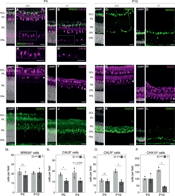
Figure 2—source data 1. Numerical source data for retinal cell type quantification in P4 and P10 KO and WT retinas.

Figure 2.

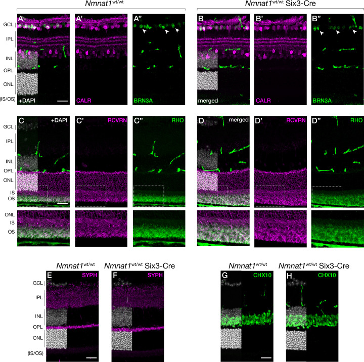
Figure 2—figure supplement 1. Six3-Cre does not cause obvious defects in the mature retina.

Figure 2—figure supplement 1.

Representative retinal sections from P65 wild-type (Nmnat1wt/wt) and Six3-Cre expressing (Nmnat1wt/wt;Six3-Cre) mice labeled with antibodies against calretinin (CALR) and BRN3A (A–B), recoverin (RCVRN) and rhodopsin (RHO) (C–D), synaptophysin (SYPH) (E,F), or CHX10 (G,H). Corresponding zoom panels are indicated with dotted rectangles. n = 3 biological replicates for all panels. Scale bars, 30 μm. Abbreviations: P, postnatal day; GCL, ganglion cell layer; IPL, inner plexiform layer; OPL, outer plexiform layer; INL, inner nuclear layer; ONL, outer nuclear layer; IS/OS, photoreceptor inner segment/outer segment layer.