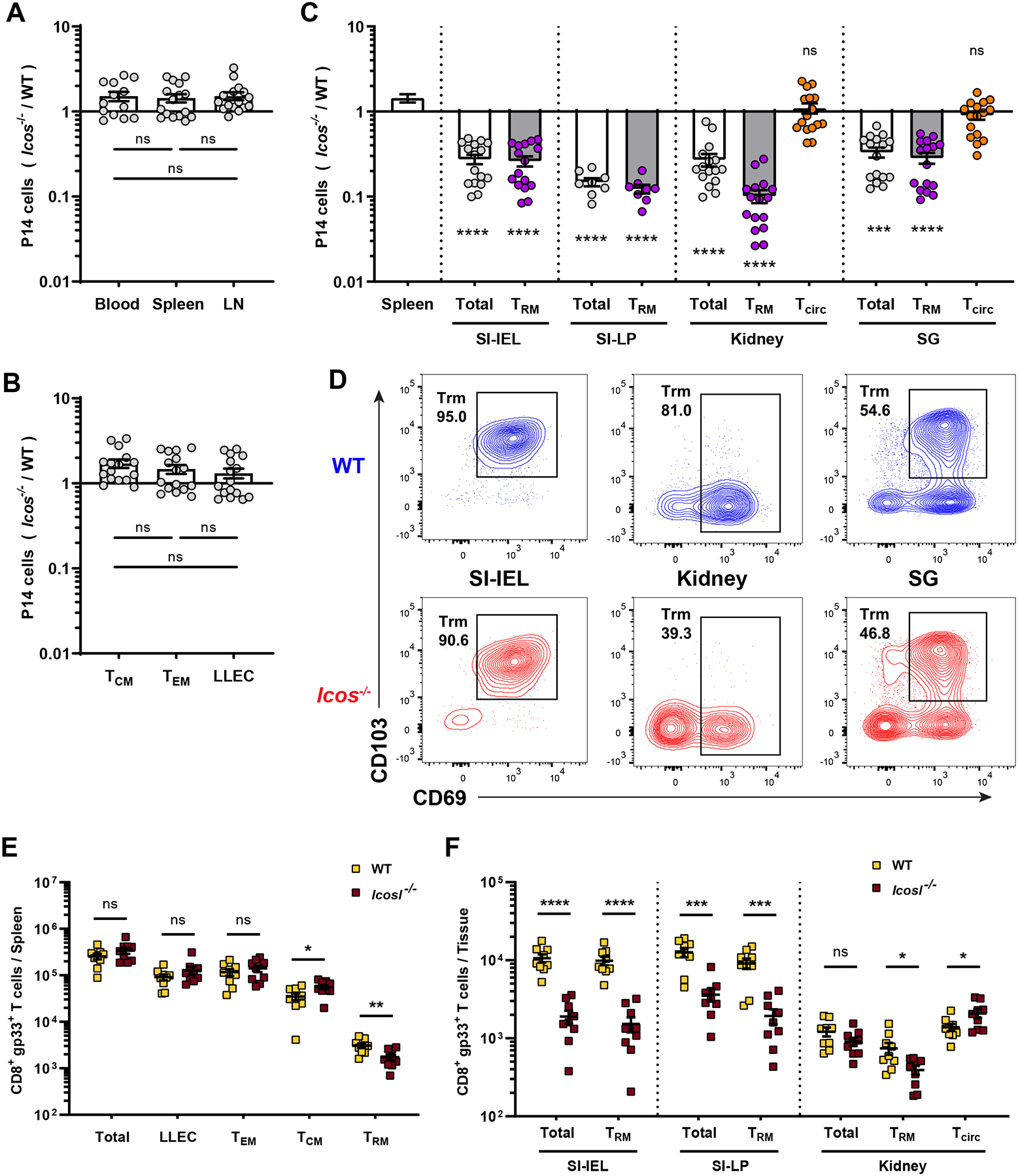
Figure 1. ICOS is critical for generation of resident but not circulating memory CD8+ T cells.

(A–D) Equal numbers of congenically distinct WT and Icos−/− P14 T cells were co-transferred into WT recipient mice, followed by LCMV-Arm infection. At least 40 days later, cells were isolated and the compiled relative frequencies of donor WT and Icos−/− P14 T cells from indicated tissues were determined. (A) shows the ratio among cells in blood and lymphoid tissues, while (B) distinct splenic circulating memory subsets, defined as Tcm (CD62L+), Tem (CD62L− KLRG1− CD127Hi) and LLEC (CD62L− KLRG1+ CD127Lo). In (C),Trm defined as CD8αiv−CD69+CD103+ (kidney Trm as CD8αiv−CD69+) while vascular contaminants (“Tcirc”) were defined as CD8αiv+CD69−. (D) shows representative phenotypic analysis for the indicated tissues. (E–F) WT and Icosl−/− mice were infect with LCMV-Arm. At least 35 days post infection, the indicated tissues were analyzed for the number of gp33/Db-specific CD8+ T cells. Data are from 2–5 independent experiments with a total 8–16 mice. Error bars represent mean ± SEM. Statistical significance was calculated with: One-way ANOVA with multiple comparisons in (A and B), Kruskal-Wallis test with multiple comparisons relative to spleen in (C), and unpaired t-test in (E and F): ns, not significant (p>0.05); * p<0.05; ** p<0.01; *** p<0.001; **** p<0.0001.
