Abstract
A 24-year-old man presented with a dumbbell-shaped right posterior mediastinal mass. The patient was placed in the prone position following general anaesthesia and intubation. After laminectomy and dissection of the dorsal part of the tumour using a posterior approach were performed, the tumour was completely resected using a robotic approach in the thoracic cavity without repositioning. This is the first report of robotic resection for posterior mediastinal tumour in the prone position as well as a novel combined posterior approach and robotic resection for dumbbell tumours.
Keywords: Mediastinal tumour, Prone position, Robotic resection
Resection of Eden type 2 or 3 dumbbell thoracic spinal tumours requires both anterior and posterior approaches.
INTRODUCTION
Resection of Eden type 2 or 3 dumbbell thoracic spinal tumours requires both anterior and posterior approaches. The standard approach for such dumbbell tumours is combined laminectomy in the prone position and video-assisted thoracoscopic surgery (VATS) resection in the lateral position. Recently, robot-assisted thoracic surgery (RATS) performed with the da Vinci surgical system (Intuitive Surgical, Sunnyvale, CA, USA) has also been used for posterior mediastinal tumours, but the standard approach uses the lateral decubitus position [1]. In this article, we report the first combined posterior approach and robot-assisted resection for dumbbell tumours in the prone position to avoid repositioning and reduce procedure time.
CASE PRESENTATION
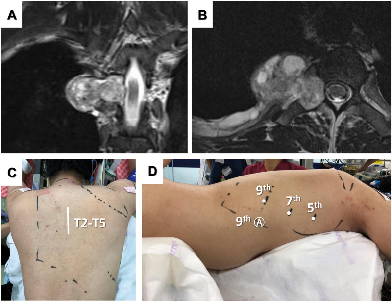
The patient was a 24-year-old man diagnosed with a dumbbell tumour. Magnetic resonance imaging showed a dumbbell-shaped right foraminal and paravertebral tumour at the T4 level (Fig. 1A). The tumour was 3 cm in diameter and extended to the lateral side along the fourth intercostal nerve (Fig. 1B). After induction of general anaesthesia, the patient was placed in the prone position. First, a posterior skin incision was made (Fig. 1C), and pedicle screws at right T3 and T5 were inserted using an O-arm navigation system. After right hemi-laminectomy and right hemi-facetectomy of the right transverse process at T4, the tumour was exposed and dissected from the surrounding tissue. The nerve root was ligated and sectioned at the proximal portion of the tumour. After the back wound was temporally closed with a gauze, 8-mm da Vinci ports were placed in the seventh intercostal space on the posterior axillary line for a camera, in the fifth intercostal space on the mid-axillary line for the first arm and in the ninth intercostal space on the back for the third arm (Fig. 1D). A 12-mm assist port was placed in the ninth intercostal space on the mid-axillary line, and CO2 was insufflated at a pressure of 8 mmHg. The fenestrated bipolar forceps was manipulated using the first arm of the robot, and the Maryland bipolar forceps was manipulated by the third arm. The parietal pleura around the tumour was opened, and the tumour was resected from the surrounding tissue (Fig. 2A). The tumour extended along the fourth intercostal nerve, and the nerve was cut at the level with normal findings. The tumour resection proceeded to the back and the tumour was extirpated completely and removed (Fig. 2B). The back wound was closed after the right T3–T5 posterior fusion. The total operation time was 4 h 19 min, and the console time was 36 min. The estimated blood loss was 660 ml, and most of it was from the neurosurgical part. The operation was completed without any complications, and the postoperative course was uneventful. The resected specimen was histologically diagnosed as a schwannoma.
Figure 1:
Magnetic resonance image showing a posterior mediastinal dumbbell tumour in the sagittal plane (A) and the axial plane (B). Posterior incision (C) and port placement for robotic tumour resection (D).
Figure 2:
Intraoperative view of the tumour (A) and intrathoracic view of the tumour (B).
COMMENT
To the best of our knowledge, this is the first report of robotic resection for a posterior mediastinal tumour in the prone position as well as a novel combined posterior approach and robotic resection for dumbbell tumours. However, the operation was safely and promptly performed without any complications. CO2 insufflation provided an optimal view, and the port setting enabled the robotic arms to move smoothly. This procedure will be possible with adequate port setting even if the tumour locates at a different level from the present case. The console surgeon felt no stress during the procedure.
The most important advantage of this approach is that repositioning is not necessary during the procedure. We can choose back or robotic approaches anytime, which make it possible to approach from both directions at the same time. Furthermore, this method can avoid intraoperative accidents related to repositioning, e.g. dislocation of intubation, and unfavourable effects on respiratory and circulatory dynamics. We were able to use the back wound as an outlet for the tumour and confirmed hemostasis from the back incision after robotic tumour resection, indicating that this approach is safe and feasible. Furthermore, robotic procedure time was only 36 min and would be much faster than the time for position change and operative field disinfection when we use decubitus position for the residual tumour resection from thoracic cavity. McKenna et al. [2] first reported the use of VATS to treat posterior mediastinal dumbbell tumour with the patient in the prone position, but this procedure has not been widely used, possibly due to the technical difficulties of VATS in the prone position. VATS instruments, particularly the back port, are sometimes restricted by the chest wall. In contrast, RATS with flexible wrist mechanisms provided an easy and quick resection.
It may be disputed whether this approach is the best for all posterior mediastinal tumours because the prone position may injure organs such as the skin, eye and peripheral nerves due to pressure effects and make reintubation and chest compression difficult [3]. However, the risk of them is minimum during RATS procedure in the present approach because it takes quite short time. Furthermore, the safety and feasibility of prone position for oesophageal cancer surgery have been widely accepted. The present approach will be the best if the back approach in the prone position is necessary for laminectomy.
CONCLUSION
We reported the first technique using combined posterior approach and robot-assisted resection for dumbbell tumours in the prone position, and this approach may be the best for Eden type 2 and 3 dumbbell-shaped tumours.
Conflict of interest: none declared.
Reviewer information
Interactive CardioVascular and Thoracic Surgery thanks Olgun Kadir Aribas, Mohsen Ibrahim and the other, anonymous reviewer(s) for their contribution to the peer review process of this article.
REFERENCES
- 1.Li XK, Cong ZZ, Xu Y, Zhou H, Wu WJ, Wang GM. et al. Clinical efficacy of robot-assisted thoracoscopic surgery for posterior mediastinal neurogenic tumors. J Thorac Dis 2020;12:3065–72. [DOI] [PMC free article] [PubMed] [Google Scholar]
- 2.McKenna RJ Jr, Maline D, Pratt G.. VATS resection of a mediastinal neurogenic dumbbell tumor. Surg Laparosc Endosc 1995;5:480–2. [PubMed] [Google Scholar]
- 3.Feix B, Sturgess J.. Anaesthesia in the prone position. Contin Educ Anaesth Crit Care Pain 2014;14:291–7. [Google Scholar]


