FIG 5.
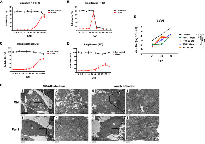
Ferroptosis inhibitors reduce CV-A6 replication. RD cells were challenged with CV-A6 in the presence of increasing concentrations of the ferroptosis inhibitor Fer-1 (A), TRO (B), ROSI (C), or PIO (D) for 36 h. Mock-infected cells were used as a control. Cell viability was measured using the CellTiter96 Aqueous One solution cell proliferation assay (Promega). Data are presented as means ± SD from three wells of a 96-well plate. (E) Ferroptosis inhibitors (Fer-1, TRO, ROSI, and PIO) reduced viral titers of CV-A6. RD cells were treated with Fer-1 (100 μM) (green line), TRO (20 μM) (red line), ROSI (80 μM) (blue line), or PIO (40 μM) (orange line) and infected with CV-A6 at an MOI of 0.01. Viruses were collected at 24, 36, and 48 h postinfection, and viral titers were measured using a plaque assay. Data are presented as means ± SD from three independent experiments: ****, P < 0.0001. Statistical significance was determined by two-way ANOVA. (F) RD were mock-infected or infected with CV-A6 at an MOI of 20 in the presence of Fer-1 (100 μM), and images were obtained by transmission electron microscopy at 7 h postinfection. Low-magnification images are shown in images 1, 3, 5, and 7. High-magnification images are shown in images 2, 4, 6, and 8. N, nucleus. Scale bar: 5 μm in images 1, 3, 5, and 7 and 1 μm in images 2, 4, 6, and 8.

