Abstract
Soft tissue calcifications are common findings in patients with various diseases, such as malignant tumors, collagen diseases, trauma, and chronic kidney disease. The majority of these lesions are not clinically significant; however, they can cause specific disorders within a limited space, such as the spinal canal. Here, we report the case of a patient undergoing fusion surgery for lumbar canal stenosis due to degenerative spondylolisthesis and multiple intraspinal canal calcifications associated with psoriatic arthritis (PsA). A 55-year-old female patient presented with pain in the left leg and intermittent claudication for 1 month. One year ago, she was diagnosed with PsA and received outpatient treatment, including biological medication, at the Division of Rheumatology, Department of Internal Medicine of our institution. She was referred to our department, and radiological examination revealed lumbar canal stenosis caused by spondylolisthesis and multiple calcifications in the lumbar spinal canal. We performed posterior lumbar interbody fusion (PLIF) with percutaneous pedicle screw fixation concomitant with removal of the calcifications. The postoperative course was uneventful, and her neurological symptoms improved. Although several prior case reports have noted intraspinal canal calcifications due to collagen disease or chronic kidney disease, calcifications associated with PsA are rare. We discuss the diagnosis of PsA and its relationship with intraspinal canal calcifications by reviewing the previous relevant literature.
Keywords: intraspinal canal calcifications, psoriatic arthritis, axial involvement, degenerative spondylolisthesis, lumbar canal stenosis
Introduction
Ectopic soft tissue calcifications related to metabolic disorders of calcium phosphate, chronic kidney disease, or other causes are incidentally detected on radiological examination in daily clinical practice,1) with most of them being asymptomatic. In contrast, symptomatic intraspinal canal calcifications are relatively uncommon, except for partial calcifications of herniated intervertebral discs, the ligamentum flavum, and tumors.
Psoriatic arthritis (PsA) is a rare autoimmune disorder that was first described by Moll et al. in 1973.2) PsA is reported to occur in up to 30% of patients with psoriasis,3) who exhibit psoriatic skin lesions accompanied by joint inflammation. Since PsA causes various symptoms similar to those of rheumatoid arthritis (RA), differentiation between these two entities is a diagnostic challenge. Physicians treating patients with autoimmune diseases or arthritis should properly evaluate joint manifestations with sufficient knowledge and keep in mind that the symptoms can be caused by PsA. Medical treatment for PsA includes administration of antitumor necrosis factor (TNF) or anti-interleukin (IL)-17 agents, cyclosporine, and methotrexate (MTX). Especially for patients with associated spinal lesions who have failed therapy with non-steroidal anti-inflammatory drugs (NSAIDs) and physiotherapy, infliximab, etanercept, or adalimumab are recommended.4)
Axial lesions, such as spondyloarthritis or sacroiliitis, are considered to be signs of PsA; however, little is known about the relationship between calcifications around the spinal canal and PsA. Literature reviews on symptomatic paraspinal or intraspinal canal calcifications have revealed a limited number of reported cases, which were primarily associated with synovial cysts and systemic sclerosis.5–7) We present a case of a patient presenting with low back pain and intermittent claudication. Her radiological study revealed lumbar spondylolisthesis and spinal canal stenosis associated with multiple intraspinal canal calcifications.
Case Report
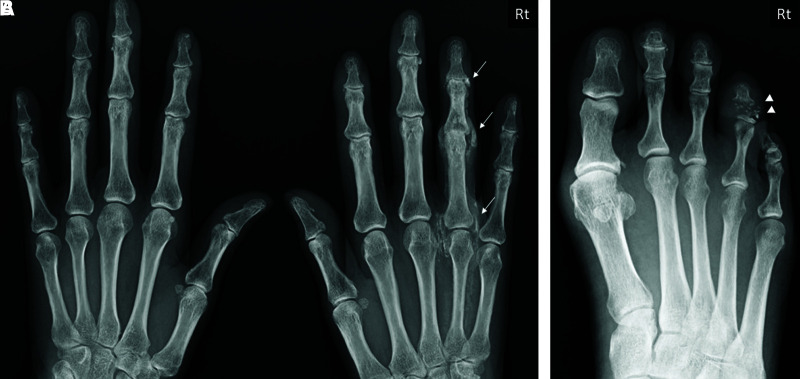
A 55-year-old woman presented with intermittent claudication, progressive muscle weakness of the lower extremities, and pain in the bilateral hips and dorsal aspects of the thighs. She had been suffering from low back pain, and her family doctor diagnosed her with lumbar spondylolisthesis by lumbar spine X-rays 2 years before. Additionally, she was diagnosed with PsA 10 months ago at the Division of Rheumatology, Department of Internal Medicine of our hospital, receiving outpatient drug therapy, including biological agents. The diagnosis was based on swelling of her right 4th finger and toe, asymmetric ossification and joint space narrowing of the PIP/DIP joints of the right 4th finger, osteolysis of the 4th toe on X-ray (Fig. 1A and 1B), and negative rheumatoid factor (RF). The laboratory data showed a matrix metalloproteinase-3 level of 456 ng/ml and CRP level of 0.82 mg/dl. The only past medical history of note was vocal cord polyps and asthma. For the treatment of PsA, she had been administered ixekizumab, an IL-17 inhibitor, 80 mg subcutaneously once every 2 weeks; prednisolone, 15 mg daily; and MTX 8 mg once a week.
Fig. 1. Hand X-ray (A) showing ossification and joint space narrowing of the joints of the right 4th finger (white arrow), and foot X-ray (B) showing osteolysis of the right 4th toe (white arrowhead).
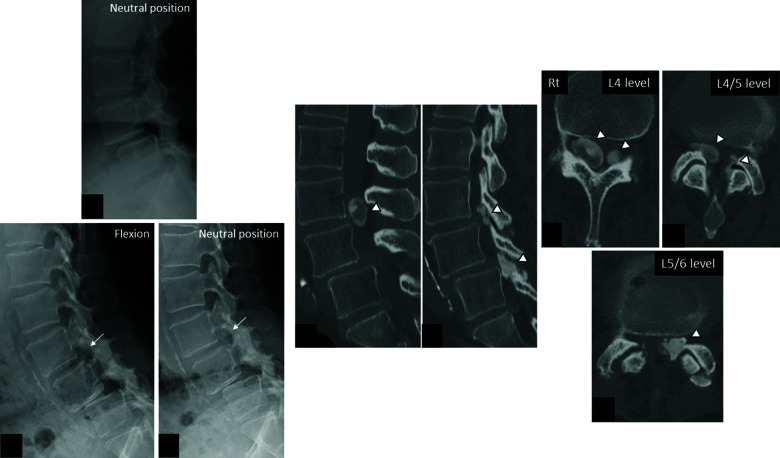
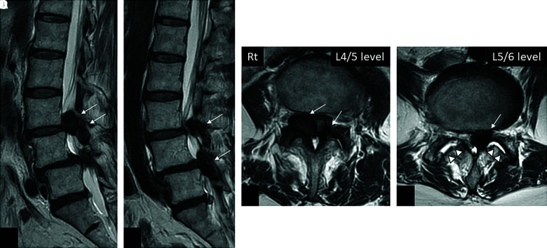
She was referred to our department for further examination and treatment of her lumbar lesions. On neurological examination, she had pain and numbness in the bilateral L5 and S1 nerve root territories, and the bilateral Achilles and patellar tendon reflexes were almost absent. Motor weakness of the bilateral lower extremities was also observed, especially on the left side, and the muscle strength of the quadriceps femoris, hamstrings, tibialis anterior, and gastrocnemius was approximately 3–4/5 in terms of the manual muscle test. She denied bowel or bladder incontinence. The clinical diagnosis was bilateral L4-S1 radiculopathy. Lumbar spine X-rays demonstrated de novo development of calcification in the L4/5 intervertebral foramen and worsened L5/6 spondylolisthesis compared to the X-ray images 2 years before (Fig. 2A–2C). The dynamic X-rays showed 2.1 mm translation of L5 over L6 in the flexed position (Fig. 2B and 2C). A preoperative lumbar computed tomography (CT) scan showed multiple calcifications in the spinal canal at the level of L4-6, and some calcified lesions extended into the intervertebral foramen or lateral recesses at the right L4/5 and left L5/6 levels (Fig. 2D–2H). Lumbar magnetic resonance imaging (MRI) demonstrated severe canal stenosis at the L4/5 and L5/6 levels caused by a combination of spondylolisthesis and multiple intraspinal canal calcifications (Fig. 3A–3D). Axial T2-weighted MRI revealed facet joint opening and high signal intensity within the bilateral facet joints at L5/6 (Fig. 3D). X-rays and MRI of the lumbar spine suggested lumbar segmental instability at L5/6.
Fig. 2. Lumbar spine X-ray lateral view 2 years prior to surgery (A) demonstrating grade 1 spondylolisthesis at L5/6. Preoperative lumbar spine dynamic X-rays (B, C) showing calcification in the L4/5 intervertebral foramen (white arrow), grade 2 spondylolisthesis/angulation at L5/6, and mild translation of L5/6 in flexion. Preoperative lumbar CT showing sagittal views (D, E) and axial views (F–H) demonstrating intraspinal canal calcifications at the levels of L4/5 and L5/6 (white arrowheads). CT: computed tomography.
Fig. 3. T2-weighted lumbar MRI sagittal views (A, B) and axial views (C, D) showing compression of the common dural sac by multiple intraspinal canal calcifications at the levels of L4/5 and L5/6 (white arrows). Axial T2-weighted MRI (D) demonstrating hyperintensity facet joint effusion at L5/6 (white arrowheads). MRI: magnetic resonance imaging.
Given the rapidly progressive intermittent claudication and muscle weakness of the lower extremities, we decided to perform lumbar surgery. Rt. L4/5 facetectomy was necessary to remove the calcification that extended into the Rt. L4/5 intervertebral foramen, and grade 2 spondylolisthesis with instability at L5/6 was observed on preoperative lumbar images. Therefore, we planned a two-level fusion surgery at L4-6. Considering the possible complications caused by surgery under ixekizumab use, it should be withheld for a certain period prior to surgery. We discussed with the attending physician specializing in rheumatic diseases that neither prednisolone nor MTX should be suspended perioperatively, and surgery was performed on the intermediate day of MTX administration.
She underwent posterior lumbar interbody fusion (PLIF) at the L4/5 and L5/6 levels 12 days after the last ixekizumab injection. The procedure included L4/L5 laminectomy, L4/5-L5/6 facetectomy, removal of the calcified intraspinal canal masses, interbody fusion using Stryker titanium cages, and pedicle screw fixation. All six pedicle screws were inserted percutaneously under navigation guidance. During surgery, direct gross observation revealed that the intraspinal canal lesions were partially calcified soft tissue masses originating from the facets or ligamentum flavum and were firmly attached to the dura mater, making it difficult to remove the lesion at the L4 level. Gross total removal of the calcified lesions at the other levels was performed, and we confirmed the decompression of the common dural sac and bilateral L5/S1 nerve roots. No intraoperative complications were noted. She was discharged from the intensive care unit the next day.
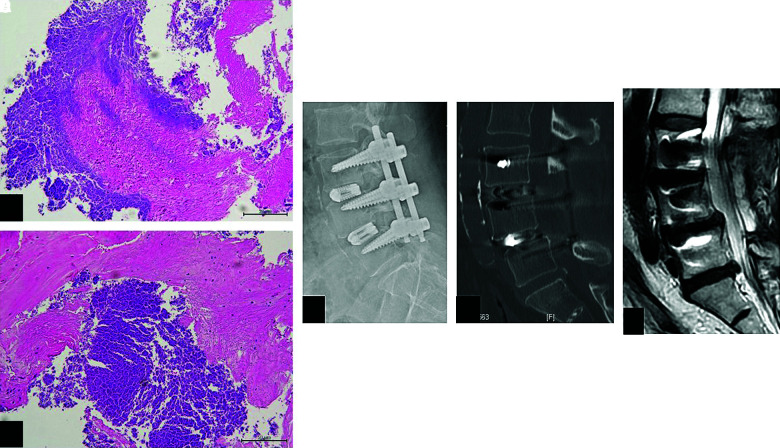
Histopathology of the specimens revealed highly degenerated ligamentous tissues with focal calcification, and no cystic lesions or neoplastic processes were observed (Fig. 4A and 4B). The background PsA and histologic findings of the intraspinal canal lesions were indicative of a diagnosis of axial involvement of PsA.
Fig. 4. (A, B) Histopathologic examination of an intraspinal canal lesion showing a degenerated ligament with focal calcification. Hematoxylin and eosin staining, original magnification: ×40 (A), ×100 (B), scale bar = 50 μm. Postoperative lumbar spine X-ray lateral view (C) and CT sagittal view (D) showing suitable positioning of the cages and pedicle screws. The alignment of the lumbar spine was corrected. Three-month postoperative lumbar MRI sagittal view (E) showing decompression of the dural sac. CT: computed tomography.
Postoperative radiographs showed appropriate positioning of the cages and no apparent loosening of the pedicle screws (Fig. 4C and 4D). The sagittal alignment of the lumbar spine was improved after surgery. She received physical therapy postoperatively and made an uneventful recovery, without any complications. Perioperative administration of prednisolone, MTX, and biological agents did not give rise to surgical site infection or protracted wound healing. Her lower extremity symptoms gradually improved, and she was discharged from the hospital with a walking cane 2 weeks after surgery. Lumbar MRI performed 3 months after surgery showed preservation of the dural sac decompression (Fig. 4E). The improvement in her symptoms was maintained at follow-up 10 months after surgery.
Discussion
Lumbar canal stenosis (LCS) resulting from intraspinal canal calcifications without ossification of the posterior longitudinal ligament or yellow ligament is rare. Intraspinal canal calcification is not uncommon in clinical situations, and the lesion by itself is not an indication for surgical removal unless it causes symptoms. In this case, she presented with typical symptoms of LCS, and the radiological examination showed unusual multiple lumbar calcifications coexisting with spondylolisthesis, which was consistent with the neurological findings.
Although PsA shares various symptoms with RA, patients with PsA usually show skin changes and are negative for RF in most cases. The common signs of PsA include asymmetric multiple arthritis, psoriasis skin rash, enthesitis, and dactylitis.3) Several studies found that entheseal abnormalities on ultrasound assessment were observed only among patients with PsA and not among those with RA.8,9) The cause of PsA is still unclear, and both genetic and environmental factors are considered to be involved in the pathogenesis of this disorder.10)
In the present case, the CASPER criteria, commonly used for a clinical diagnosis of PsA, scored 3 points, namely, negative RF, evidence of dactylitis, and radiologic findings of poorly marginated osteogenesis of the articular surface.11) Moreover, joint erosion and joint space narrowing of the finger on X-ray strongly supported the diagnosis. Enthesitis is also observed in ankylosing spondylitis; however, apparent sacral arthritis or bamboo spine was not observed radiologically, and human leukocyte antigen B27 was negative in this case.
Axial involvement is a significant spinal complication of PsA, which was first reported by Wright et al. in 1961.12) The defining characteristics of axial involvement have not been established, and it is reported to occur in 25–70% of patients with PsA.13) The clinical manifestation of axial involvement in spondylitis is inflammatory back pain, and the frequency of spinal symptoms in PsA is reported to be approximately 60%.14,15) Additionally, there are no guidelines for the pathologic diagnosis of the spinal lesions of PsA. In this case, pathologic evidence of inflammation was not found in the intraspinal canal lesions; however, the calcifications may be secondary to inflammation, and the origin of the calcification appeared to be the attachment of the ligamentum flavum to the lamina according to the intraoperative observations and pathological findings. Hence, we speculated that the spondylolisthesis was attributed to facet joint arthritis and the calcifications were caused by enthesitis of the ligamentum flavum.
Her past medical history included neither metabolic disorders nor chronic kidney disease; furthermore, her intraspinal canal calcified lesions were radiologically exceptional among commonly detected ectopic calcifications. Thus, it is presumed that the calcified intraspinal canal lesions in the present case represent axial disease related to PsA.
There are many different types of agents used to treat PsA. Among them, TNF inhibitors, IL-17 inhibitors, MTX, and NSAIDs are frequently administered for active PsA. Recently, Mease et al. reported that brodalumab, an IL-17 receptor subunit A inhibitor, is associated with significant improvements of both joint and skin symptoms in patients with PsA without any substantial adverse effects.16) Ixekizumab is a monoclonal antibody that inhibits IL-17A, which was administered preoperatively in the present case. Perioperative continuation of biological agents (e.g., TNF inhibitors or IL-17 inhibitors) can lead to postoperative wound complications, such as surgical site infection and wound dehiscence. According to the American College of Rheumatology/American Association of Hip and Knee Surgeons Guideline in 2017, it is recommended to stop biological agents prior to surgery and to plan for surgery at the end of the dosing cycle.17) However, the appropriate management of the discontinuation of these agents is not clarified. Currently, the perioperative withdrawal period of biological drugs or MTX is dependent on the physician’s preferences. In this case, she underwent PLIF with a 12-day preoperative withdrawal time of ixekizumab without discontinuation of prednisolone and had no postoperative wound-related complications.
To our knowledge, this is the first reported surgical case of LCS with spondylolisthesis and multiple intraspinal canal calcifications associated with PsA. Although the standard management for axial involvement of PsA is medication therapy, our patient was treated surgically due to rapidly worsening symptoms accompanied by worsening imaging findings. Accumulation of these rare cases and the establishment of clinical guidelines for the diagnosis of axial involvement of PsA are necessary.
Acknowledgments
We wish to thank Dr. T Ozaki, Director of Ozaki Orthopedic Clinic, for generously providing us with the lumbar X-ray images 2 years prior to surgery.
Footnotes
Conflicts of Interest Disclosure
None declared.
References
- 1). Locatelli F, Cannata-Andía JB, Drüeke TB, et al.: Management of disturbances of calcium and phosphate metabolism in chronic renal insufficiency, with emphasis on the control of hyperphosphataemia. Nephrol Dial Transplant 17: 723– 731, 2002 [DOI] [PubMed] [Google Scholar]
- 2). Moll JM, Wright V: Psoriatic arthritis. Semin Arthritis Rheum 3: 55– 78, 1973 [DOI] [PubMed] [Google Scholar]
- 3). Ritchlin CT, Colbert RA, Gladman DD: Psoriatic arthritis. N Engl J Med 376: 957– 970, 2017 [DOI] [PubMed] [Google Scholar]
- 4). Soriano ER: Treatment guidelines for psoriatic arthritis. Int J Clin Rheumatol 4: 329– 342, 2009 [Google Scholar]
- 5). Almefty R, Arnautovic´ KI, Webber BL: Multilevel bilateral calcified thoracic spinal synovial cysts. J Neurosurg Spine 8: 473– 477, 2008 [DOI] [PubMed] [Google Scholar]
- 6). Arginteanu MS, Perin NI: Paraspinal calcinosis associated with progressive systemic sclerosis. Case report. J Neurosurg 87: 761– 763, 1997 [DOI] [PubMed] [Google Scholar]
- 7). Liberato ACP, Amaral LLFD, Marussi VHR: Tumoral calcinosis in the lumbar spine secondary to systemic sclerosis: a rare cause of radiculopathy in an adult with advanced disease. BJR Case Rep 2: 20150435, 2016 [DOI] [PMC free article] [PubMed] [Google Scholar]
- 8). Fournié B, Margarit-Coll N, Champetier de Ribes TL, et al.: Extrasynovial ultrasound abnormalities in the psoriatic finger. Prospective comparative power-doppler study versus rheumatoid arthritis. Joint Bone Spine 73: 527– 531, 2006 [DOI] [PubMed] [Google Scholar]
- 9). Zabotti A, Salvin S, Quartuccio L, De Vita S: Differentiation between early rheumatoid and early psoriatic arthritis by the ultrasonographic study of the synovio-entheseal complex of the small joints of the hands. Clin Exp Rheumatol 34: 459– 465, 2016 [PubMed] [Google Scholar]
- 10). Talotta R, Atzeni F, Sarzi-Puttini P, Masala IF: Psoriatic arthritis: From pathogenesis to pharmacologic management. Pharmacol Res 148: 104394, 2019 [DOI] [PubMed] [Google Scholar]
- 11). Taylor W, Gladman D, Helliwell P, et al. : Classification criteria for psoriatic arthritis: development of new criteria from a large international study. Arthritis Rheum 54: 2665– 2673, 2006 [DOI] [PubMed] [Google Scholar]
- 12). Wright V, Psoriatic arthritis. A comparative radiographic study of rheumatoid arthritis and arthritis associated with psoriasis. Ann Rheum Dis 20: 123– 132, 1961 [DOI] [PMC free article] [PubMed] [Google Scholar]
- 13). Thumboo J, Tham SN, Tay YK, et al.: Patterns of psoriatic arthritis in orientals. J Rheumatol 24: 1949– 1953, 1997 [PubMed] [Google Scholar]
- 14). Sadek HA, Abdel-Nasser AM, El-Amawy TA, Hassan SZ: Rheumatic manifestations of psoriasis. Clin Rheumatol 26: 488– 498, 2007 [DOI] [PubMed] [Google Scholar]
- 15). Gottlieb AB, Merola JF: Axial psoriatic arthritis: An update for dermatologists. J Am Acad Dermatol 84: 92– 101, 2021 [DOI] [PubMed] [Google Scholar]
- 16). Mease PJ, Helliwell PS, Hjuler KF, Raymond K, McInnes I: Brodalumab in psoriatic arthritis: results from the randomised phase III AMVISION-1 and AMVISION-2 trials. Ann Rheum Dis 80: 185– 193, 2021 [DOI] [PMC free article] [PubMed] [Google Scholar]
- 17). Goodman SM, Springer B, Guyatt G, et al.: 2017 American College of Rheumatology/American Association of Hip and Knee Surgeons Guideline for the perioperative management of antirheumatic medication in patients with rheumatic diseases undergoing elective total hip or total knee arthroplasty. Arthritis Rheumatol 69: 1538– 1551, 2017 [DOI] [PubMed] [Google Scholar]


