Primary Sjogren's syndrome (pSS), a systemic autoimmune disorder, mainly involves the epithelium of exocrine glands that leads to organ swelling, inflammation, and dysfunction. The most common clinical manifestations are dryness of eye and mouth due to the involvement of the salivary gland and lacrimal gland, and nearly one-quarter of patients would have salivary gland enlargement; just like the patient with painless swelling of bilateral lacrimal glands, parotid glands, and submandibular glands described by Johann von Mikulicz-Radecki in 1888; or the three patients with dry mouth, dry eyes, and salivary gland atrophy described by Henri Gougerot in 1925. Because salivary glands are the organ been most violated, its pathological and morphological changes could be used as the diagnostic factors or follow-up indicators of pSS. However, tissue proven is an invasive procedure, which is difficult to be performed repeatedly in clinical practice; the morphology changes can be obtained by noninvasive imaging examinations, which are harmless and easy to be accepted by patients, such as ultrasound, sialography, computed tomography, and magnetic resonance imaging. Recently, given the evolution of ultrasound and its advantages of nonradiation, low price, and easy to use, ultrasonography represents the most frequently used to evaluate the morphological changes of salivary glands, even only tiny pathological changes. The standard salivary gland ultrasonography (SGUS) of parotid glands can be assessed in transverse plane and longitudinal plane with the landmark of the mandibular ramus; submandibular glands can be assessed in the longitudinal plane and transverse planes at the posterior part of submandibular triangle.
There are available SGUS semiquantitative scoring systems[1] for the morphological change of pSS. Among them, the most common one been used is the scoring system developed by De Vita et al.[2] in 1992. The scoring system divides the severity of parenchymal inhomogeneity of a single salivary gland into 0 points (normal) to 3 points (most severe) with a total score of 0–12 points. In 2008, Salaffi et al.[3] modified the De Vita scoring system and divided the abnormalities of each salivary gland (parenchymal homogeneity, echogenicity, gland size, and posterior glandular boundary) into 0 points (normal) to 4 points (most severe), with a total score of 0–16 points, using the cutoff value of 6, to be assessed in 77 pSS versus 79 non-pSS patients. The sensitivity for detecting pSS is 75.3% and the specificity is 83.5%. The reproducibility of the SGUS is good. Furthermore, in the same study,[3] two radiologists who participated in this study had a high interobserver agreement (kappa 0.71–0.83) in the measurement of the presence/absence of homogeneity, echogenicity, gland size, and posterior glandular border; and the intraobserver agreement (kappa 0.80–0.85) is also good. However, it is quite common that the agreement of the parotid gland SGUS is often much better than that of the submandibular gland SGUS.
Because of the different scoring systems been used in various literature, it is difficult to compare with each other and makes it difficult to review and integrate the data. Therefore, the subtask force of the Outcome Measures in Rheumatology Clinical Trials (OMERACT) working group under the European Alliance of Associations for Rheumatology was created in June 2016, to validate the use of SGUS as a possible outcome measurement instrument. After three Delphi rounds, 25 experts from 14 countries established a consensus on SGUS definitions [Table 1],[4] which was published in 2019. This is also the first time that fatty echo structures (Graded 1) and fibrous echo structures (Graded 3) have been included in the scoring system. A total of 18 experts read all of 199 SGUS gray scale video clips (from pSS to non-pSS patients) for reliability evaluation, and there is an excellent intrareader reliability (Light's kappa 0.81), and good interreader reliability (Light's kappa 0.66). However, the reliability of the submandibular gland SGUS is still lower than parotid gland SGUS, which is the same as the previous scoring method. In 2020, by Fana et al.,[5] a cross-sectional, observational study of all patients referred to the Center for Rheumatology and Spine Diseases, Rigs Hospitalet, Copenhagen, Denmark, who were suspected of having pSS, using the OMERACT SGUS scoring system to analyze totally 134 patients. The SGUS was done by three rheumatologists with more than 10 years of experience in musculoskeletal ultrasound and more than 5 years of experience in scanning salivary glands. If we defined cutoff as >1 gland has score >2, the sensitivity of detecting pSS is 72% and the specificity is 91%. Among them, there are 43 patients with pSS, who have the highest Score 0 in any salivary gland were two patients, highest Score 1 were 10 patients, highest Score 2 were 15 patients, and highest Score 3 were 16 patients. So far, the sensitivity and specificity of this SGUS scoring system for pSS seem to be good, but we need more studies to verify it.
Table 1.
Outcome measures in rheumatology clinical trials four-grade semiquantitative scoring system for parotid glands and submandibular glands in primary Sjogren’s syndrome[4]
| Grade | Definitions |
|---|---|
| Grade 0 | |
| Normal parenchyma | Normal parenchyma |
| Grade 1 | |
| Minimal change | Mild inhomogeneity without anechoic/hypoechoic areas and/or fatty change |
| Grade 2 | |
| Moderate change | Moderate inhomogeneity with focal anechoic/hypoechoic areas |
| Grade 3 | |
| Severe change | Diffuse inhomogeneity with anechoic-hypoechoic area occupying the entire gland surface and/or fibrous change |
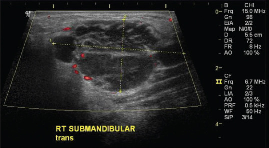
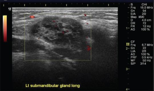
Based on the personal experience of Dr. Po-Hao Huang, the use of SGUS in daily clinical practice is very often. Dr. Huang has ever reviewed the patients who have salivary gland diseases during the period between 2014 and 2015 in outpatient clinics. There were 27 patients with pSS and other nine patients with other diseases (sarcoidosis, IgG4-related disease, Rosai-Dorfman disease, and lymphoma). After rescoring of these patients using the 2019 OMERACT SGUS scoring system, the most patients with pSS whose scores fall between 1 (48%) and 2 (40%) [Figure 1], and only <10% of patients with the Score 0, consistent with the findings of other research, but it should be noticed that 56% of nine patients with disease other than pSS have scored above 2 [Figure 2]. Therefore, we should remember that many diseases could mimic the features of pSS on SGUS image, especially lymphoma, which also tends to occur in patients with pSS.
Figure 1.

Ultrasound image of submandibular gland in primary Sjogren's syndrome
Figure 2.

Ultrasound image of submandibular gland in sarcoidosis
Financial support and sponsorship
Nil.
Conflicts of interest
Prof. Der-Yuan Chen, an editorial board member at Journal of Medical Ultrasound, had no role in the peer review process of or decision to publish this article. Dr. Po-Hao Huang declared no conflict of interest in writing this paper.
REFERENCES
- 1.Delli K, Dijkstra PU, Stel AJ, Bootsma H, Vissink A, Spijkervet FK. Diagnostic properties of ultrasound of major salivary glands in Sjögren's syndrome: A meta-analysis. Oral Dis. 2015;21:792–800. doi: 10.1111/odi.12349. [DOI] [PubMed] [Google Scholar]
- 2.De Vita S, Lorenzon G, Rossi G, Sabella M, Fossaluzza V. Salivary gland echography in primary and secondary Sjögren's syndrome. Clin Exp Rheumatol. 1992;10:351–6. [PubMed] [Google Scholar]
- 3.Salaffi F, Carotti M, Iagnocco A, Luccioli F, Ramonda R, Sabatini E, et al. Ultrasonography of salivary glands in primary Sjögren's syndrome: A comparison with contrast sialography and scintigraphy. Rheumatology (Oxford) 2008;47:1244–9. doi: 10.1093/rheumatology/ken222. [DOI] [PubMed] [Google Scholar]
- 4.Jousse-Joulin S, D’Agostino MA, Nicolas C, Naredo E4, Ohrndorf S, Backhaus M, et al. Video clip assessment of a salivary gland ultrasound scoring system in Sjögren's syndrome using consensual definitions: An OMERACT ultrasound working group reliability exercise. Ann Rheum Dis. 2019;78:967–973. doi: 10.1136/annrheumdis-2019-215024. [DOI] [PubMed] [Google Scholar]
- 5.Fana V, Dohn UM, Krabbe S, Terslev L. Application of the OMERACT Grey-scale Ultrasound Scoring System for salivary glands in a single-centre cohort of patients with suspected Sjögren's syndrome. RMD Open. 2021 Apr;7(2):e001516. doi: 10.1136/rmdopen-2020-001516. [DOI] [PMC free article] [PubMed] [Google Scholar]


